386
Corresponding author: Prof. Geraldo Bezerra da Silva Junior e-mail: [email protected]
Received 9 September 2015 Accepted 19 November 2015
Chronic kidney disease related to renal tuberculosis:
a case report
Geraldo Bezerra da Silva Junior
[1],[2], Luiz David Salles Brito
[1],
Samia Thabida de Oliveira Rabelo
[1]and Zenar Maria Ribeiro Mendes de Saboia
[2][1]. Curso de Medicina, Centro de Ciências da Saúde, Universidade de Fortaleza, Fortaleza, Ceará, Brasil. [2]. Programa de Pós-Graduação em Saúde Coletiva, Universidade de Fortaleza, Fortaleza, Ceará, Brasil.
Abstract
Genitourinary tuberculosis (TB) is the third most common form of extrapulmonary TB. A 34-year-old man with severe kidney function loss secondary to renal TB initially presented with urinary symptoms, including dysuria and polacyuria. The diagnosis was based on clinical history and laboratory tests; the urinalysis revealed acid-fast bacilli. The patient’s condition stabilized
after beginning TB-speciic treatment, but the right kidney function loss persisted. Renal TB can lead to irreversible loss of renal
function. As such, renal function should be considered in all patients from TB-endemic areas who present with urinary symptoms and whose urine cultures are negative for common pathogens.
Keywords: Renal tuberculosis. Kidney disease. Chronic kidney disease. Rev Soc Bras Med Trop 49(3):386-388, May-June, 2016
doi: 10.1590/0037-8682-0310-2015
Case Report
INTRODUCTION
Tuberculosis (TB) is a stigmatizing and highly contagious disease that is transmitted by Mycobacterium tuberculosis
through respiratory droplets.It is more prevalent in developing countries, where socioeconomic issues and ineffective public health systems are unable to control the spread of the disease despite the availability of effective treatments(1) (2). The
acquired immunodeiciency syndrome (AIDS) epidemic and
drug resistance contributes to the disease burden(2). TB affects millions of people worldwide. The disease is endemic in Latin America, where incidence rates vary from 25 to 149 cases per 100,000 inhabitants(2). Brazil is one of the 20 countries with the highest number of cases(1).
Tuberculosis includes a variable clinical spectrum and may affect different organs. Extrapulmonary involvement occurs in 10 to 42% of cases(2), and the most common extrapulmonary forms are pleural, lymph node, and renal TB, in this order(1) (3) (4). The most frequent presentation of renal TB is sterile pyuria, which is frequently associated with hematuria and may present with or without urinary symptoms such as dysuria and polacyuria(1). Evidence suggests that granulomatous tubulointerstitial nephritis is the most frequent histopathologic manifestation of renal TB(5).
After obtaining the patient’s consent, we describe a case of renal TB that initially presented with urinary symptoms and that was detected via laboratory examinations and imaging
tests. This case is notable because it resulted in signiicant and
permanent renal function loss.
CASE REPORT
A 34-year-old man sought medical attention due to a 6-month history of dysuria, macroscopic hematuria, polacyuria, and supra-pubic pain associated with sporadic vespertine fever, sometimes with chills. He had also experienced weight loss, but he attributed this to intentional dietary restriction because he was obese. His symptoms continued after receiving different antimicrobial treatments (including cephalexin and
ciproloxacin), despite persistent negative urine cultures. He
reported a 10-year history of alcohol abuse, and he denied smoking or coming into contact with people with tuberculosis.
At the time of physical examination, he was in good general health with normal vital signs and no edema. Cardio-pulmonary auscultation was normal and no abnormalities were found upon abdominal examination. Laboratory tests showed the following results: hemoglobin, 14mg/dL; hematocrit, 41%; white blood count, 5,100/mm³; platelets, 233,000/mm³; creatinine, 1.0mg/
dL [estimated glomerular filtration rate (GFR) = 98mL/
min/1.73m2]; urea, 39mg/dL; glycemia, 76mg/dL; uric acid, 5mg/dL; the patient tested negative for hepatitis B, hepatitis C,
human immunodeiciency virus (HIV) infection, and syphilis.
Urinalysis showed the following results: pH 5.0, leukocyturia
387
Silva Junior GB et al. - Renal tuberculosis: a case report
TABLE 1 - Laboratory tests from a patient with renal tuberculosis and kidney function loss.
Presentation (before Treatment initiated Treatment end
Tests treatment) 1 month after 2 months after 2 months after 6 months after
Urinalysis pH = 5.0 pH = 6.0 pH = 6.0 - pH = 5.0
Leukocyturia Leukocyturia Leukocyturia Hematuria (1+)
(14/hpf) (15/hpf) (30/hpf) Hematuria (10/hpf) Hematuria (15/hpf) Hematuria (4+)
Protein (traces)
Acid-fast bacilli in urine Positive - - Negative Negative
Urea (mg/dL) 39 39 50 50 42
Creatinine (mg/dL) 1.0 1.0 1.6 1.3 1.6
GFR (mL/min/1.73m2)* 98 98 55 71 55
Hematocrit (%) 41 41 - -
-Hemoglobin (g/dL) 14 14 - -
-pH: potencial hidrogênico; hpf: high power ield. GFR: glomerular iltration rate; CKD-EPI: chronic kidney disease epidemiology collaboration. *Estimated
through CKD-EPI formula
FIGURE 1 - Abdominal computed tomography image showing pyelocaliceal dilation and thinning of the cortical region in the right kidney due to renal tuberculosis.
traces of protein. A routine urine culture did not yield any bacteria. Abdominal ultrasound revealed moderate dilation of the right pyelocaliceal system. Laboratory test results are presented in Table 1.
Due to suspected renal TB, we performed a tuberculin skin
test (which was negative) and screened for acid-fast bacilli in the urine (which was positive in 5 of the 10 samples), and positive culture for M. tuberculosis. Sputum examination
yielded negative indings. Abdominal computed tomography
demonstrated right pyelocaliceal dilation but no evidence of obstructive disease (Figure 1). A renal scintigraphy showed a severe reduction in right kidney function with accentuated decrease in radioisotope caption, resulting in only 16% of renal function. The dynamic test indicated normal glomerular function
in the left kidney and conirmed severe loss of function in the
right kidney (Figure 2).
Based on these results, the patient began a 6-month course of rifampin, isoniazid, pyrazinamide, and ethambutol according to World Health Organization current guidelines. The patient stabilized, but continued to experience dysuria for two months
after treatment. Renal function continued to decrease after
treatment, and 10 months after treatment ended, the creatinine
level was 1.6mg/dL (GFR = 55mL/min/1.73m2), likely due to
continued right kidney decline.
DISCUSSION
This is an unusual case of renal TB that resulted in nearly complete loss of right kidney function. There were no other
detected risk factors for chronic kidney disease (CKD) in this patient, suggesting that the patient developed CKD as a direct
result of TB. In a study of 25 patients with renal TB, 9 developed
CKD and required renal replacement therapy in the irst 6 months
after the symptoms began. The remaining 16 patients had variable degrees of kidney function loss in the following 36 months(5).
Renal TB is a commonly overlooked genitourinary disease,
but it must be considered in patients from TB-endemic regions who present with urinary symptoms and who do not respond to
typical antibiotic treatment. Renal TB is the most likely diagnosis
in patients who present with pyuria and hematuria and who have negative urine cultures(1) (3) (6). Renal TB occurs secondary to a primary pulmonary infection after the bacilli reaches other organs, typically through hematologic dissemination, although in many cases this primary infection can be asymptomatic or
388
Rev Soc Bras Med Trop 49(3):386-388, May-June, 2016
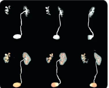
FIGURE 2 - Renal scintigraph showing the right kidney with reduced dimensions, with heterogeneous distribution of radioisotope caption and no evidence of excretion (obstructive pattern).
In the present case, the patient reported chronic dysuria with hematuria and supra-pubic pain. These symptoms call attention to an unusual cause of urinary infection. The other symptoms (vespertine fever and chills) suggested TB. The usual course of TB is insidious, particularly for renal TB. The time between primary infection and initial symptoms can be long. Some patients are asymptomatic, and sterile pyuria, with or without hematuria, can be the only sign that is found during routine tests. These urinary abnormalities are found in more than 90% of renal TB cases(1).
The gold standard for diagnosing renal TB is based on urine culture(1). As bacilli are not shed continuously in urine, at least three urine samples from different occasions should be collected, preferentially in different days. The urine should be cultivated in the Lowenstein-Jensen medium in order to isolate M. tuberculosis.
The urine is classically negative in routine culture media, but sometimes there can be an associated infection from common pathogens. Urine pH is typically acidic in patients with renal TB, which is an important parameter to keep in mind when performing differential diagnoses(1). In the present case, urine pH was acidic, and there were the classic signs of renal TB, including leukocyturia, hematuria, and a negative urine culture for usual
urinary tract pathogens. The diagnosis of TB was conirmed with
the presence of acid-fast bacilli in 5 different urine samples that resulted in positive cultures for M. tuberculosis.
Radiologic diagnosis of renal TB depends on the infection
stage(3) (4). Tuberculous granulomas develop in the renal pyramids, which can cause ulcer formation, leading to shedding of Mycobacterium bacilli in the urine as well as the formation of purulent secretions. As non-treated lesions increase, abscesses may form. In the late stages of infection, caseous material associated with
calciications may develop and lead to renal failure. The collecting
system is the most common site for genitourinary TB(1) (3) (4). In the present case, ultrasound and computed tomography revealed pyelocaliceal dilation in the right kidney without
evidence of obstructive factors. Renal scintigraphy indicated
severe function loss in this kidney, and the patient experienced worsening kidney function even after TB treatment ended. There have been no previous reports of renal TB leading to end-stage kidney disease(6), which may also lead to death(7). Notably, this patient experienced chronic kidney disease due to severe loss of function only in the right kidney, which highlights the unilateral TB involvement in this kidney.
Tuberculosis treatment consists of combined pharma-cotherapy with rifampin, isoniazid, pyrazinamide, and ethambutol, which is the suggested protocol for both pulmonary and extrapulmonary TB(2). These agents effectively eradicate infection in most cases, but it can cause nephrotoxicity.
Rifampin is a known nephrotoxic drug that can cause signiicant
kidney damage, including interstitial nephritis and acute kidney injury(8), but these effects are rare. In the present case, the patient received treatment for 6 months, and reported continued dysuria
that persisted for 2 months after treatment. Renal function was
stable owing to regular left kidney function. We believe our patient did not experience drug toxicity, although he did not achieve complete renal function recovery after TB treatment.
Damage related to rifampin toxicity could have been ruled out
with a renal biopsy, but this was not preformed because the patient remained stable after treatment and exhibited no signs of drug toxicity.
This case highlights the need for physicians to understand
how to diagnose TB and, if necessary, institute TB-speciic treatments. Renal TB must be considered in patients from
TB-endemic areas who present with urinary symptoms, such as leukocyturia and hematuria, who have negative urine cultures, and in whom treatment for common urinary infections have failed.
Conlict of interest
The authors declare that there is no conlict of interest.
REFERENCES
1. Daher EF, Silva Jr GB, Guardão Barros EJ. Renal tuberculosis in
the modern era. Am J Trop Med Hyg 2013; 88: 54-64.
2. Zumla A, Raviglione M, Hafner R, von Reyn CF. Tuberculosis.
N Engl J Med 2013; 368:745-755.
3. Eastwood JB, Corbishley CM, Grange JM. Tuberculosis and the kidney. J Am Soc Nephrol 2001; 12:1307-1314.
4. Gibson MS, Puckett ML, Shelly ME. Renal tuberculosis.
Radiographics 2004; 24:251-256.
5. Chapagain A, Dobbie H, Sheaff M, Yaqoob MM. Presentation,
diagnosis, and treatment outcome of tuberculous-mediated
tubulointerstitial nephritis. Kidney Int 2011; 79:671-677.
6. Lima NA, Vasconcelos CC, Filgueira PH, Kretzmann M, Sindeaux
TA, Feitosa Neto B, et al. Review of genitourinary tuberculosis with focus on end-stage renal disease. Rev Inst Med Trop
Sao Paulo 2012; 54:57-60.
7. Daher EF, Silva Jr GB, Damasceno RT, Santos GM, Corsino GA, Silva
SL, et al. End-stage renal disease due to delayed diagnosis of renal
tuberculosis: a fatal case report. Braz J Infect Dis 2007; 11:169-171. 8. Min HK, Kim EO, Lee SJ, Chang YK, Suh KS, Yang CW, et al.
Rifampin-associated tubulointerstitial nephritis and Fanconi syndrome