268
Rev. Col. Bras. Cir. 2009; 36(3): 268-270
L u n a L u n a L u n a L u n a L u n a Transoral stapled diverticulotomyTechnical NoteTechnical NoteTechnical NoteTechnical NoteTechnical Note
Transoral stapled diverticulotomy
Transoral stapled diverticulotomy
Transoral stapled diverticulotomy
Transoral stapled diverticulotomy
Transoral stapled diverticulotomy
Diverticulotomia transoral grampeada
Diverticulotomia transoral grampeada
Diverticulotomia transoral grampeada
Diverticulotomia transoral grampeada
Diverticulotomia transoral grampeada
RENATO ABRANTES LUNA, TCBC-RJ1; JEAN-MARIE COLLARD2
From the Hospital Universitário Gafrèe e Guinle - HUGG - UNIRIO - RJ - BR.
1. Surgery Unit B – Hospital Universitário Gafrée e Guinle – UNIRIO – HUGG – UNIRIO – Rio de Janeiro – RJ – BR; 2. Upper G-I Surgery Unit, Cliniques Universitaires Saint-Luc, Université catholique de Louvain, Bruxelas, Bélgica.
INTRODUCTION
INTRODUCTION
INTRODUCTION
INTRODUCTION
INTRODUCTION
S
ince its description in 1796 by Ludlow1, and the first report of successful surgical treatment by Wheeler in 1886, many treatment options have been described for the treatment of Zenker’s diverticulum. Schimid, in 1912, described the diverticulopexy with lower rates of morbidity and mortality than resection. Mosher in 1917 introduced the treatment by rigid endoscopy, sectioning the septum of the diverticulum. In 1958, Harrison added a myotomy as part of the treatment of Zenker’s diverticulum. In 1960 Dohlman incorporated to the technique of rigid endoscopy2, a speculum with esophageal cleft in the distal end. Collard in 1993 published a technical modification of this procedure3, which uses a speculum with independent shafts, using a stapler for section of the septum. In Brazil, the flexible endoscopic technique was presented by Prof. Sakai, member of Prof. Ishioka´s team, in 1982.Their experience was internationally published in 19954 . In 2004 Evrard et al.5, published a technical amendment with the use of an overtube, bilabiate at its end, facilitating the division of the diverticular septum. Since then, fewer cases reach the general surgeon for treatment.This note aims to provide the minimally invasive procedure, developed by Collard3, as an alternative for the treatment of Zenker’s diverticulum, bringing the disease back to the action of the general surgeon. This procedure performed since the 90s abroad, yet failed to achieve appropriate dissemination among us. In Pubmed and Bireme database, no reference was found regarding this technique in portuguese. In 2006, the Brazilian Society of Digestive Endoscopy published in its textbook, a chapter of authorship of Dr. Colaiacovo, with a series of 18 cases with this technique6. After the first case in HUGG-UNIRIO, this note was sent for publication, seeking greater disclosure of the technique in our environment.
Surgical technique Surgical technique Surgical technique Surgical technique Surgical technique
The patient is on supine position, with the neck fully extended (Figure 1). The surgeon stays at the patient´s head with video laparoscope monitor on the patient´s right, facing the surgeon, at a comfortable distance.
With the patient under general anesthesia, a diverticuloscope is inserted in the closed position and advanced to the esophagus under direct vision or, better, with the aid of a video camera with 5 mm scope. The tracheal intubation can be oral or nasal.
The diverticuloscope is then slowly withdrawn until the visualization of the septum between the esophagus and diverticulum, and then carefully opened and advanced, so that one blade is positioned inside the diverticulum and the other inside the esophagus. Care must be taken, because in larger diverticula, the diverticuloscope preferably enters in the diverticular lumen, leading to the risk of perforation. In small diverticulum, the diverticuloscope can obliterate the lumen , hindering it.
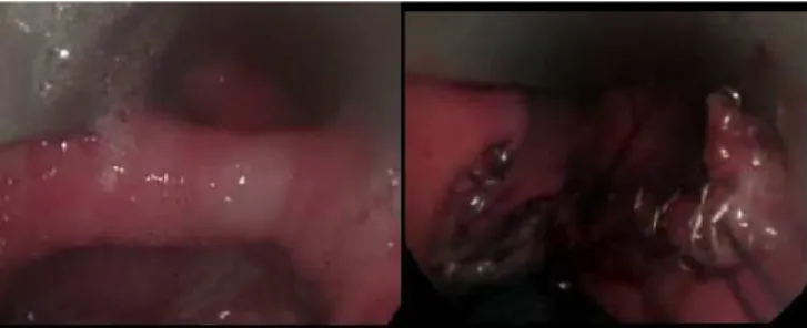
Once the septum is exposed between the blades (Figure 2), the diverticuloscope is then stabilized at the patient’s chest wall, allowing the surgeon to work with his hands free (Figure 1). An endoscopic linear stapler is then inserted through mouth and closed with the septum between
Figura 1 - Patient positioned with the diverticuloscope inserted.
Figure 2 - a) diverticulum-esophageal septum. b) aspect of the septum after stapple.
L u n a L u n aL u n a L u n aL u n a
Transoral stapled diverticulotomy 269
Rev. Col. Bras. Cir. 2009; 36(3): 268-270
the jaws. The firing is followed by the section of the septum and by anastomosis between the diverticulum and the esophagus. There is then a V-shaped cleft between the esophagus and the diverticulum (Figure 2). As many cartridges as necessary are used, to achieve complete section of the septum. Especial attention is necessary to avoid leaving a significant residual septum, especially because the cutting line ends before the stapling line. This is especially relevant in smaller diverticula. Some authors advice to saw the tip of the anvil, to reduce the length of the residual spur (Figure 3). Another key aspect is that small diverticula have smaller septum, which does not allow a complete section of the cricopharyngeal muscle and the proximal muscles of the esophagus, limiting the use of this technique in these patients. Nowadays, this technique is mostly reserved for diverticula with three or more centimeters7-9.
The hemostasis is revised and the procedure is finished.
A contrast swallow is performed in the next morning (Figure 4), and if no leakage is observed, a full liquid diet is initiated and the patient is discharged. This diet is maintained for seven days and then progressed to normal diet.
The main advantages of this procedure are: shorter operative time, less surgical trauma, lower morbidity rate (less injury of recurrent laryngeal, fistula, esophageal perforation), reduced length of hospitalization. On the other hand, there are more tooth injuries than open approach7-9. It is especially useful in patients who had previous cervicotomy. Although there are no randomized trials comparing the various techniques, the stapled diverticulotomy is potentially safer than endoscopic techniques, which offers only the division of the septum, without any sutures or fixation between the diverticulum and cervical esophagus.
It is important to note that the results of the technique are related to the proper selection of patients, with special attention to the size of the diverticulum, mouth opening and neck mobility.
Figure 3 - Modified anvil of cartridge.
Figure 4 - Pre and post operative esophagogram.
REFERENCES
REFERENCES
REFERENCES
REFERENCES
REFERENCES
1. Ludlow A. A case of obstructed deglutition from a preter natutal bag formed in the pharynx. In: Johnson W, Caldwell T, editors. Medical observations and inquiries. 2nd ed. London: Society of
Physicians; 1769. p. 85-101.
2. Dohlman G, Mattsson O. The endoscopic operation for hypopharyngeal diverticula: a roentgencinematographic study. AMA Arch Otoralyngol. 1960; 71:744-52.
3. Collard JM, Otte JB, Kenstens PJ. Endoscopic stapling technique of esophagodiverticulostomy for Zenker´s diverticulum. Ann Thorac Surg. 1993; 56(3):573-6.
4. Ishioka S, Sakai P, Maluf Filho F, Melo JM. Endoscopic incision of Zenker´s diverticula. Endoscopy. 1995; 27(6):433-7.
5. Evrard S, Le Moine O, Hassid S, Devière J. Zenker´s diverticulum: a new endoscopic treatment with a soft diverticuloscope. Gastrointest Endosc. 2003; 58(1):116-20.
6. Colaiacovo W. Divertículos faringoesofagicos de Zenker – Stapler endoluminal. In: Endoscopia gastrointestinal terapêutica. 1ª ed. SOBED; 2006. p. 324-6.
7. Gutshow CA, Hamoir M, Rombaux P, Otte JB, Goncette L, Collard JM. Management of pharyngoesophageal (Zenker´s) diverticulum: which technique? Ann Thorac Surg. 2002; 74(5):1677-82; discussion 1682-3.
8. Lerut TE, Luketich JD, Bizekis C. Esophageal diverticula. In: Patterson GA, Pearson FG, Cooper JD, Deslauriers J, Rice TW, Luketich JD et al, editors. Pearson´s thoracic and eophageal surgery. 3rd ed. Philadelphia: Churchill Livingstone; 2008. p.
702-13.
9. Bonavina L, Bona D, Abraham M, Saino G, Abate E. Long term results of endosurgical and open surgical approach for Zenker diverticulum. World J Gastroenterol. 2007; 13(18):2586-9.
270
Rev. Col. Bras. Cir. 2009; 36(3): 268-270
L u n a L u n a L u n a L u n a L u n a Transoral stapled diverticulotomy
Received in 15/11/2008
Accepted for publication in 17/01/2009 Conflict of interest: None
Financial source: None
How to cite: How to cite:How to cite: How to cite: How to cite:
Renato Abrantes Luna RA, Collard JM. TRANSORAL STAPLED DIVERTICULOTOMY. Rev Col Bras Cir. [periódico na Internet] 2009; 36(3). Disponível em URL: http://www.scielo.br/rcbc
Correspondence address: Correspondence address: Correspondence address: Correspondence address: Correspondence address: Renato Abrantes Luna