Morphological alterations of upper gastrointestinal tract
in patients with new onset-dermatomyositis: correlation
with demographic, clinical and laboratory features
Thammi de Matos Amorim,I Carlos Kiyoshi Furuya Junior,II Sergio Barbosa Marques,II Samuel Katsuyuki ShinjoI
DOI: 10.5935/MedicalExpress.2017.02.04
I Division of Rheumatology, Hospital das Clinicas HCFMUSP, Faculdade de Medicina, Universidade de Sao Paulo, Sao Paulo;
II Unit of Endoscopy, Division of Gastroenterology, Hospital das Clinicas HCFMUSP, Faculdade de Medicina, Universidade de Sao Paulo, Sao Paulo, Brazil (BR)
OBJECTIVE: To endoscopically assess the upper digestive tract of adult patients with newly diagnosed
dermatomyositis; to correlate possible changes in the gastrointestinal tract with demographic, clinical and laboratory features in this population.
METHOD: A cross-sectional study evaluating 65 newly diagnosed dermatomyositis cases from 2004 to 2015 was carried out. We excluded patients with clinically amyopathic dermatomyositis, overlap dermatomyositis, polymyositis, liver diseases, prior gastric surgery, upper gastrointestinal tract symptoms (except for upper dysphagia), systemic infections, alcohol consumption and smoking.
RESULTS: Mean age of patients was 44.9 years, with disease duration of four months. Endoscopic indings were observed in 70.8% of patients. (1) Esophageal disease/gastric distress was documented in 18.5% of patients: erosive distal esophagitis (16.9%) and non-erosive distal esophagitis distal (1.5%); (2) gastric distress in 63.1% of cases: antral gastritis (42.3%) and pangastritis (27.8%); (3) duodenal involvement in 15.4% of patients: bulbar duodenitis (10.9%) and duodenal ulcers (7.7%). There were no neoplasic lesions. On multivariate analysis, erosive distal esophagitis was less associated with older patients. Males had a higher prevalence of erosive gastritis. Enanthematous pangastritis was less associated with lesions with “V-neck” sign lesions.
CONCLUSIONS: This study provides the irst estimates of the prevalence of high endoscopic indings in adult patients with newly diagnosed dermatomyositis. The results may be relevant to guide conduct in digestive disorders with upper digestive endoscopy, and point to the need for pharmacological prevention of digestive tract lesions in these patients. Further studies are needed to validate this data and evaluate patients with dyspeptic symptoms.
KEYWORDS: Dermatomyositis, dyspepsia, gastrointestinal endoscopy, myositis.
Amorim TM, Furuya-Junior CK, Marques SB, Shinjo SK. Morphological alterations of upper gastrointestinal tract in patients with new onset-dermatomyositis: correlation with demographic, clinical and laboratory features. MedicalExpress (São Paulo, online). 2017 Apr;4(2)M170204
Received for Publication on February 6, 2017; First review on March 13, 2017; Accepted for publication on March 20, 2017; Online on March 28, 2017
E-mail: [email protected]
■
INTRODUCTIONDermatomyositis (DM) is a rare systemic autoimmune disease characterized by the presence of proximal, symmetrical and progressive muscle weakness of limbs and the presence of typical skin lesions, such as heliotrope rash and/or Gottron’s papules.1-5 In addition,
patients with DM may present with constitutional
symptoms as well as joint, cardiac, pulmonary and gastrointestinal tract involvement.1-6
DM is an immune-mediated microangiopathy which affects both the skin and skeletal muscles
diagnosed by confirming the presence of inflammatory infiltrate composed of B lymphocytes, macrophages and
CD4+ lymphocytes, especially in the perivascular region of muscle tissue biopsy.3-5,7
(dyspnea and computed tomography scan with evidence of interstitial lung disease and/or lung disease in “ground-glass” abnormality), muscle strength of limbs (grade 0: no muscle contraction; grade I: trace of contraction; grade II: normal amplitude movements but incapable of countering the action of gravity; grade III: normal amplitude movements against gravity; grade IV: full mobility against gravity and degree of resistance; grade V: complete mobility against strong resistance against the action of gravity).19
Serum creatine phosphokinase levels (normal range: 24 - 173 IU/L) and aldolase (1.0 - 7.5 IU/L) were determined by the automated kinetic method. Autoantibodies against cellular components (antinuclear antibodies - ANA) were
determined by indirect immunofluorescence using Hep-2
cells as a substrate. Analyses of the Mi-2 and anti-Jo-1 were performed by using commercial solid-phase immunoblotting kit, a qualitative immunoassay line for detection of human immunoglobulin G autoantibodies
against specific myositis antigens in serum (Euroimmun, Lübeck, Germany). In order to increase the specificity of
the method, the manufacturer’s protocol was followed.
Reaction positivity was defined according to a previously
published study.20
Information on the UDE was obtained from
electronic reports issued by the endoscopy service of the institution. Furthermore, the Los Angeles ratings were
used to define gastroesophageal reflux disease,21 while the current Sydney classification was employed for endoscopic findings of the gastric mucosa.22 This study was approved
by the institutional ethics committee.
Statistical analysis. The Kolmogorov-Smirnov test was used to evaluate the distribution of each continuous variable. The data are expressed as: mean ± standard deviation (SD) or median (25th - 75th interquartile) for
continuous variables, and frequencies (%) for categorical variables. Comparisons between the different parameters were made using Student’s t-test or the Mann-Whitney test for continuous variables. Pearson’s chi-squared test or Fisher’s exact test was used to evaluate the categorical variables. The measurements (univariate and multivariate)
were expressed as an odds ratio (OR) with 95% confidence
interval (CI) by using a non-conditional logistic model. For gastrointestinal tract data in patients with new onset-dermatomyositis values of p < 0.05 were considered
significant. All of the analyses were performed with the
SPSS 15.0 statistics software (Chicago, USA).
■
RESULTSA total of 65 consecutive DM patients were included in the study, with 75.4% female gender and 76.9% white ethnicity. The mean age of patients was 44.9 ± 15.5 years, with a median duration of symptoms attributed to DM of
regurgitation, heartburn, nausea, vomiting, abdominal distension and pain in the upper abdominal region.8-13
Despite this diversity of digestive symptoms, there are currently no studies analyzing the possible anatomical alterations found in the upper gastrointestinal tract of patients with DM. In cases of juvenile DM, vascular abnormalities in gastroduodenal mucosa, as well as bioelectric activity of gastric muscles, have been described.14-17 Although serious, the presence of vascular
disease of the gastrointestinal tract in juvenile DM is rare.18
Thus, the primary objective of this study was to
evaluate the upper digestive endoscopy (UDE) of adult DM
patients. All patients were newly-diagnosed DM and had no upper digestive tract symptoms (except high dysphagia). Secondarily, we correlated gastrointestinal tract changes with demographic, clinical and laboratory data in this sample.
■
MATERIALS AND METHODSThis retrospective, cross-sectional, single-center study consecutively evaluated 65 adult patients that met at
least four of the five criteria of Bohan and Peter, including
mandatorily, typical cutaneous lesions (heliotrope rash and/or Gottron’s papules).4
The patients were initially admitted to our tertiary service from January 2004 to January 2015 for investigation of symmetrical and proximal progressive muscle weakness of limbs associated with classic skin lesions (heliotrope rash and/or Gottron’s papules) and elevated muscle enzymes (e.g. creatine phosphokinase and aldolase) with no apparent cause. As part of the internal service protocol,
these patients were also submitted to UDE to rule out any
neoplasia lesions. To characterize DM, patients underwent electromyography and muscle biopsy (vastus lateralis
muscle). In this investigation, these patients were defined
as new-onset DM and subsequently included in the study.
Exclusion criteria: patients with polymyositis,
amyopathic DM, DM associated with other systemic autoimmune diseases, liver disease, previous gastric surgery, cancer, upper gastrointestinal tract symptoms (except high dysphagia), systemic infections, history of alcohol abuse and smoking.
Demographic, clinical, laboratory and therapeutic data were obtained from a review of electronic medical records, containing previously standardized and parameterized data. The laboratory tests presented in this study were
performed at the time of UDE. The following parameters
were analyzed: constitutional symptoms, skin changes (heliotrope, Gottron’s papules, ulcers, photosensitivity, “V-neck” sign, “shawl” sign, calcinosis, ulcers and vasculitis - skin biopsy: histopathological analysis with perivascular
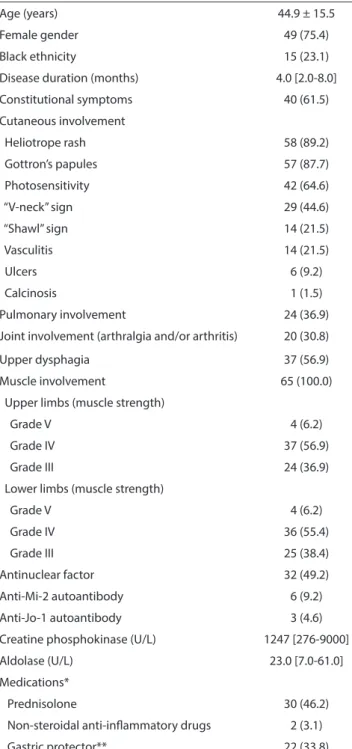
Table 1 - General data from 65 patients with new-onset dermatomyositis
Age (years) 44.9 ± 15.5
Female gender 49 (75.4)
Black ethnicity 15 (23.1)
Disease duration (months) 4.0 [2.0-8.0]
Constitutional symptoms 40 (61.5)
Cutaneous involvement
Heliotrope rash 58 (89.2)
Gottron’s papules 57 (87.7)
Photosensitivity 42 (64.6)
“V-neck” sign 29 (44.6)
“Shawl” sign 14 (21.5)
Vasculitis 14 (21.5)
Ulcers 6 (9.2)
Calcinosis 1 (1.5)
Pulmonary involvement 24 (36.9)
Joint involvement (arthralgia and/or arthritis) 20 (30.8)
Upper dysphagia 37 (56.9)
Muscle involvement 65 (100.0)
Upper limbs (muscle strength)
Grade V 4 (6.2)
Grade IV 37 (56.9)
Grade III 24 (36.9)
Lower limbs (muscle strength)
Grade V 4 (6.2)
Grade IV 36 (55.4)
Grade III 25 (38.4)
Antinuclear factor 32 (49.2)
Anti-Mi-2 autoantibody 6 (9.2)
Anti-Jo-1 autoantibody 3 (4.6)
Creatine phosphokinase (U/L) 1247 [276-9000]
Aldolase (U/L) 23.0 [7.0-61.0]
Medications*
Prednisolone 30 (46.2)
Non-steroidal anti-inlammatory drugs 2 (3.1)
Gastric protector** 22 (33.8)
Data expressed as mean ± standard deviation, median (interquartile range 25%-75%) or percentage (%). * At time of upper digestive endoscopy; ** Proton pump inhibitor (omeprazole) or anti-H2 blocker (ranitidine).
Constitutional symptoms were present in 61.5% of patients at diagnosis; dermatologic manifestations such as heliotrope, Gottron’s papules and photosensitivity were common, with a prevalence of 89.2%, 87.7% and 64.6%, respectively (Table 1).
Pulmonary involvement, articular problems, and high dysphagia were present in 36.9%, 30.8% and 56.9% of patients, respectively.
All but four of the patients had some degree of objective muscle weakness.
Approximately half of patients tested ANA positive (Table 1). Anti-Mi-2 and anti-Jo-1 were found in 6 (9.2%) and 3 (4.6%) patients, respectively. Median levels of serum creatine kinase and aldolase were 1,247 U/L and 23.0 U/L, respectively (Table 1).
At the time of the UDE, 46.2% of patients were in
use of prednisone (0.5 - 1.0 mg/kg/day) and 3.1% had been using non-steroidal anti-inflammatory drugs for at least one week.
Approximately one third of patients had been using a gastric protector (omeprazole or ranitidine) for at least one week.
About 70% of patients presented endoscopic changes of the upper digestive tract (esophagus, stomach and/or duodenum). Of these, 47.7% exhibited changes in one of the upper digestive tract areas, 20.0% in two areas and 3.1% concomitantly in all three areas (esophagus, stomach and duodenum).
Table 2 shows endoscopic findings for the upper gastrointestinal tract of patients. Esophageal involvement was documented in 18.5% of patients, distal non-erosive esophagitis in 1.5% of cases, and distal erosive esophagitis in 16.9%. In the latter group, 9 patients were Grade A and 2 patients Grade B, according to the Los Angeles classification. Gastric changes were identified in 63.1% of patients and were primarily characterized by the presence of antral gastritis (42.3%), followed by pangastritis (27.8%). There were no cases of corpus or severe gastritis, according to the Sydney classification. Endoscopy results revealed that 15.4% had duodenal involvement, 10.9% bulbar duodenitis and 7.7% duodenal ulcer.
Table 2 - Percentage of patients with new-onset dermatomyositis by type of endoscopic change in upper gastrointestinal tract.
Region Number of patients (%)
B Esophageal involvement 12 (18.5)
Distal esophagitis Erosive Mild 11 (16.9)
Moderate 0
Non-erosive Mild 1 (1.5)
Moderate 0
Gastric involvement 44 (63.1)
Antral gastritis Enanthematous Mild 3 (4.9)
Moderate 4 (6.6)
Erosive Mild 13 (20.0)
Moderate 4 (6.2)
Atrophic 3 (4.6)
Pangastritis Enanthematous Mild 10 (15.4)
Moderate 2 (3.1)
Erosive Mild 3 (4.6)
Moderate 2 (3.1)
Atrophic 1 (1.6)
Duodenal involvement 10 (15.4)
Duodenal ulcer Healed 5 (7.7)
Bulbar duodenitis Enanthematous Mild 4 (6.2)
Moderate 0
Erosive Mild 1 (1.6)
Moderate 2 (3.1)
Concomitant involvement of: 46 (70.8)
One area (esophagus, stomach or duodenum) 31 (47.7)
Two areas (esophagus, stomach and/or duodenum) 13 (20.0)
Three areas (esophagus, stomach and duodenum) 2 (3.1)
Table 3 - Univariate and multivariate analysis of endoscopic indings of upper gastrointestinal tract in new-onset dermatomyositis.
Endoscopic indings Parameters Univariate Multivariate
OR 95%CI OR 95%CI
Erosive distal esophagitis Age 0.95 0.91-0.99 0.94 0.88-0.99
Erosive antral gastritis Male gender 4.45 1.22-16.16 4.78 1.35-16.96
Enanthematous antral gastritis Black ethnicity 5.70 1.03-31.70 5.52 1.02-29.97
Enanthematous pangastritis “V-neck” sign 0.21 0.04-0.99 0.08 0.007-0.91
OR: Odds ratio; CI: conidence interval.
Moreover, endoscopic changes did not correlate (p > 0.05) with all parameters showed in the Table 1, including
drug therapy (prednisone, nonsteroidal anti-inflammatory
and/or gastric protector).
No neoplasia lesions were identified in this study.
■
DISCUSSIONThe present study found that approximately 70%
gastrointestinal tract symptoms (except high dysphagia) had endoscopic changes in the upper digestive tract.
Although DM is a rare disease and strict exclusion criteria were employed in the present study, this
analysis included a sample of 65 patients with defined
DM. Information on patients was based on previously standardized and parameterized data, ensuring reliable
study data. Endoscopic examinations were performed at the
Unlike other skin lesions analyzed in this study, only the presence of the cutaneous type “V-neck” sign injury was
associated with UDE findings. In this case, there was an inverse
relationship between presence of the injury and enanthematous
pangastritis. Further studies are needed to confirm this
association between the two parameters and to assess a possible cause-consequence relationship.
Duodenal disease was documented in 15.4% of the cases, with duodenal ulcer present in 7.7% of the sample analyzed. Although the study was conducted at a reference center, Suzuki
et al.28 found a 6% prevalence of duodenal ulcer in patients with
dyspeptic symptoms. This result may suggest an increased prevalence of duodenal ulcer in our patients, since the present study only included asymptomatic dyspeptic cases.
The prevalence of neoplasms in patients with newly-diagnosed DM is 8.6%,29 where the most common primary
sites in descending order are: lung, ovaries, uterus, thyroid, hematologic, colon, skin and prostate. Corroborating the results of the cited study,28 no progressive neoplasia was found on the present UDE analysis.
Study limitations: no control group was included; only a small sample was involved, given the rarity of the disease and the strict exclusion criteria applied. Some changes, such as gastric ulcer, require a greater number of patients for prevalence analysis; no data analysis of infection by Helicobacter pylori, known to be linked to some gastroduodenal changes presented by patients was carried out; the cross-sectional evaluation of the patients analyzed allowed evaluation of possible associations between the parameters found but precluded deduction of
possible cause-effect relationships; finally, specific muscle and
cutaneous measures validated in DM were not used in the retrospective study.
■
SUMMARYThe estimated prevalence of gastrointestinal disorders in the study sample analyzed may be relevant to
guide conduct in digestive disorders with UDE examination
and points to the need for pharmacological prevention of esophageal and gastroduodenal lesions in these patients.
■
CONFLICT OF INTERESTAll authors declare no conflict of interest
■
AUTHOR CONTRIBUTIONAmorim TM: planning, reviewing literature, executing and writing the article. Furuya Junior CK: reviewing literature, executing and writing the article.
Marques SB: reviewing literature, executing and writing the
article. Shinjo SK: planning, reviewing literature, executing and writing the article.
as UDE examinations were performed in newly-diagnosed
patients, the cases were evaluated at the same time and at a similar phase, rendering the sample more homogeneous for subsequent analysis.
We included only cases without upper gastrointestinal tract symptoms that were newly-diagnosed and therefore in a fully active phase of the disease. The objective was to
exclude or reduce many parameters that can influence
the endoscopic examination, such as drug therapy (glucocorticoids, immunosuppressives, nonsteroidal
anti-inflammatory drugs, gastric protectors).
Although well-documented in juvenile DM,14-18 there are currently no descriptions of endoscopic findings of the
upper digestive tract in adult patients with DM.
Approximately 70% of our patients had endoscopic changes in at least one area of the upper digestive tract. Gastric involvement occurred in approximately two-thirds of the patients analyzed, while changes in the esophagus and duodenum were present in 18.5% and 15.4% of cases, respectively.
In the case of esophageal involvement, the majority of patients had an erosive distal esophagitis picture, corresponding to 16.9% of all our cases, and therefore comparable to the rate reported in the literature of 12.0 - 17.3%.23,24 In addition, the frequency of erosive distal
esophagitis decreased with patient age. Further studies are warranted to evaluate this possible inverse relationship between esophageal injury and age.
Gastric changes were present in two thirds of patients
with DM. Erosive antral gastritis and erosive pangastritis
accounted for half of all gastric changes. The presence of this erosive gastritis is not associated with the use of
medications, especially nonsteroidal anti-inflammatory drugs. At the time of UDE, half of the patients were in use of non-steroidal anti-inflammatory drugs or prednisone.
Nevertheless, the use of these medications, as well as gastric
protectors, had no impact on endoscopic findings, suggesting that the changes observed were inherent to DM. Because
DM is a vascular disease, in which vascular depletion leads to tissue ischemia, the subsequent compensatory stimulus may result in neoangiogenesis. This phenomenon is clearly demonstrated on nailfold capillaroscopy, which discloses capillary ectasia, micro-hemorrhages, vessel bushes, and areas of capillary dropout. One hypothesis for the present
findings is that such vascular changes also occur within the
wall of the gastrointestinal tract.
ALTERAÇÕES MORFOLÓGICAS DO TRATO INTESTINAL SUPERIOR EM PORTADORES DE DERMATOMIOSITE DE INSTALAÇÃO RECENTE: CORRELAÇÕES COM CARACTERÍSTICAS DEMOGRÁFICAS, CLÍNICAS E LABORATORIAIS
OBJETIVOS: Avaliar os exames de endoscopia
digestiva alta (EDA) de pacientes adultos com DM
(dermatomiosite) recém-diagnosticados; correlacionar eventuais alterações do trato gastrintestinal com dados
demográficos, clínicos, e medicamentosos desta população.
MÉTODO: Estudo transversal, em que foram
avaliados 65 casos de DM recém-diagnosticados, no
período entre 2004 a 2015. Foram excluídos casos de DM clinicamente amiopática, sobreposição com DM,
hepatopatias, cirurgia gástrica prévia, sintomas do trato gastrointestinal (exceto disfagia alta), quadros infecciosos sistêmicos, etilismo e tabagismo.
RESULTADOS: A média idade dos pacientes foi de
44,9 anos, com um tempo de sintomas atribuídos a DM de
quatro meses. Alterações endoscópicas foram encontradas em 70,8% dos pacientes. O acometimento esofágico/ gástrico foi documentado em 18,5% dos pacientes: esofagite
distal erosiva (16,9%) e esofagite distal não-erosiva (1,5%);
alterações gástricas em 63,1% dos casos: gastrite antral (42,3%) e pangastrite (27,8%); o acometimento duodenal em 15,4% dos pacientes: bulboduodenite (10,9%) e úlcera
duodenal (7,7%). Não foram detectadas lesões malignas. Em análise multivariada, a esofagite distal erosiva esteve menos associada a indivíduos de idade maior. Sexo
masculino apresentava mais diagnóstico de gastrite erosiva. A pangastrite enantemática esteve menos associada a lesões em “V” do decote.
CONCLUSÕES: O presente estudo estima, pela primeira vez, a prevalência de alterações endoscópicas altas em pacientes adultos com DM recém-diagnosticada. Os resultados podem ser relevantes para guiar potenciais
alterações digestivas com exame de EDA, bem como apontar para necessidade de prevenção medicamentosa de lesões
do trato digestivo nestes pacientes.
PALAVRAS-CHAVE: Dermatomiosite, dispepsia, endoscopia gastrointestinal, miosite.
■
REFERENCES1. Callen JP, Wortmann RL. Dermatomyositis. Clin Dermatol. 2006;24(5): 363-73. DOI: 10.1016/j.clindermatol.2006.07.001
2. Briani C, Doria A, Sarzi-Puttini P, Dalakas MC. Update on idiopathic inflammatory myopathies. Autoimmunity. 2006;39(3):161-70. DOI: 10.1080/08916930600622132
3. Dalakas MD, Hohlfeld R. Polymyositis and dermatomyositis. Lancet. 2003;362(9388):971-82. DOI: 10.1016/S0140-6736(03)14368-1 4. Bohan A, Peter JB. Polymyositis and dermatomyositis (first part). N Engl
J Med. 1975;292(7):344-7. DOI: 10.1056/NEJM197502132920706 5. Euwer RL, Sontheimer RD. Dermatologic aspects of myositis. Curr Opin
6. Souza FHC, Levy-Neto M, Shinjo SK. Adult dermatomyositis: experience of a Brazilian tertiary care center. Rev Bras Reumatol. 2012; 52(6):
897-902. DOI: 10.1590/S0482-50042012000600008
7. Dalakas MC. Molecular immunology and genetics of inflammatory muscle diseases. Arch Neurol. 1998; 55(12):1509-12.DOI: 10.1001/ archneur.55.12.1509
8. Dietz F, Logeman JA, Sahgal V, Schmid FR. Cricopharyngeal muscle dysfunction in the differential diagnosis of dysphagia in polymyositis. Arthritis Rheum. 1980;23(4):491-5. DOI: 10.1002/ art.1780230412
9. Ertekin C, Secil Y, Yuceyar N, Aydogdu I. Oropharyngeal dysphagia in polymyositis/dermatomyositis. Clin Neurol Neurosurg. 2004;107(1):32-7. DOI: 10.1016/j.clineuro.2004.02.024
10. De Merieux P, Verity MA, Clements PJ, Paulus HE. Esophageal abnormalities and dysphagia in polymyositis and dermatomyositis. Arthritis Rheum. 1983;26(8):961-8.
11. O’Hara JM, Szemes G, Lowman RM. The esophageal lesions in dermatomyositis. A correlation of radiologic and pathologic findings. Radiology. 1967;89(1):27-31.
12. Horowitz M, McNeil JD, Maddern GJ,Collins PJ, Shearman DJ. Abnormalities of gastric and esophageal emptying in polymyositis and dermatomyositis. Gastroenterol. 1986;90(2):434-9.
13. Machado WM, Freire BFA, Rocha OM, Azambuja CAP, Oliveira MEC. Proposal of a questionnaire for prevalence’s characterization of the digestive symptoms in the conective tissue diseases. Arq Gastroenterol. 2004;41(1):64-70. DOI: 10.1590/S0004-28032004000100013 14. Musaev SN, Novikova AV, Klimanskaia EV, Shershevskaia AI.
Clinico-endoscopic and morphometric characteristics of gastric and duodenal mucosa in children with dermatomyositis. Revmatologiia (Mosk). 1991;4(5):21-5.
15. Mamyrova G, Kleiner DE, James-Newton L, Shaham B, Miller FW, Rider LG. Late-onset gastrointestinal pain in juvenile dermatomyositis as a manifestation of ischemic ulceration from chronic endarteropathy. Arthritis Rheum. 2007;57(5):881-4. DOI: 10.1002/art.22782 16. Banker BQ, Victor M. Dermatomyositis (systemic angiopathy) of
childhood. Medicine. 1966;45(4):261-89. DOI: 10.1097/00005792-196607000-00001
17. Lowry CA, Pilkington CA. Juvenile dermatomyositis: extramuscular manifestations and their management. Curr Opin Rheumatol.
2009;21:575-80. DOI: 10.1097/BOR.0b013e328331927e
18. Ebert EC. Review article: the gastrointestinal complications of myositis.
Aliment Pharmacol Ther. 2009;31(3):359-65. DOI: 10.1111/j.1365-2036.2009.04190.x.
19. Medical Research Council. Aids to the examination of the peripheral
nervous system, Memorandum no. 45. Her Majesty’s Stationery Office,
London, 1981.
20. Cruellas MG, Viana V dos S, Levy-Neto M, Souza FH, Shinjo SK.
Myositis-specific and myositis-associated autoantibody profiles and their
clinical associations in a large series of patients with polymyositis and dermatomyositis. Clinics. 2013;68(7):909-14. DOI: 10.6061/ clinics/2013(07)04
21. Lundell LR, Dent J, Bennett JR, Blum AL, Armstrong D, Galmiche JP,
et al. Endoscopic assessment of oesophagitis: clinical and functional
correlates and further validation of the Los Angeles classification. Gut.
1999;45(2):172-80.
22. Tytgat GN. The Sydney system: endoscopic division. Endoscopic appearances in gastritis/duodenitis. J Gastroenterol Hepatol. 1991;
6(3): 223-33. DOI: 10.1111/j.1440-1746.1991.tb01469.x
23. Wang FW, Tu MS, Chuang HY, Yu HC, Cheng LC, Hsu PI. Erosive
esophagitis in asymptomatic subjects: risk factors. Dig Dis Sci. 2010;55(5):1320-4. DOI: 10.1007/s10620-009-0888-z
24. Ou JL, Tu CC, Hsu PI, Pan MH, Lee CC, Tsay FW, et al. Prevalence
and risk factors of erosive esophagitis in Taiwan. J Chin Med Assoc. 2012;75(2):60-4. DOI: 10.1016/j.jcma.2011.12.008
25. Nogueira C, Figueiredo C, Carneiro F, Gomes AT, Barreira R, Figueira
26. Lee I, Lee H, Kim M, Fukumoto M, Sawada S, Jakate S, et al. Ethnic
difference of Helicobacter pylori gastritis: Korean and Japanese gastritis is characterized by male- and antrum-predominant acute foveolitis in comparison with American gastritis. World J Gastroenterol. 2005; 11(1): 94-8.
27. Snyder JD, Hardy SC, Thorne GM, Hirsch BZ, Antonioli DA. Primary antral gastritis in Young American children. Low prevalence of
Helicobacter pylori infections. Dig Dis Sci. 1994;39(9):1859-63. DOI:
10.1007/BF02088115
28. Suzuki RB, Cola RF, Cola LT, Ferrari CG, Ellinger F, Therezo AL, et al.
Different risk factors influence peptic ulcer disease development in a Brazilian population. World J Gastroenterol. 2012; 18(38): 5404-11.
DOI: 10.3748/wjg.v18.i38.5404.
29. Souza FHC, Shinjo SK. Newly diagnosed dermatomyositis in the elderly as predictor of malignancy. Rev Bras Reumatol. 2012;52(5):713-21.