Evaluation of the association between indicators of oral
health and sociodemographic variables in children with
orofacial clinical signs of chronic mouth breathing
Avaliação da associação entre os indicadores de saúde bucal, os
aspectos socioeconômicos e crianças com sinais clínicos orofaciais
indicativos de respiração oral crônica
Fabiane Piva1, Juliana Kern de Moraes2, Vitor Rezende Vieira2, Alexandre Emídio Ribeiro Silva3, Raquel
Massotti Hendges4, Gilberto Timm Sari1
ABSTRACT
Purpose: To evaluate whether clinical and sociodemographic indi-cators of oral health in school-age children (from 8 to 12 years) with mixed dentition are associated with oral breathing. Methods: Fifity-five children selected from a public school in the city of Cachoeira do Sul (RS) were evaluated. After obtaining parental consent, a trained speech pathologist performed examinations to identify children with orofacial clinical signs indicative of oral breathing. For the oral health exams, dental students underwent training and calibration according to the cri-teria of the World Health Organization for epidemiological surveys. In addition to the intra-oral examinations, socioeconomic and demographic data were collected from the parents of the children using a questionnai-re. Results: Although the prevalence of children with mouth breathing symptoms was notable, it was lower than that reported in the literature. No difference was observed between the groups (mouth breathers and nose breathers) regarding socioeconomic variables associated with the tested dental aspects. Conclusion: Oral breathing was not associated with clinical indicators of oral health such as caries, visible plaque, and gingival bleeding. Further, no association was found between sociode-mographic variables and oral breathing.
Keywords: Epidemiology; Mouth breathing; Dental caries; Gingivitis; Speech, language and hearing sciences
RESUMO
Objetivo: Avaliar se os indicadores clínicos de saúde bucal e socio-demográficos de crianças em idade escolar (8 a 12 anos), em fase de dentição mista, estão associados com a respiração oral. Métodos: Foram avaliadas 55 crianças, selecionadas em uma escola estadual do município de Cachoeira do Sul (RS). Após a autorização dos responsáveis, foram realizados exames por uma fonoaudióloga treinada, para a identificação de crianças com sinais clínicos orofaciais, indicativos de respiração oral. Para a realização dos exames de saúde bucal, os estudantes de odon-tologia foram treinados e calibrados, de acordo com os critérios para levantamentos epidemiológicos da Organização Mundial de Saúde. Além dos exames intrabucais, foram coletadas informações socioeconômicas e demográficas com os responsáveis pelas crianças, por meio de um ques-tionário. Resultados: A prevalência de crianças com sinais de respiração oral foi relevante, porém, menor do que descreve a literatura. Não houve diferença entre os grupos (respiradores orais e respiradores nasais), tanto em relação às variáveis socioeconômicas, como em relação aos aspectos odontológicos testados. Conclusão: Não houve associação da respiração oral com nenhum indicador clínico de saúde bucal (cárie, placa visível e sangramento gengival). Também não foi encontrada associação entre as variáveis sociodemográficas e a respiração oral.
Descritores: Epidemiologia; Respiração bucal; Cárie dentária; Gengi-vite; Fonoaudiologia
Study conducted at the School of Dentistry, Universidade Luterana do Brasil – ULBRA – Cachoeira do Sul, Rio Grande do Sul (RS), Brazil. (1) School of Dentistry, Universidade Luterana do Brasil – ULBRA – Cachoeira do Sul, Rio Grande do Sul (RS), Brazil.
(2)Dentist, Cachoeira do Sul (RS), Brazil.
(3)School of Dentistry, Universidade Federal de Pelotas – UFPel – Pelotas (RS), Brazil. (4)Speech-therapist, Cachoeira do Sul (RS), Brazil.
Conflict of interests: No
Author’s contribution: FP advisor, responsible for the conception and design of the study; JKM, VRV, and RMH performed sample collection, tabulation, and data analysis; AERS coordinator, responsible for statistical analysis; GTS collaboration regarding correction and data tabulation.
Correspondence address: Fabiane Piva. R. Martinho Lutero, 301, Universitário, Cachoeira do Sul (RS), Brazil, CEP: 96501-595. Email: [email protected]
INTRODUCTION
The breathing of an individual is predominantly nasal under resting conditions, which leads to sealing of the lips; however, this pattern is disrupted when the individual is exercising or has a nasal obstructive condition(1). The nasal breathing model is ideal for maintaining the balance of the stomatognathic system, including harmony of the intraoral and extraoral structures. However, when mouth breathing becomes predominant, it can affect the health and daily lives of children, particularly during the period of increased development and growth(1,2).
Mouth breathing leads to the introduction of cold, dry, and unprepared air, which insults the buccal mucosa, nasopharynx, and lungs(1,3).
The term “oral breathing” can be used to define a condi-tion in which the pattern of nose or mixed respiratory (nasal and oral) breathing is replaced with a physiological pattern of predominantly mouth breathing. The mouth tends to remain slightly open, resulting in predominantly parched lips, a hy-potonic recessed tongue, and a flat nose with small nostrils(4). This change in the breathing pattern may be associated with several causes that generally have a physical obstructive origin. One of the most common causes of mouth breathing is allergic rhinitis, followed by hypertrophy of the adenoids(5,6).
The prevalence of mouth breathing is high in children, ranging between 53.35% and 55%(2,5). Dentists should become more aware of this condition, because it can cause various intra and extraoral changes, such as maxillary atresia, protrusion of the upper dental arch, cross bite, poor lip seal, a long face, and other general changes(7,8).
Mouth breathing can cause various oral cavity changes, including pathologies commonly found in children, such as caries and gingivitis. Studies that have attempted to show a positive relationship between higher accumulation of bacterial biofilms, increased susceptibility to decay, and a higher degree of gingival inflammation in oral breathers have reported con-flicting results(9,10).
Several studies found associations between oral breathing and a higher prevalence of caries and gum disease in chil-dren(3-11-16). One study counted Streptococcus mutans, the main bacteria related to dental caries, and analyzed the salivary flow in children with oral and nasal breathing. The authors concluded that the salivary flow was higher in mouth breathers than in nasal breathers (the control group), indicating a compensatory mechanism for dryness of the mucosa. In addition, less salivary immunoglobulin IgA and IgM anti-Streptococcus mutans were found in mouth breathers, suggesting that these individuals had lower resistance to tooth decay than the control group. Although mouth breathers had a higher microorganism count than nose breathers, this difference was not significant(16).
Another study found a higher number of early caries lesions in the posterior teeth of children who breathe through the mouth. The authors found no differences in the rates of initial
caries in the anterior teeth or in the count of Streptococcus mutans, although the salivary levels of Streptococcus mutans
were higher in mouth breathing children than in nasal breathing children. However, these data showed no difference in children with deciduous teeth, which may indicate that the changes caused by mouth breathing develop over time(10).
Children who breathe through their mouths tend to keep their mouths slightly open, producing adverse effects on the mucosal and gingival tissues. Because saliva contains factors that inhibit caries activity, a decrease in the saliva level can in-crease the risk of caries, especially if the diet is cariogenic; this may lead to worse oral states in children who breathe through the mouth continuously(14).
Salivary flow provides mechanical cleaning of residues pre-sent in the mouth, such as non-adherent bacteria, cellular debris, and food particles. Therefore, the lack of saliva flow results in accumulation of these waste products and plaque, promoting oral acidosis and increased microflora, which contribute to the development of caries. Saliva also has rheological properties (viscosity and elasticity) as a result of its chemical, physical, and biological composition, and these features are essential for maintaining the equilibrium conditions of the oral cavity. The complaint of oral dryness is very common in mouth breathers, and usually this symptom is associated with decreased salivary flow(3). Together, these factors also cause inflammation, edema, and a bright gum surface that bleeds easily(14).
Higher levels of plaque were found in individuals with a dry mouth caused by keeping the mouth open during sleep; further, these individuals showed a prevalence of parted lips at rest and decreased upper-lip(11).
Both the dentist and the speech-therapist must be aware of the symptoms as well as the socioeconomic and environmental variables that lead to mouth breathing in children.
Many health professionals are unaware that obstruction of the upper airways has an impact on facial growth and general health in children. If untreated, mouth breathing can cause a narrow face and mouth, dental malocclusion, a gummy smile, and other health problems. Children with nasal obstruction usually sleep with their mouths open, which affects sleep and could adversely affect school activities(3).
Given the divergence of opinions on the subject in the lite-rature, the present study aimed to evaluate whether clinical and sociodemographic indicators of oral health in school-age children with mixed dentition are associated with major clinical signs of mouth breathing. This analysis favors the development of the activities of the various health professionals, such as multidis-ciplinary team, focusing both on prevention of injuries caused by respiratory disorder such as better identify these patients.
METHODS
epidemiological surveys, with some adjustments proposed by other authors(15).
The initial study population consisted of 155 students, when estimating 10% refusal out of a total of 167 individuals needed for the study, in children aged 8-12 years enrolled in a state school the city of Cachoeira do Sul (RS).
The final sample consisted of 55 children without ortho-dontic treatment and who delivered the statement of consent signed by the parents or a responsible adult. All 55 children were examined between August and December 2012, and their personal information was removed from the composition in order to fulfill ethical guidelines for research.
Training and calibration for oral health tests
Initially, a theoretical meeting was held with four pairs of examiners (dental students and their annotators) the methods of each exam and the codes to be recorded on data collection forms according to the proposal of the WHO(17) for epidemio-logical surveys. Subsequently, a training session was held in which 10 students were examined, followed by the calibration with 15 age-matched children who satisfied the same inclusion criteria. Both training and calibration were performed in a municipal school consisting of 1st to 8th grade levels in the city of Cachoeira do Sul (RS). The percentage of agreement for the gingival bleeding index (GBI) and visible plaque index (VPI) was calculated. For the dental caries index (deciduous and permanent teeth decayed, missing, and filled [DMFT/dmft]), the weighted kappa coefficient for intra and inter dental caries was calculated. The weighted kappa was between the minimum value of 0.66 and the maximum value of 0.85.
Study outcome
The identification of the main clinical signs of chronic mou-th breamou-thing was performed by speech mou-therapist who classified each subject into 1 of 2 categories: children with the characte-ristics of a mouth breather (MB) or with the charactecharacte-ristics of nasal breathing (NB). The criteria for classifying an individual as a mouth breather were as follows: dark circles under the eyes hypotonic orofacial muscles, a long narrow face, parched parted lips, an apparently short upper lip with functional alteration of lip closing, atypical swallowing, and labioverted bulky, small nose, atrophied, tapered, positioned behind the jaw, with lack of movement and/or smaller than the ideal size, visible facial asymmetry, maxillary atresia, or in a “V” shape(15).
The speech therapist conducted a visual examination of the changes in the dental office; the evaluator dentist was pre-sent at this evaluation, and recorded the findings in ambient light while seated in a chair facing the speech therapist. The children were in close contact with the speech therapist who assessed the physical conditions by observing the muscles and the appearance of the face and oral cavity.
The following materials were used for this procedure: disposable latex gloves (Supermax™, Brazil Importing S/A), masks (Metallurgical Fava, São Paulo, Brazil), clear protective eyewear (PPE’S Fênix™ São Paulo, Brazil), trowels disposable timber, and a glass cup (200 ml) filled halfway with water.
The speech therapist inspected carefully to the patient’s face at rest, with upright posture, as well as the oral cavity, with the aid of wooden spatulas. Once in front of the child, the professional asked her to take a sip of water and swallow without, however, explaining the reason for the child was not induced.
Clinical examination of oral health
The following materials were used for the intraoral exami-nations: periodontal probes recommended by the WHO, with a 0.5-mm ball; clinical clamps (Duflex™, São Paulo/Brazil of SSWHITE™ Dental Manufacturing, PA, USA); autoclaved, disposable, latex examination gloves (Supermax™, Brazil Importing S/A); masks and hats (Metallurgical Fava™, São Paulo, Brazil); and clear protective eyewear (PPE’S Fênix™ São Paulo, Brazil). Clipboards, pencils, erasers, and a data collection form were used for note taking.
The dental examinations were performed by three dental students who had undergone training and calibration in order to determine the index of DMFT/dmft, VPI and GBI. The examinations were performed in the dental office set up in the school chosen for the study.
For the determination of DMFT/dmft, the criteria proposed by the WHO (1997) were used; the data collection instrument was adapted from the proposed survey according to the Oral Health (SB) Brazil, 2010(14) model. The recommendations of the WHO were adopted for evaluation of primary and perma-nent teeth codes.
The teeth were examined in quarters in the following se-quence: 17–11, 21–27, 37–31, and 41–47. If permanent and deciduous teeth occupied the same space, the condition was considered permanent.
A periodontal probe recommended by the WHO, with a 0.5 mm ball at the tip, was used for the gingival examination. The mouth was divided into sextants, and the teeth were examined in following sequence: 16, 11, 26, 36, 31, and 46. If no teeth were present, the primary corresponding molars or premolars in the sextant end were examined, or the corresponding tooth was examined in the case of incisors.
expected response after probing followed the recommendation of the SB Brazil (10–30 seconds while holding the probe). Further, the codes and criteria for periodontal examination adhered to those recommended by the SB Brazil 2010(14), with some adaptations.
Variables
A questionnaire designed specifically for this study was administered to the parents or responsible adult to obtain infor-mation regarding family income, the number of people living in the household, and mother’s education level.
Statistical analysis
The data were tabulated and analyzed by using the Stata 12.0 statistical package. Descriptive analyzes were performed by using chi-squared and Fischer statistical tests. The signi-ficance level was set at 5% to verify the association between the exhibition of characteristics and outcome variables applied in the study.
Ethical considerations
This study was approved by the Research Ethics Committee of the Universidade Luterana do Brasil, through the Platform Brazil: approval number, 183 939.
If a child tested positive for mouth breathing and/or tooth decay, the need to seek necessary treatment was adequately communicated to the parents or responsible adult/s.
RESULTS
The prevalence of oral breathing in this study was 29.9%, which is lower than that of 53.35% and 55% reported in the literature(2,5). Regarding clinical oral health information, 61.8% of the children had at least one DMFT/dmft of the deciduous and/or permanent type. The gingival examination revealed lack of bleeding at any face in 67.3% of the children (Table 1).
The DMFT/dmft data, which refers to the permanent dentition and the decidua, respectively, revealed a prevalence of decayed teeth; this confirmed the findings of the epidemio-logical survey of the Ministry of Health in Brazil in 2010(14), which reported that decay exceeded the other components of the DMFT/dmft. Further, in agreement with the results of this survey, the deciduous teeth had more decay than the perma-nent teeth in our study. These data are important because they reflect the population’s limited access to oral health services as well as the insufficient attention that is given to the deciduous teeth (Table 2).
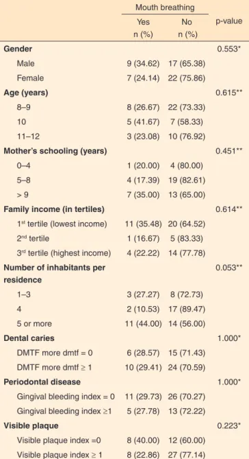
Regarding socioeconomic factors, a lack of association between mouth breathing and variables evaluated in the study was evident (Table 3).
The predominant maternal educational level was between 5 and 8 years (47.9%) and the majority of the subjects (56.3%) had a household income belonging to the first tertile (lowest income). The predominant number of residents in the household was 5 or more residents in this study (45.4%), compared with 1–3 residents and four residents.
DISCUSSION
Chronic mouth breathing is a common symptom in chil-dhood, and its prevalence has been demonstrated in different studies to range from 53.3% to 55.0%(2,5,18) in schoolchildren. The high prevalence of this condition patients indicates the need for better observation of signs and symptoms among dentists, speech therapists, and other health professionals because the lack of an early diagnosis can lead to worsening of the sequelae and a decrease in the quality of life of children.
The present study found that 29.9% of children had clini-cally characteristic signs of mouth breathing syndrome among a sample of 55 school-age children (8–12 years), which is a
Table 1. Description of the children evaluated in this study
n %
Gender
Male 26 47.3
Female 29 52.7
Age (years)
8–9 30 54.5
10 12 21.8
11–12 13 23.6
Mother’s schooling (years)
0–4 5 10.4
5–8 23 47.9
> 9 20 41.7
Family income (in tertiles)
1st tertile (lowest income) 31 56.3
2nd tertile 6 10.9
3rd tertile (highest income) 18 32.7 Number of inhabitants per residence
1-3 11 20.0
4 19 34.6
5 or more 25 45.4
Dental caries
DMTF more dmtf = 0 21 38.2
DMTF more dmtf ≥ 1 34 61.8
Periodontal Disease
Gingival Bleeding Index = 0 37 67.3
Gingival Bleeding Index ≥1 18 32.7
Mouth breathing 16 29.9
lower prevalence than that reported in the literature(2,5). This may be related to the non-random selection process of the study subjects, and this limitation may have led to a lower than expected result for children with mouth breathing. Further, the method of evaluating the symptoms of mouth breathing may have contributed to the low incidence of mouth breathing in this study; although our methods were consistent with the criteria described in the reviewed literature(15), these criteria have been too restrictive for identifying the positive signs of mouth breathing.
A recent study found a higher number of initial carious lesions in the posterior teeth of children who breathe through the mouth. In the same study, no difference in the rates of ini-tial caries in the anterior teeth or in the count of Streptococcus mutans was found. However, the authors determined that the salivary levels of Streptococcus mutans were higher in mouth breathing children than in nasal breathing children(12).
In the present study, the prevalence of caries was 70.59% in nasal breathers and was 29.41% in subjects with symp-tomatic oral breathing. In the sympsymp-tomatic group, 10 out of 16 children (62.5%) had dental caries, whereas 24 out of 39 children (61.53%) who were nasal breathers had caries. This finding confirms the observation that children with a possible diagnosis of mouth breathing have more tooth decay than na-sal breathing children, but this difference was not statistically significant. This result is consistent with that of another study that evaluated 63 students with mixed dentition(15).
Regarding the presence of visible plaque, 22.86% of the
group with positive signs of mouth breathing and 77.14% of the nasal breathing group had at least one face with visible plaque; however, no difference between oral and nasal breathers. A si-milar study found a higher presence of plaque in oral breathers 10–12 years of age in the region of the anterior teeth(15), which is inconsistent with the findings of the present study. However, the method of evaluation regarding the teeth-indices used in this study followed the SB Brazil 2010(17); this differs from that used by the authors of the previous study because their method included fewer anterior teeth. The evaluation of the presence of bleeding gums revealed that mouth breathers had an index of 5 (27.78%) ≥1 and nasal breathers had an index of 13 (72.22%), in agreement with the current literature(15).
In a case-control study, oral health conditions including caries, oral hygiene, the gingival index, and physical changes in the saliva count of Streptococcus mutans were assessed in 30 subjects, 18–22 years of age, diagnosed as mouth breathers and in 30 age-matched individuals who were considered nasal breathers. The authors concluded that the group of mouth breathers had more plaque and higher counts of Streptococcus mutans than the control group. Further, the research group had lower salivary flow rates and lower (more acidic) pH than nasal breathers, as described in the literature(3).
Regarding socioeconomic factors, only one study correla-ting the mouth-breathing pattern with family life was identified in the literature. The authors stressed that the frequency of mouth breathing decreased as the educational level of the father and mother increased, but this correlation was not significant.
Table 2. Description of the clinical variables of oral health in the evaluated children
n Mean Standard deviation
(SD) Minimum Maximum
Permanent caries - DMFT 55 0.60 1.20 0 6
Decay component 55 0.53 1.17 0 6
Restored component 55 0.07 0.26 0 1
Lost component 55 0 0 0 0
Deciduous caries - DMFT 55 1.47 1.94 0 7
Decay component 55 1.20 1.68 0 6
Restored component 55 0.02 0.14 0 1
Lost component 55 0.25 0.95 0 6
Carious deciduous and permanent 55 2.07 2.60 0 12
Decay component 55 1.72 2.16 0 9
Restored component 55 0.09 0.29 0 1
Lost component 55 0.25 0.95 0 6
Teeth with gingival bleeding - upper and lower jaw 55 0.60 0.97 0 4
Teeth with gingival bleeding - upper arch 55 0.24 0.47 0 2
Teeth with gingival bleeding - lower teeth 55 0.36 0.62 0 2
Teeth with visible plaque - upper and lower jaw 55 1.53 1.76 0 6
Teeth with visible plaque - upper arch 55 0.78 1.10 0 3
Teeth with visible plaque - lower teeth 55 0.75 0.99 0 3
The authors also found no association between mouth breathing and the socioeconomic and demographic variables studied, such as parental education, parental separation, the number of people living in the home, family medical care, the number of rooms in the house, and responsible for the child(8).
The majority of families in this study belonged to the first tertile (low income), and 89.6% of the mothers had 5 or more years of education. However, these variables were not associa-ted with the group of symptomatic mouth breathers. A tendency was observed between the group of possible mouth breathing subjects and the “number of people who lived in the same house” to be in the category of 5 or more inhabitants. Thus, the tendency for a child to be an oral breather increased as the
number of inhabitants of the residence (68.75%) increased; however, this association was not significant.
CONCLUSION
Based on the data obtained by using the methodology des-cribed herein, we found no association between children iden-tified as possible chronic mouth breathers and the DMFT/dmft index, VPI, gingival bleeding, mother’s education, gender, age, family income, and the number of people in the household.
REFERENCES
1. Lima JG, Diamante C. Síndrome do respirador bucal: abordagem fisioterapêutica [trabalho de conclusão de curso]. Cascavel: Universidade Estadual do Oeste do Paraná; 2003.
2. Menezes VA, Leal RB, Pessoa RS, Pontes RMES. Prevalência e fatores associados à respiração oral em escolares participantes do projeto Santo Amaro-Recife, 2005. Rev Bras Otorrinolaringol. 2006;72(3):394-9. http://dx.doi.org/10.1590/S0034-72992006000300017
3. Al-Awadi RN, Al-Casey M. Oral health status, salivary physical properties and salivary Mutans Streptococci among a group of mouth breathing patients in comparison to nose breathing. J Bagh Coll Dent. 2003;25(special 1):152-9.
4. Ministério da Saúde. Secretaria de Atenção à Saúde. Departamento de Atenção Básica. Doenças respiratórias crônicas. Brasília: Ministério da Saúde; 2010. (Série A. Normas e manuais técnicos. Cadernos de Atenção Básica; vol. 25).
5. Abreu RR, Rocha RL, Lamounier JA, Guerra AFM. Etiologia, manifestações clínicas e alterações presentes nas crianças respiradoras orais. J Pediatr. 2008;84(6):529-35. http://dx.doi.org/10.1590/S0021-75572008000700010
6. Leite RMS, Leite AAC, Friedman H, Friedman I. A síndrome do respirador bucal como fator de risco para queilite actínica. An Bras Dermatol. 2003;78(1):73-8. http://dx.doi.org/10.1590/S0365-05962003000100007
7. Petry C, Pereira MU, Pitrez PM, Jones MH, Stein RT. The prevalence of symptoms of sleep-disordered breathing in Brazilian schoolchildren. J Pediatr (Rio J). 2008;84(2):23-9. http://dx.doi.org/10.2223/JPED.1770 8. Menezes VA, Leal RB, Moura MM, Granville-Garcia AF. Influência de fatores socioeconômicos e demográficos no padrão de respiração: um estudo piloto. Rev Bras Otorrinolaringol. 2007;73(6):826-34. http:// dx.doi.org/10.1590/S0034-72992007000600014
9. Ashley FP, Usiskin LA, Wilson RF, Wagaiyu E. The relationship between irregularity of the incisor teeth, plaque, and gingivitis: a study in a group of schoolchildren aged 11-14 years.Eur J Orthod. 1998;20(1):65-72. http://dx.doi.org/10.1093/ejo/20.1.65
10. Nascimento Filho E, Mayer MPA, Pontes PAL, Pignatari ACC, Weckx LLM. A respiração bucal é fator de risco para cárie e gengivite? Rev Bras Alerg Imunopatol. 2003;26(6):243-9.
11. Gulati MS, Grewal N, Kaur A. A comparative study of effects of mouth breathing and normal breathing on gingival health in children. J Indian Soc Pedod Prev Dent. 1998;16(3):72-83.
Table 3. Relationship between expository study variables and mouth
breathing
Mouth breathing
p-value Yes
n (%)
No n (%)
Gender 0.553*
Male 9 (34.62) 17 (65.38)
Female 7 (24.14) 22 (75.86)
Age (years) 0.615**
8–9 8 (26.67) 22 (73.33)
10 5 (41.67) 7 (58.33)
11–12 3 (23.08) 10 (76.92)
Mother’s schooling (years) 0.451**
0–4 1 (20.00) 4 (80.00)
5–8 4 (17.39) 19 (82.61)
> 9 7 (35.00) 13 (65.00)
Family income (in tertiles) 0.614**
1st tertile (lowest income) 11 (35.48) 20 (64.52)
2nd tertile 1 (16.67) 5 (83.33)
3rd tertile (highest income) 4 (22.22) 14 (77.78) Number of inhabitants per
residence
0.053**
1–3 3 (27.27) 8 (72.73)
4 2 (10.53) 17 (89.47)
5 or more 11 (44.00) 14 (56.00)
Dental caries 1.000*
DMTF more dmtf = 0 6 (28.57) 15 (71.43) DMTF more dmtf ≥ 1 10 (29.41) 24 (70.59)
Periodontal disease 1.000*
Gingival bleeding index = 0 11 (29.73) 26 (70.27) Gingival bleeding index ≥1 5 (27.78) 13 (72.22)
Visible plaque 0.223*
Visible plaque index =0 8 (40.00) 12 (60.00) Visible plaque index ≥ 1 8 (22.86) 27 (77.14)
12. Nascimento Filho E, Mayer MPA, Pontes P, Pignatari ACC, Weckx LLM. Caries prevalence, levels of mutans streptococci, and gingival and plaque indices in 3.0- to 5.0- year-old mouth breathing children. Caries Res. 2003;38(6):572-5. http://dx.doi.org/10.1159/000080589
13. StenssonM, Wendt LK, Koch G, Oldaeus G, Birkhed D. Oral health in preschool children with asthma. Int J Paediatr Dent. 2008;18(4):243-50. http://dx.doi.org/10.1111/j.1365-263X.2008.00921.x
14. Fior RA. Risco à cárie e gengivite em respiradores bucais [monografia de conclusão de especialização em odontopediatria]. Piracicaba: Faculdade de Odontologia da Universidade Estadual de Campinas; 2000.
15. Oliveira LR, Cortelli SC, Kogo C, Cortelli JR, Aquino DR, Franco GCN et al. Prevalência de cárie, presença de biofilme e inflamação gengival em pacientes com síndrome da respiração bucal. Periodontia. 2009;19(2):118-23.
16. Koga Ito CY, Umterkircher CS, Fantinato V, Watanabe H, Jorge AO. Influência da síndrome do respirador bucal na presença de estreptococos do grupo mutans e imunoglobulinas anti-streptococcus mutans na saliva. Rev Odontondol UNESP. 1996;25(2):207-16.
17. Ministério da Saúde. Secretaria de Atenção à Saúde. Secretaria de Vigilância à Saúde. SB: Brasil 2010: pesquisa nacional de saúde bucal: resultados principais. Brasília: Ministério da Saúde; 2012. [acesso em: 10 nov 2013] Disponível em: http://bvsms.saude.gov.br/bvs/publicacoes/ pesquisa_nacional_saude_bucal.pdf