Defibrinogenating Activities of Acutobin and Its
Recombinant Glycoforms
Ying-Ming Wang1., Inn-Ho Tsai1,2
*., Jin-Mei Chen1
, An-Chun Cheng1, Kay-Hooi Khoo1,2
1Institute of Biological Chemistry, Academia Sinica, Taipei, Taiwan,2Institute of Biochemical Sciences, National Taiwan University, Taipei, Taiwan
Abstract
Acutobin isolated from Deinagkistrodon acutus venom has been used to prevent or treat stroke in patients. This defibrinogenating serine protease is a 39 kDa glycoprotein containing terminal disialyl-capped N-glycans. After sialidase treatment, the enzyme showed similar catalytic activities toward chromogenic substrate, and cleaved the Aa chain of fibrinogen as efficiently as the native acutobin did. However, the level of fibrinogen degradation products in mice after i.p.-injection of desialylated-acutobin was significantly lower than the level after acutobin i.p.-injection, suggesting that the disialyl moieties may improve or prolong the half-life of acutobin. Two recombinant enzymes with identical protein structures and similar amidolytic activities to those of native acutobin were expressed from HEK293T and SW1353 cells and designated as HKATB and SWATB, respectively. Mass spectrometric profiling showed that their glycans differed from those of acutobin. In contrast to acutobin, HKATB cleaved not only the Aachain but also the Bbandcchains of human fibrinogens, while SWATB showed a reduceda-fibrinogenase activity. Non-denaturing deglycosylation of these proteases by peptide N-glycosidase F significantly reduced their fibrinogenolytic activities and thermal stabilities. Thein vivodefibrinogenating effect of HKATB was inferior to that of acutobin in mice. Taken together, our results suggest that the conjugated glycans of acutobin are involved in its interaction with fibrinogen, and that the selection of cells optimally expressing efficient glycoforms and further glycosylation engineering are desirable before a recombinant product can replace the native enzyme for clinical use.
Citation:Wang Y-M, Tsai I-H, Chen J-M, Cheng A-C, Khoo K-H (2014) Correlation between the Glycan Variations and Defibrinogenating Activities of Acutobin and Its Recombinant Glycoforms. PLoS ONE 9(6): e100354. doi:10.1371/journal.pone.0100354
Editor:Jose´ Marı´a Gutie´rrez, Universidad de Costa Rica, Costa Rica
ReceivedJanuary 10, 2014;AcceptedMay 26, 2014;PublishedJune 19, 2014
Copyright:ß2014 Wang et al. This is an open-access article distributed under the terms of the Creative Commons Attribution License, which permits unrestricted use, distribution, and reproduction in any medium, provided the original author and source are credited.
Funding:NSC grant 101-2319-B-001-003 from National Science Council of Taiwan. The funders had no role in study design, data collection and analysis, decision to publish, or preparation of the manuscript.
Competing Interests:The authors have declared that no competing interests exist.
* Email: [email protected]
.These authors contributed equally to this work.
Introduction
Viperid snake venoms usually contain serine protease isoforms with different specificities toward plasma proteins. Snake venom thrombin-like enzymes (SVTLEs) exhibit specific fibrinogenolytic activities but do not activate factor XIII, plasminogen or platelets. These enzymes generate abnormal fibrin polymers and are useful for lowering fibrinogen concentration and blood viscosity in patients [1–3]. For past decades, SVTLEs have been used as therapeutics to prevent or treat stroke and other cardiovascular diseases [4–6]. The amino acid sequences and glycan structures of SVTLE glycoproteins from different species are not similar [7] and the number and positions of their N-glycosylation sites are not conserved [8–10].
Most SVTLEs cleave fibrinogens either at the Aa-chain only, or at the Aa- followed by the Bb-chain [6]. Acutobin, the major SVTLE isolated from the venom ofDeinagkistrodon acutus(formerly namedAgkistrodon acutus) [7,11] is a specifica-fibrinogenase. It has been commercialized under the names of Defibrase or Acutin, and has been applied on many Chinese patients over the past three decades. Previously, we cloned the cDNA and solved the acutobin sequence [7], and showed that its complex type N-glycan structures were capped almost exclusively by terminal NeuAca
2-8NeuAc disialyl units, with the largest detectable structure consisted of as many as four such disialylated antennae [8]. Compared to ancrod [4], acutobin appears to be more effective in treating thrombosis because of its higher specific activity [12] and may have fewer adverse side effects [13]. It is unclear whether the differences in their clinical performances are due to the distinct structures of the proteins, the glycans, or both. To better understand the roles of the unusual N-glycans of acutobin, the following questions must be addressed: (a) How do the disialyl glycans affect acutobin’s functions? (b) Are the recombinant acutobins (abbreviated as ATBs) prepared from mammalian cells as good as or better than the native acutobin in terms of therapeutic efficiency? (c) How does deglycosylation by peptide N-glycosidase (PNGase F) affect the fibrinogenolytic activities, chain specificities, and stabilities of acutobin and ATBs?
degradation products (FDP) in mice. The present study sheds light on the glycobiology of SVTLEs and should contribute toward the design and development of better defibrinogenating and anti-thrombotic agents.
Materials and Methods
Animals
Six-week-old albino mice [Bltw: CD1(ICR)] were purchased from the National Laboratory Animal Center, Taipei, Taiwan, and bred in our institutional animal facility. Permission for the animal study was obtained from the Institutional Animal Care and Use Committee (IACUC) of the National Laboratory Animal Center (Permit Number IACUC2010-096). This study was carried out in strict accordance with the recommendations in the Guide for the Care and Use of Laboratory Animals of the National Institutes of Health (USA).
Cell Lines
Human embryonic kidney epithelial cells (HEK293T), chon-drosarcoma cells (SW1353), and Chinese hamster ovary cells (CHO-K1), were obtained from American Type Culture Collec-tion (Rockville, MD). HEK293T and SW1353 cells were cultured in Dulbecco’s modified Eagles media (DMEM) (HyClone/ Thermo Scientific, USA) supplemented with 10% final concen-tration of fetal bovine serum (FBS) (HyClone/Thermo Scientific, USA). CHO-K1 cells were cultured in Dulbecco’s modified
Eagles/F12 media (DMEM/F12) (HyClone/Thermo Scientific, USA) supplemented with 10% FBS. All the cells were cultured at 37uC in a humidified atmosphere of 5% CO2.
Enzymes and Reagents
Native acutobin was purified from D. acutus venom (Hunan province, China) [7]. Restriction enzymes were from Promega (Madison, WI, USA). N-Tosyl-Gly-Pro-Arg- p-nitroanilide was from Sigma Aldrich (St Louis, MO, USA). Recombinant PNGase F ofFlavobacterium meningosepticnmandArthrobacter ureafacienssialidase (neuraminidase) were purchased from Roche (Mannheim, Ger-many) and Bio-Rad Laboratories (Hercules, CA, USA), respec-tively. Pre-cast NuPAGE Novex Bis-Tris mini gels and buffers were obtained from Invitrogen Inc. (Carlsbad, CA, USA).
Glycoprotein Modification by Sialidase and PNGase F
Removal or modification of N-glycans in acutobin and ATBs was carried out by sialidase or PNGase F treatment under mild or non-denaturing conditions such that their amidolytic activities on chromogenic substrate were not affected. Desialylation (DS-) of acutobin with 20 mU ofArthrobacter ureafacienssialidase was carried out in 100ml of 50 mM ammonium acetate (pH 6.5) at 37uC for 24 h. Deglycosylation of the enzymes by PNGase F (1.0 unit) was performed in 50ml of 40 mM sodium phosphate (pH 7.2) at 37uC for 24 h. The molecular masses and homogeneities of the proteins, with or without the enzyme treatment, were analyzed by 4–12% NuPage SDS-PAGE.
Vector Construction and Expression of HKATB and SWATB
The DNA sequences encoding myc epitope, His-Tag, Factor Xa cleavage site and acutobin were amplified by PCR and ligated into pSecTag2/Hygro A plasmid (Invitrogen, USA). Successful construction was verified by DNA sequencing. Three mammalian cells, HEK293T, SW1353 and CHO-K1 were used to express the ATBs. The cells were maintained in DMEM, DMEM/F12 media supplemented with glutamine, FBS and non-essential amino acids, respectively. Before transfection, HEK293T and SW1353 cells were washed at least twice with DMEM and suspended in 1 ml of DMEM, and CHO-K1 was maintained in 1 ml of DMEM/F12. About 2mg of plasmids and 5ml of Lipofectamine 2000 (Invitrogen, USA) were mixed and incubated at room temperature for 20 min. Then the mixture was added into the cell suspension and incubated at 37uC for 16 h. After 48 h of cultivation in fresh complete growth medium (DMEM or DMEM/F12 supplemented with 10% FBS and 200mg/ml Hygromycin B), the cells were collected. Likewise, the control cells were transfected with blank vectors pSecTag2/Hygro A. After further cultivation for 72 h, the culture medium was harvested and the secreted proteins were analyzed by SDS–PAGE and western blotting using anti-Myc antibodies.
The culture was also scaled-up to 1 L and the secreted ATB fusion proteins were purified and concentrated using a nickel affinity column for chromatography. Factor Xa was added to the fusion proteins to a final concentration of 1% (w/w) and incubated at room temperature for 24 h to remove the tag peptide. Subsequently, the processed ATBs was injected into a Mono Q column (5/50 GL; GE Healthcare) pre-equilibrated with 50 mM Tris-HCl (pH 8.0), and then eluted with a 0–1.0 M gradient of NaCl at a flow rate of 1 ml/min. The purified ATBs were also digested by trypsin and subjected to MS/MS analysis for protein identification [14].
Mass Spectrometry Analyses of Permethylated Glycans
The glycans were released by PNGase F from reduced, alkylated and proteases-digested acutobin, HKATB and SWATB, and then were permethylated using a modified NaOH/DMSO method before subjecting to MALDI-TOF/TOF (4700 Proteo-mics Analyzer, Applied Biosystems) MS mapping, as described previously [8].
Amidase Assay using Chromogenic Substrate
Enzyme concentrations of acutobin and other derivatives were determined by absorbance at 280 nm assuming a percent extinction coefficient of 1.0 for a 1.0 mg/ml solution, and confirmed with BCA protein assay (BioRad, USA). To measure the amidolytic activities using a SpectraMax M2e Microplate Reader (Molecular Devices, CA, USA), 0.5mg of the protease was added to final volume of 0.2 ml, containing 50mM of Tosyl-Gly-Pro-Arg-p-nitroanilide in 0.1 M Tris-HCl (pH 8.0), in the well of a microtiter plate (Nalge Nunc International, Rochester, NY, USA). Absorbance at 405 nm was followed immediately at 25uC and the specificity constants, kcat/Km, were calculated using
GraphPad Prism. Thermal denaturation of the enzymes was studied by incubating the enzymes in 0.1 M Tris-HCl buffer for 1 h at 37uC, 55uC and 65uC. At the end of incubation, 0.5mg of
the enzymes were added to the substrate solution (50mM) and the
absorbance changes were monitored at 25uC for 20 min.
SDS-PAGE Analyses of the Fibrinogenolytic Products
Purified acutobin, ATBs, or PNGase-treated (PNG-) enzymes (0.2mg each) was added to 8mg of human or rabbit fibrinogen
(Sigma Aldrich, MO, USA) dissolved in 50 mM Tris-HCl (pH 8.0) in a total volume of 10ml and incubated for different time at 37uC.
The fibrinogen cleavage products were mixed with concentrated sample buffers and analyzed by SDS-PAGE. After staining and destaining, the Aaband intensity in each lane was quantified by a VersaDoc imaging system (Bio-Rad 4000MP).
Analyses of Fibrinopeptides Released from the Clot
To detect the release of fibrinopeptide A (FpA) from fibrinogen, 1 NIH unit of a-thrombin or 0.1mg of acutobin, HKATB, SWATB or the PNG-enzymes were added to 10 mg of human fibrinogen dissolved in 1 mL of 50 mM Tris-HCl (pH 8.0) and incubated for 1 h or 20 h at 37uC. The reactions were terminated by boiling for 5 min and the clots formed were removed by centrifuging at 12000 rpm for 15 min. The supernatants were lyophilized, re-dissolved in A buffer (0.07% trifluoroacetic acid) and subjected to reverse phase high performance liquid chroma-tography (RP-HPLC) using a Vydac C18 column (0.46625 cm). A
linear gradient of 10–100% of acetonitrile containing 0.07% trifluoroacetic acid was applied for elution over a period of 45 min and the absorbance at 280 nm was monitored. Peptide peaks were pooled and lyophilized. The peptides were analyzed by ESI-MS using a PE-Sciex API100 mass analyzer (Perkin-Elmer). Samples were dissolved in 20ml of 50% CH3CN containing 0.1% acetic
acid and injected into the analyzer for positive-mode analysis.
Coagulation Kinetics and Clotting Timein vitro
Normal coagulation control plasma (ACCUCLOT Control I, Trinity Biotech, Ireland) or 2.6 mg/ml human fibrinogen in 100ml of the buffer (25 mM Tris pH7.4/0.15 M NaCl/4 mM CaCl2) was kept at 37uC in a flat-bottomed microtiter plate. The
enzyme in 100ml was added (final 0.25mg/ml) and the plate was read in the SpectraMax 340PC384Microplate Reader (Molecular Devices, Sunnyvale, USA) which had been pre-warmed at 37uC. The polymerization of fibrin was measured spectrophotometrically at 405 nm every 30 seconds for 30 min [15].
Coagulation times at higher protease concentrations were also measured by an automatic coagulation analyzer (Hemostasis Analyzer KC-1; Sigma Diagnostics, St. Louis, MO, USA). Aliquots of 75ml ACCUCLOT Control I were incubated at 37uC for 3 minutes. After incubation, 75ml of the protease stock solution was added to trigger coagulation. Pre-warmed thrombin solution (3 NIH U/ml) was used as control.
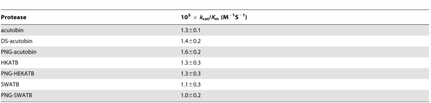
Table 1.Catalytic efficiencies of acutobin, DS-acutobin, ATBs, and PNG-enzymes toward chromogenic substrate Tosyl-Gly-Pro-Arg-p-nitroanilide.
Protease 1056kcat/Km(M21S21)
acutobin 1.360.1
DS-acutobin 1.460.2
PNG-acutobin 1.660.2
HKATB 1.360.3
PNG-HEKATB 1.360.3
SWATB 1.160.3
PNG-SWATB 1.060.2
doi:10.1371/journal.pone.0100354.t001
Euglobulin Clot Lysis Assay
The assay was performed using a modified version of the method described by Smith et al. [16]. Euglobulin fraction was prepared by adding 350ml of normal coagulation control plasma (Trinity Biotech., St. Louis, USA) to 6.3 ml of working acetic acid prepared by adding 1.5 ml of 1% acetic acid to 90 ml reagent grade H2O. The solution was placed on ice for 20 minutes and
then centrifuged at 3500 rpm for 15 min. The supernatant was discarded and the pellet was resuspended in 700ml of a solution containing 150 mM NaCl and 2.6 mM sodium borate, pH 9.0 and kept at 37uC. Acutobin or the indicated proteases were then added to 150ml of the resuspended euglobulin fraction in a
microplate. Finally 150ml of 25 mM CaCl2was added to initiate
clotting. The plate was then placed in SpectraMax M2e
Microplate Reader (Molecular Devices, CA, USA) that had been pre-warmed at 376C, and the absorbance was measured at 340 nm for 10 h.
Plasma Fibrinogen and FDP Levels in Mice
Experimental mice were kept in a 12/12 h light/dark cycle with food and water ad libitum. To determine the plasma fibrinogen concentration, venous blood was collected from the mice in 3.8% sodium citrate (9:1 v/v) and centrifuged at 2500gfor 15 minutes. The test solutions were prepared by diluting plasma 10-fold with a buffer containing 30 mM imidazole, 125 mM sodium chloride and 0.1% sodium azide (pH 7.35). 100ml of the diluted plasma
was then mixed with thrombin (33.3 U/ml) and the clotting time was measured [17]. The plasma fibrinogen concentration was then
determined by intrapolating or extrapolating from a calibration curve prepared by mixing serially-diluted plasma (0.1,2.4 mg/
ml) with thrombin.
Acutobin or DS-acutobin (in 100ml of PBS) was injected intraperitoneally (i.p.) into male ICR mice (6 weeks old weighing 28–30 g). Control mice were similarly injected with PBS. Blood was extracted from the jugular vein of the animals at 1, 2, 3, 4 or 24 h after enzyme injection and anticoagulated with 3.8% sodium citrate (9:1 v/v). FDP concentrations were determined using NANOPIA P-FDP kit (Daiichi Pure Chemicals Co Ltd., Tokyo, Japan) according to the manufacturer’s protocol. Two way analysis of variance (ANOVA) with repeated measures followed by Bonferroni’s post-hoc analysis was used for statistical comparisons. In another experiment, FDP concentrations were determined 4 h after the injection (i.v.) of acutobin, HKATB, SWATB or their PNG-treated forms. One way ANOVA followed by Bonferroni’s post-hoc analysis was employed in this case. GraphPad Prism software was used for statistical analyses. P values ,0.05 are considered to be statistically significant.
Results
Thein vitroandin vivoEffects of DS-acutobin
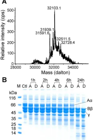
Acutobin was incubated with sialidase for 24 h under a mild condition such that its enzyme activity remained intact (Table 1). The ESI-MS results of DS-acutobin revealed a mixture of glycoproteins distributed at around 32103 Da (Fig. 1A), whereas the masses of native acutobin was distributed at around 36476 Da [8]. The results suggest that most of the sialyl groups, or an
Table 2.Peptide mass fingerprinting analysis of PNG-HKATB.
Determ., Da Calcul., Da Score Modification Positions Amino acid sequence
500.26 1497.79 10 1 SCMCa 1–13 VIGGVECDINEHR
433.69 865.36 25 62–68 FDDEQGR
731.84 1461.67 92 62–73 FDDEQGREPIEK
777.34 1552.66 86 1 SCMCa, 1 NQb 74–85 YFYNCSNNLTTR
502.28 1002.55 57 86–93 DKDIMLIR
757.64 3026.57 55 1 SCMCa 94–121 LDRPV….PSVGSVCR
757.90 3027.55 113 1 SCMCa, 1 NQb 94–121 LDRPV….PSVGSVCR
580.79 1159.58 69 122–132 VMGWGAISPSR
1224.59 2447.16 152 2 SCMCa 133–153 DVLPD….NAECR
868.76 2603.26 115 2 SCMCa 133–154 DVLPD….NAECRR
429.92 1286.72 28 154–164 RAYPRLPATSR
377.88 1130.62 28 155–164 AYPRLPATSR
869.88 1737.76 98 2 SCMCa 165–180 TLCAGVMQGGIDSCNR
870.38 1738.74 38 2 SCMCa, 1 NQb 165–180 TLCAGVMQGGIDSCNR
877.89 1753.75 79 2 SCMCa, 1MOc 165–180 TLCAGVMQGGIDSCNR
1216.90 3647.66 119 2 SCMCa 181–214 DSGGPL….PALYTK
1222.23 3663.65 116 2 SCMCa, 1MOc 181–214 DSGGPL….PALYTK
622.29 1242.57 54 215–223 VYDYNDWIR
1243.07 2484.14 2 1 SCMCa 215–236 VYDYN….TAACPP
631.29 1260.57 40 1 SCMCa, 1 NQb 224–236 SITAGNTTAACPP
The deglycosylated HKATB was reduced and alkylated by iodoacetamide before trypsin hydrolysis. Masses determined for the resultant peptides were match with those calculated based on the amino acid sequence of acutobin.
aSCMC: S-carbamidomethyl Cys. bNQ: asparagine deamidation. cMO: methionine oxidation.
average of about 15 sialyl per acutobin molecule, were removed by sialidase. DS-acutobin showed similar catalytic activity toward chromogenic substrate as the native acutobin (Table 1). Fibrino-gen is a hexamer containing two sets of three different chains, namely Aa, Bb, andc; both acutobin and DS-acutobin at 20mg/ ml cleaved the Aa chains with similar efficacy (Fig. 1B). Additionally, 0.3mg DS-acutobin clotted the ACCUCLOT solution in 12 seconds, similar to the coagulation time elicited by 0.3mg of the native acutobin. These results suggest that although
the removal of the terminal disialyl caps of the glycans reduced the surface negative charges, it probably did not affect the catalytic center conformation of acutobin.
To evaluate the contribution of disialylated glycans for the
in vivodefibrinogenase activity, we compared the plasma levels of FDP in ICR mice afteri.p.injection of acutobin or DS-acutobin. Blood was extracted from the mice at different time points up to 24 h after the enzyme injection, and the FDP concentrations were determined (Fig. 2). Notably, the FDP level in mice injected with 250 ng/g acutobin increased rapidly in the first 3,4 h, while the
FDP level of DS-acutobin injected mice increased slowly and declined after 3,4 h. Compared to the control mice, acutobin
increased the mouse FDP level by 3,5-fold. Apparently, acutobin
hydrolyzed fibrinogen/fibrin faster than DS-acutobin in mice. After injected with high doses, some acutobin-treated mice showed temporary discomfort and decreased mobility, but DS-acutobin-treated mice showed no signs of illness throughout the experiment.
Purification and Fibrinogenase Specificities of ATBs
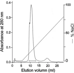
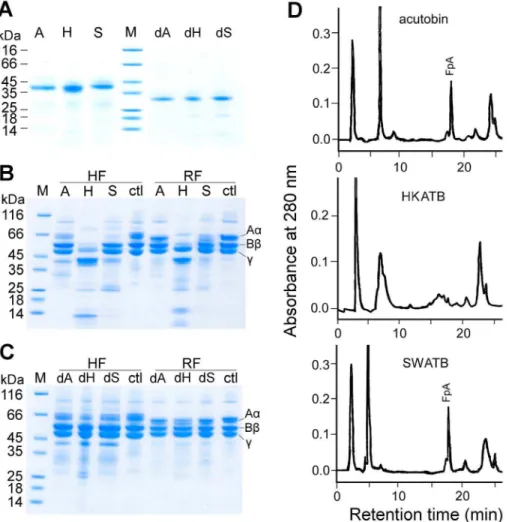
Recombinant fusion proteins of acutobin were successfully harvested from both HEK293T, SW1353 cell cultures and purified. After Factor Xa processing and purification by ion exchanger chromatography (Fig. 3), the yields of HKATB and SWATB were found to be 0.6 and 0.4 mg/L, respectively. However, the yield of the fusion protein from CHO-K1 cells was too low to be purified. Both purified HKATB and SWATB appeared as single bands on polyacrylamide gel (Fig. 4A) and had molecular masses very similar to that of acutobin (38 kDa). The peptide MS/MS analysis of HKATB revealed 58% sequence coverage when Mascot search was performed and the coverage rose to 86% if the glycoproteins were pre-treated with PNGase F before the trypsin hydrolysis step (Table 2).
Using purified fibrinogen (8mg) and 0.2mg enzyme in 10ml of Tris-HCl buffer at 37uC for 2 h, the specificities of acutobin, HKATB and SWATB for human and rabbit fibrinogens were compared. The product analyses by SDS-PAGE revealed that acutobin hydrolyzed rabbit fibrinogen much slower than human fibrinogen; this is in agreement with an earlier finding on the acutobin purified from TaiwaneseD. acutusvenom [11]. Notably, acutobin and SWATB cleaved only the Aachain of human and rabbit fibrinogens but HKATB hydrolyzed the Aa, Bb and c
chains of both fibrinogens under the same condition (Fig. 4B). The results also confirmed that HKATB cleaved the rabbit fibrinogen subunits much faster than acutobin, and SWATB also had a slightly higher activity than acutobin toward the rabbit Aa-chains. Thus, glycan alterations in the ATBs may affect either their specificity or reactivity toward fibrinogen, the native substrate of acutobin.
Effects of PNGase Treatment on the Enzymes Activities and Specificities
To investigate the functional importance of the multivalent glycosylation, a non-denaturing process was carried out to remove the majority of the N-glycans from acutobin and ATBs. Treatment of acutobin and ATBs with PNGase F for 24 h yielded sharp
bands of lower molecular weights on the SDS-PAGE gel (Fig. 4A). MALDI-TOF mass analysis of PNG-acutobin revealed a molec-ular mass of 29.5 kDa (not shown), which is slightly higher than the predicted mass of 26 kDa for the totally deglycosylated acutobin [7]. Since there is no evidence of O-glycosylation in acutobin and the masses of acutobin N-glycans are around 2.7– 4.9 kDa [8], the PNGase treatment probably removed an average of three glycans from each acutobin or ATB molecule. Similar partial resistance to PNGase F hydrolysis was also observed in the cases of other venom serine proteases, including Russelobin [5] and BJ-48 [18].
As shown in Table 1, the amidolytic activities of PNG-enzymes were similar to those of the untreated enzymes toward Tosyl-Gly-Pro-Arg-p-nitroanilide. But all the PNG-enzymes hydrolyzed only the Aa chains of human fibrinogen, and the rates of hydrolysis were also lower than those of the original enzymes (Fig. 4C vs. Fig. 4B). Notably, PNG-HKATB hydrolyzed the human or rabbit fibrinogen much slower than HKATB and could no longer hydrolyze the Bbandcchains (Fig. 4C).
HPLC and Mass Analyses of Fibrinopeptides
The fibrinopeptide A (FpA) released from the fibrin clots formed by acutobin, ATBs or their PNG-treated counterparts was analyzed by RP-HPLC (Fig. 4D). The released FpA showed a mass of 1536.6 Da corresponding to the N-terminal 16 residues of the Aachain. We found that acutobin and PNG-acutobin released FpA within 1 h of incubation, and peaks corresponding to fibrionopeptide B were not detected. In contrast, the HPLC profile of the HKATB products showed fragments that were eluted before FpA, but both HKATB and PNG-HKATB could not release FpA even after 20 h. Interestingly, FpA was detected in the clot formed by SWATB after 20 h, supporting the notion that SWATB can cleave the fibrinogen Aachain but at a much slower rate.
Figure 3. Purification of the recombinant acutobin by Mono Q column. After eluted from Ni-affinity columns and removal of the fusion peptides by Factor Xa, HKATB or SWATB was loaded into a Mono Q column (5/50 GL) that had been pre-equilibrated with 50 mM Tris-HCl buffer (pH8.0), and the enzymes were eluted with a NaCl gradient from 0,1.0 M. The fractions containing HKATB (indicated by a bar) were pooled. The purification of SWATB showed similar results.
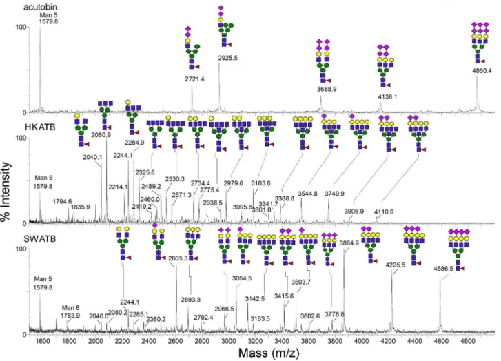
N-glycan Structures of HKATB and SWATB
The N-glycans were released from HKATB and SWATB and profiled by MALDI-MS (Fig. 5). As expected, different cells produced different glycoforms of ATBs and a higher degree of N-glycosylation heterogeneity was observed on both ATBs relative to that on native acutobin. In general, they all carried a range of bi-, tri and tetra-antennary complex type structures with varying degrees of sialylation (Fig. 5). N-glycans from HKATB alone comprise many species carrying additional HexNAc attributable to incompletely galactosylated and/or bisecting GlcNAc, whereas most antennary GlcNAc in SWATB were apparently galactosy-lated and further sialygalactosy-lated to different extent. Neither of the two ATBs carried N-glycans capped by the unique terminal disialyl units as found in the N-glycans from native acutobin. Due to the heterogeneity of the sugar chains in the glycoproteins, ESI-mass spectra of intact acutobin, HKATB and SWATB showed clusters of signals centered around 36476, 34210 and 39310 Da, respectively (not shown). After subtracting the theoretical mass (26105 Da) of their polypeptide chain [7], their average mass increments contributed by N-glycosylation are 10371, 8105 and 13205 Da, respectively. Thus, the averaged N-glycan masses of HKATB are lower than those of acutobin and SWATB. This conclusion is consistent with the MALDI-MS profile afforded by
the N-glycans released from their respective enzymes, showing that the major glycans carried on HKATB and SWATB have fewer and more antennae than acutobin, respectively (Fig. 5).
Effect of Deglycosylation on Thermostability of the Enzymes
Serine proteases are known to be thermal stable at temperatures below 50uC in neutral buffer. To assess the contribution of glycosylation to their thermal stability, acutobin, ATBs and their PNGase-treated counterparts were separately incubated at 37uC, 55uC and 65uC for 1 h, respectively. The remaining amidolytic activities were then assayed at 25uC. We found that all proteases were stable at 37uC. Unlike native acutobin and ATBs which remained stable at 55uC, the deglycosylated enzymes showed 25– 30% inactivation after incubation at 55uC. Incubation at 65uC for 1 h resulted in 63–68% inactivation for most of these enzymes, and PNG-SWATB was almost completely inactivated (Fig. 6). Thus, the glycans of acutobin and HKATB could protect the enzymes against thermal denaturation while SWATB was less stable probably because of the structure or nature of its glycan moieties.
Figure 4. Hydrolysis of human and rabbit fibrinogens by acutobin and its glycoform variants.(A) Purified acutobin, HKATB, SWATB and PNGase-treated enzymes were analyzed by SDS-PAGE. (B) Hydrolyses of human fibrinogen (HF) or rabbit fibrinogen (RF) by acutobin and ATBs. Enzyme concentrations used were 20mg/ml. (C) Hydrolysis of the two fibrinogens by PNGase-treated enzymes. Abbreviations A, H, S, dA, dH, and dS: acutobin, HKATB, SWATB, and their PNGase-deglycosylated forms, respectively; M, molecular weight markers; ctl, control with fibrinogen only. (D) HPLC isolation and identification of FpA in the supernatants above the clot generated by acutobin, HKATB and SWATB, respectively.
Coagulation Kinetics of Acutobin and Other Glycoforms
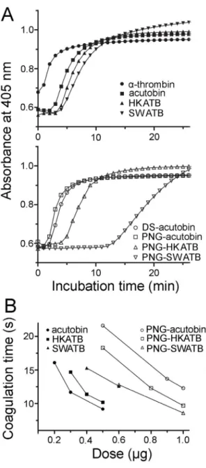
The tertiary and quaternary structural changes of fibrin/ fibrinogen molecules during hydrolysis and polymerization could be monitored spectrophotometrically. As shown in Fig. 7A, human plasma was coagulated by 1 NIH unit ofa-thrombin (as a positive control) or 0.25mg/ml (final concentration) of acutobin or other
enzymes, and these enzymes elicited an increase in absorbance at 405 nm within 12 min. The polymerization rates followed the order of acutobin/PNG-acutobin.HKATB/PNG-HKATB.
SWATB/PNG-SWATB (Fig. 7A). Thisin vitroassay also revealed that at the same enzyme concentration, both DS-acutobin and acutobin exhibit higher fibrinogenolytic activities than PNG-HKATB and PNG-SWATB. Additionally, using purified human fibrinogen at a physiological concentration of 2.6 mg/ml, we found a similar order of the polymerization rate, i.e.acutobin.
HKATB.SWATB.PNG-ATBs (data not shown). Remarkably, the polymerization rates were faster in the fibrinogen solution than in human plasma, suggesting that the activities of ATBs could be suppressed by some plasma components.
The measurement of plasma coagulation time has been a standard protocol for quality control in the manufacture of acutobin as therapeutics. Fig. 7B shows the results of coagulation time measured on a coagulation analyzer and using enzyme
Figure 5. MALDI-MS mapping of the permethylated N-glycans released from acutobin, HKATB and SWATB.Glycosyl compositions
were assigned based on the detected m/z values of the [M+Na]+molecular ion signals and the annotated sugar structures were deduced based on further biosynthesis considerations. Only the detailed structures for the acutobin glycans have been reported [8]. The symbols used for annotation of the assigned peak conform to the standard representation recommended by the Consortium for Functional Glycomics. In MS terms, circle represents hexose, square for N-acetyl-hexosamine, triangle for fucose and diamond for sialic acid.
doi:10.1371/journal.pone.0100354.g005
Figure 6. Thermal stability of different glycoforms of acutobin.
The indicated enzymes were incubated at 37uC, 55uC and 65uC for 1 h. The hydrolytic activities toward Tosyl-Gly-Pro-Arg-p-nitroanilide were assayed at 25uC and an enzyme concentration of 2.5mg/ml. The remaining activity of each enzyme after incubation at 37uC was taken as 100%.
concentrations higher than those used in Fig. 7A. To achieve the same clotting time of 12 seconds, the required amounts of acutobin, HKATB, and SWATB under the assay conditions were 0.30, 0.38 and 0.66mg (in 150ml), while those of PNG-acutobin,
PNG-HKATB, and PNG-SWATB were 1.1, 0.81 and 0.68mg,
respectively. Thus, partial (about 75%) deglycosylation significant-ly reduced the clotting rates of acutobin and HKATB, reflecting the contribution of the glycans to the clotting abilities of both fibrinogenases, although PNG-SWATB showed similar low clotting rate as SWATB. Our results also suggest that the clotting time assay (Fig. 7B) is more sensitive than the polymerization assay (Fig. 7A) to reflect the effects of glycan variations.
Results of Euglobulin Clot Lysis Assay
Euglobulin clot lysis assay was performed to compare the lysis rate of the clots formed. Like the thrombin control (1U), clots formed by 50 nM acutobin or PNG-acutobin lysed between 6– 10 h (Fig. 8). Similar results were obtained with clots induced by SWATB and PNG-SWATB (data not shown). However, the clot formed by 50 nM HKATB lysed significantly faster than the clots formed by other proteases, and when the HKATB concentration was increased to 200 nM, no clot was formed. The results could be explained by the non-specific cleavage of fibrinogen by HKATB, consistent with the HPLC results of the fibrinogen cleavage product (Fig. 4D). Notably, deglycosylation by PNGase F corrected the abnormality of HKATB in the euglobulin clot lysis assay.
Plasma Fibrinogen and FDP Level in Mice After Injected the Enzyme
In an early report, rabbit blood became incoagulable within 1– 4 h when the rabbit wasi.v.injected with 50 ng/g body weight of acutobin [11]. In our study, when 50 ng/g of acutobin or HKATB was i.v. injected into ICR mice, the mouse fibrinogen level dropped rapidly and was too low to be measured within 1 h. When a lower injected dose (5 ng/g) of acutobin or HKATB was injected, the decline of plasma fibrinogen could be measured. We found that the plasma fibrinogen level in HKATB-injected mice decreased at a slower rate compared to acutobin-injected mice (Fig. 9A). After 24 h, the fibrinogen level in HKATB-injected mice was replenished but the fibrinogen level in acutobin-injected mice remained undetectable. Thus, acutobin is a more potent and robust defibrinogenating agent compared to HKATBin vivo.
Elevation of FDP level has been frequently observed in the blood of pitviper envenomed victims, indicating systemic fibrino-genolysis and fibrinolysis [19]. We compared the FDP concentra-tions in mice 4 h afteri.v.injection of acutobin and other enzymes. The plasma FDP levels of all the mice increased after the animals were injected with 50 ng/g body weight of the enzymes, relative to the control (Fig. 9B). Notably, the FDP increases after acutobin and HKATB injection were significantly higher than those after PNG-acutobin and PNG-HKATB injection, confirming the contribution of the glycans to thein vivodefibrinogenating effects. Relative to HKATB, SWATB elicited only a small increase in plasma FDP levels (as did PNG-SWATB), especially when injected via thei.p.route.
Figure 7. Coagulation of human plasma by acutobin and its various glycoforms. (A) Coagulation of human plasma was moni-tored spectrophotometrically at 37uC after addition of the indicated enzymes (final 0.25mg/ml). The curves are averaged results of three experiments. (B) Coagulation times of human plasma in the presence of various amounts of the enzymes in a total volume of 150ml were determined by a coagulation analyzer. Data shown are based on the averaged results of two or three experiments.
doi:10.1371/journal.pone.0100354.g007
Figure 8. Euglobulin-clot-lysis assay.The stabilities of clots formed by the indicated enzymes were compared based on the results of this assay. The curves are averaged results of two experiments.
Discussion
N-glycosylation often contributes to proper protein folding and secretion during its biosynthesis in the Golgi and endoplasmic reticulum [20]. The N-glycans of Russelobin, and the sialyl groups of batroxobin were found to play a role in protecting the enzymes against neutralization by physiologically relevant inhibitors such as
a2-macroglobulin [5]. Our results showed that the removal of
three out of the four N-glycans from acutobin and ATBs decreased their fibrinogenolytic activities and stability at elevated tempera-tures. The thermal protectant effects of N-glycans have also been observed in many other SVTLEs [18,21,22]. The lower stability of SWATB relative to acutobin and HKATB nevertheless suggests that glycans with increased size or branching (Fig. 5) may destabilize acutobin. It is likely that different glycan structures may confer differential thermal protection or other physiological effects of a glycoprotein.
Most of the four N-glycosylation sites of acutobin carry the disialylated glycans, and the full range of 1 to 4 disialyl LacNAc antenna [8] is distinct from the sugars in ancrod and batroxobin [9,10]. It has been well known that sialylation of otherwise exposed termini may extend the circulatory half-life of many serum glycoproteins by reducing their clearance through hepatic
asialoglycoprotein receptor [23]. Chemically linking polysialic acids to proteins was shown to improve the half-life of the protein without adversely affecting their function [24,25]. Results in Fig. 2 and Fig. 9 support the notion that the disialyl-capped N-glycans may increase or improve the in vivo half-life of acutobin. The mechanism possibly involves the binding interaction of acutobin with certain immune cells carrying specific siglecs that may preferably bind the disialyl epitopes [26]. Such pharmacokinetic advantage would not be observed for DS-acutobin (Fig. 2) and HKATB (Fig. 9A), which do not contain similarly disialylated N-glycans. Notably, the removal of sialyl residues from acutobin (Fig. 1B) and other SVTLEs (e.g. bilineobin [27] and okinaxobin II [28]) did not affect their specificities or reactivities toward fibrinogen in vitro. The sialyl or disialyl units of these SVTLEs therefore probably do not play a direct role in the interactions with fibrinogen.
Previous preparations of venom serine proteases fromEscherichia coliinclusion bodies suffered either from improper folding [29], or from poor clotting activities [30,31]. Recombinant SVTLEs with ‘‘high mannose type’’ of N-glycans have been prepared fromPichia
yeast [32,33], but their specificities and pharmacological activities were not investigated. Current glycoprotein therapeutics are usually produced by particular human or mammalian cells, and their N-glycans vary according to the activities of glycosyltrans-ferases in their ER-Golgi systems. CHO cells can express more extensively sialylated glycoproteins with molecular weights higher than those produced by HEK293, and in some cases may retard the secretion of the recombinants [34]. The present study found that ATB fusion proteins were not secreted from CHO-K1 cells that could be attributed to the unfavorable glycosylation in the cells.
We have, for the first time, expressed recombinant venom fibrinogenases with different N-glycans from animal cells, while conserving the protein sequences and the glycosylation sites (Fig. 5). HKATB cleaved the Aa, the Bb, and thecchains of human and rabbit fibrinogen (Fig. 4B), but its cleavage sites on human fibrinogen became less specific and the enzyme did not release FpA (Fig. 4D). Moreover, HKATB clotted human plasma slower than acutobin did (Fig. 7B), and was less effective than acutobin in reducing fibrinogen levels in mice (Fig. 9A). SWATB showed lower stability and lower activities for human fibrinogen. The N-glycans of SWATB possibly are too bulky than is optimal for the enzyme to bind and cleave human fibrinogen. Thus, the fibrinogen specificities and enzymatic activities of ATBs could be affected by the size and branching of their glycan structures, the safety and clinical efficacy of using ATBs in place of native acutobin are questionable at present.
The amino acid sequence identities among SVTLEs are about 60% or higher, but their sequence identities to thrombin (with one N-glycosylation site) are less than 30% [27,28,35,36]. Most SVTLEs are fibrinogenases and do not show multiple biological activities as thrombin does, and their clotting efficacies are much lower than thrombin [21,35]. Moreover, each SVTLE could show rather different chain-selectivities or preferences for fibrinogens from different species [11,36]. Previous attempts to correlate the amino acid sequences of SVTLEs with their fibrinogen-chain specificities had only limited success [7,35–37]. It was postulated that the charge distribution profiles together with the topographic variations around the catalytic interface define the selectivity of SVTLEs for various plasma proteins [7,38]. The interaction of SVTLEs with macromolecular substrates is certainly not only dependent on their catalytic sites. Notably, most of the SVTLEs are highly glycosylated relative to other venom enzymes. This may imply certain associations between fibrinogen and the glycans of a
Figure 9. Comparison of the plasma fibrinogen and FDP levels in vivo.(A) ICR mice were i.v.injected with 5 ng/g body weight of acutobin or HKATB and the plasma fibrinogen concentrations were determined. (B) The indicated enzymes (50 ng/g body weight) werei.v.
injected into ICR mice. After 4 h, the plasma FDP levels were determined and compared with those of the control mice (injected PBS only). Values shown are means6SEM (n = 3). * indicatesP,0.05, ** indicatesP,0.01, *** indicatesP,0.001.
SVTLE during the venom evolution. Thrombin possesses charged exosites and so possibly also the venom enzymes. The contribution of sugars to the interactions between a glycoprotein enzyme and its substrate could be achieved by specific structural component of the glycans or the sugar clustering effects [39]. Acutobin and the SVTLEs from Bothrops pauloensis and Trimeresurus elegans [22,36] showed reduced fibrinogenolytic activities (Fig. 4C) and reduced clotting activities (Fig. 7B) after PNGase treatment. However, after PNGase treatment, the human FpA generation rate of bilineobin andBothropsprotease A was increased [27,40]. Both bilineobin and protease A, with 6 and 8 potential N-glycosylation sites, respectively, appear to be hyper-glycosylated, the reduction of their glycans possibly facilitated the hydrolysis of a-fibrinogen
in vitro. Taken together, the N-glycans of SVTLEs possibly participate in the interactions with fibrinogen, as also postulated by the results of the crystallographic studies on SVTLEs [41,42]. Acutobin contains four glycosylation sites at N77, N81, N100 and N231 (the acutobin numbering [8]); among them only the N231 is conserved among most of the SVTLEs which release only FpA [9,10,27]. The role of each N-glycan site in acutobin remains to be explored by site directed mutagenesis. Another serine protease isoform Da-36 has been isolated from D. acutusvenom from Central-Western provinces of China. The amino acid sequence of Da-36 is 63% identical to that of acutobin and contains only two potential glycosylation sites at N57 and N100 (acutobin numbering system) [21]. Da-36 can hydrolyze all the three subunits of fibrinogen. Acutobin, ancrod, batroxobin and many other SVTLEs [35] released only FpA from fibrinogens, but AhV TL-I [41], chitribrisin [31], bilineobin [27], okinaxobin II [28],Lachesis muta mutaSVTLEs [35], and Russelobin [5] released both FpA and FpB from fibrinogens. The structural determinants for the fibrinogen specificities of these SVTLEs remain elusive.
Fibrinogen depleting agents may help to remove the blood clot blocking the artery and re-establish blood flow to the affected areas of the brain after an ischemic stroke [4,13]. The acutobin therapeutics contain about 5mg (or 5–10 Units) per vial [43], and the effective doses used (about 5–15 Units) to treat Chinese patients are around 0.1mg/kg body weight, or 0.01–0.03 nM in
the blood of patients after i.v.injection. Our study revealed that acutobin and HKATB at 2–5 ng/g body weight could efficiently
reduce the fibrinogen level in vivo. In contrast, much higher concentrations of acutobin or ATBs were used in vitro to demonstrate similar effects in human plasma. Whether and how the glycans in acutobin and other SVTLEs may facilitate their interactions with fibrinogen under thein vivovascular circulation condition remains to be clarified.
Conclusion
Knowledge of the structure-function relationships of SVTLE glycoproteins is essential to advance our understanding of their reaction mechanisms and potential clinical use. Using acutobin as a model molecule, we have confirmed that the multivalent glycans of SVTLEs are important not only for maintaining structural stability but also for substrate specificity and possibly pharmaco-kinetics. We also showed that the disialyl-caping of acutobin is beneficial for the in vivo function of this therapeutic enzyme. HEK293T cells can be used to prepare reactive ATBs with good yields. However, in vitro coagulation and fibrinogenolysis assays andin vivostudies revealed that the clinical efficacy and specificity of HKATB may not be as good as that of native acutobin. Apparently, the specificities of SVTLE for fibrinogens from different mammalian species could be affected by the locations, numbers, and structures of their glycans. Thus, optimization of the expression cell lines and the enzyme protein engineering are required before recombinant SVTLEs become suitable for clinical applications.
Acknowledgments
We thank Academia Sinica, Taiwan and the National Science Council of Taiwan for all the research supports and the Core Facilities for Protein Structural Analysis at Academia Sinica, Taiwan for the MALDI-MS analyses.
Author Contributions
Conceived and designed the experiments: IHT YMW. Performed the experiments: YMW JMC ACC. Analyzed the data: YMW JMC ACC. Contributed reagents/materials/analysis tools: YMW JMC. Wrote the paper: IHT YMW KHK.
References
1. Mackessy SP (2010) Thrombin-like enzymes in snake venoms. In: Kini RM, Clemetson KJ, Markland FS, McLane MA, Morita T, editors. Toxins and Hemostasis: From Bench to Bedside. 519–557.
2. Phillips DJ, Swenson SD, Markland FS (2010) Thrombin-like snake venom proteinases. In: Mackessy SP, editor. Handbook of Venoms and Toxins of Reptiles. CRC Press. 139–154.
3. Marsh N, Williams V (2005) Practical applications of snake venom toxins in haemostasis. Toxicon 45: 1171–1181.
4. Levy DE, del Zoppo GJ, Demaerschalk BM, Demchuk AM, Diener HC et al. (2009) Ancrod in acute ischemic stroke: results of 500 subjects beginning treatment within 6 hours of stroke onset in the Ancrod stroke program. Stroke 40: 3796–3803.
5. Mukherjee AK, Mackessy SP (2013) Biochemical and pharmacological properties of a new thrombin-like serine protease (Russelobin) from the venom of Russell’s Viper (Daboia russelii russelii) and assessment of its therapeutic potential. Biochim Biophys Acta 1830: 3476–3488.
6. Hodgson WC, Isbister GK (2009) The application of toxins and venoms to cardiovascular drug discovery. Curr Opin Pharmacol 9: 173–176.
7. Wang YM, Wang SR, Tsai IH (2001) Serine protease isoforms ofDeinagkistrodon acutusvenom: cloning, sequencing and phylogenetic analysis. Biochem J 354: 161–168.
8. Lin CW, Chen JM, Wang YM, Wu SW, Tsai IH et al. (2011) Terminal disialylated multiantennary complex type N-glycans carried on acutobin define the glycosylation characteristics of theDeinagkistrodon acutusvenom. Glycobiology 21: 530–542.
9. Pfeiffer G, Dabrowski U, Dabrowski J, Stirm S, Strube KH et al. (1992) Carbohydrate structure of a thrombin-like serine protease from Agkistrodon rhodostoma. Structure elucidation of oligosaccharides by methylation analysis,
liquid secondary-ion mass spectrometry and proton magnetic resonance. Eur J Biochem 205: 961–978.
10. Lochnit G, Geyer R (1995) Carbohydrate structure analysis of batroxobin, a thrombin-like serine protease fromBothrops moojenivenom. Eur J Biochem 228: 805–816.
11. Ouyang C, Teng CM (1976) The effects of the purified thrombin-like and anticoagulant principles ofAgkistrodon acutusvenom on blood coagulationin vivo. Toxicon 14: 49–54.
12. Nikandrov NN, Deshimaru M, Tani A, Chijiwa T, Shibata H et al. (2005) Purification, primary structures and evolution of coagulant proteases from Deinagkistrodon acutusvenom. Toxicon 46: 907–917.
13. Liu S, Marder VJ, Levy DE, Wang SJ, Yang F et al. (2011) Ancrod and fibrin formation: perspectives on mechanisms of action. Stroke 42: 3277–3280. 14. Chang HC, Tsai TS, Tsai IH (2013) Functional proteomic approach to discover
geographic variations of king cobra venoms from Southeast Asia and China. J Proteomics 89: 141–153.
15. Lovely RS, Boshkov LK, Marzec UM, Stephen R, Farrell DH (2007) Fibrinogen c’ chain carboxy terminal peptide selectively inhibits the intrinsic coagulation pathway. British J Haematol 139: 494–503.
16. Smith AA, Jacobson LJ, Miller BI, Hathaway WE, Manco-Johnson MJ (2003) A new euglobulin clot lysis assay for global fibrinolysis. Thromb Res 112: 329–337. 17. Whitton CM, Sands D, Hubbard AR, Gaffney PJ (2000) A collaborative study to
establish the 2nd
International Standard for fibrinogen, plasma. Thromb Haemost 84: 258–262.
19. Li QB, Yu QS, Huang GW, Tokeshi Y, Nakamura M et al. (2000) Hemostatic disturbances observed in patients with snakebite in south China. Toxicon 38: 1355–1366.
20. Aebi M (2013) N-linked protein glycosylation in the ER. Biochim Biophys Acta 1833: 2430–2437.
21. Zheng Y, Ye FP, Wang J, Liao GY, Zhang Y et al. (2013) Purification, characterization and gene cloning of Da-36, a novel serine protease from Deinagkistrodon acutusvenom. Toxicon 67: 1–11.
22. Costa FL, Rodrigues RS, Izidoro LF, Menaldo DL, Hamaguchi A et al. (2009) Biochemical and functional properties of a thrombin-like enzyme isolated from Bothrops pauloensissnake venom. Toxicon 54: 725–735.
23. Grewal PK (2010) The Ashwell-Morell receptor. Methods Enzymol 479: 223– 241.
24. Gregoriadis G, Jain S, Papaioannou I, Laing P (2005) Improving the therapeutic efficacy of peptides and proteins: a role for polysialic acids. Int J Pharm 300: 125–130.
25. Lindhout T, Iqbal U, Willis LM, Reid AN, Li J et al. (2011) Site-specific enzymatic polysialylation of therapeutic proteins using bacterial enzymes. Proc Natl Acad Sci USA 108: 7397–7402.
26. Nicoll G, Ni J, Liu D, Klenerman P, Munday J et al. (1999) Identification and characterization of a novel siglec, siglec-7, expressed by human natural killer cells and monocytes. J Biol Chem 274: 34089–34095.
27. Nikai T, Ohara A, Komori Y, Fox JW, Sugihara H (1995) Primary structure of a coagulant enzyme, bilineobin, from Agkistrodon bilineatusvenom. Arch Biochem Biophys 318: 9–96.
28. Nose T, Shimohigashi Y, Hattori S, Kihara H, Ohno M (1994) Purification and characterization of a coagulant enzyme, okinaxobin II, from Trimeresurs okinavensis(himehabu snake) venom which releases fibrinopeptides A and B. Toxicon 32: 1509–1520.
29. Jiang X, Xu J, Yang Q (2010) Soluble expression, purification, and characterization ofGloydius shedaoensisvenom gloshedobin inEscherichia coliby using fusion partners. Appl Microbiol Biotech 85: 635–642.
30. Yuen S, Duan H., Liu C, Liu X, Liu T et al. (2004). The role of thioredoxin and disulfide isomerase in the expression of the snake venom thrombin-like enzyme calobin inEscherichia coliBL21 (DE3). Protein Expr Purif 38: 51–60.
31. Lin Y, Yu X, He Q, Li H, Li D et al. (2009) Expression and functional characterization of chitribrisin, a thrombin-like enzyme, in the venom of the Chinese green pit viper (Trimeresurus albolabris). Protein Expr Purif 67: 48–52. 32. You WK, Choi WS, Koh YS, Shin HC, Jang Y et al. (2004) Functional
characterization of recombinant batroxobin, a snake venom thrombin-like enzyme, expressed fromPichia pastoris. FEBS Lett 571: 67–73.
33. Yu X, Li Z, Xia X, Fang H, Zhou C et al. (2007) Expression and purification of ancrod, an anticoagulant drug, inPichia pastoris.Protein Expr Purif 55: 257–261. 34. Otto VI, Schu¨rpf T, Folkers G, Cummings RD (2004) Sialylated complex-type N-glycans enhance the signaling activity of soluble intercellular adhesion molecule-1 in mouse astrocytes. J Biol Chem 279: 35201–35209.
35. Castro HC, Rodrigues CR (2006) Current status of snake venom thrombin-like Enzymes. Toxin Rev 25: 291–318.
36. Oyama E, Takahashi H (2003) Purification and characterization of a thrombin like enzyme, elegaxobin II, with Lys-bradykinin releasing activity from the venom ofTrimeresurus elegans(Sakishima-Habu). Toxicon 41: 559–568. 37. Serrano SM, Maroun RC (2005) Snake venom serine proteinases: sequence
homology versus substrate specificity, a paradox to be solved. Toxicon 45: 1115– 1132.
38. Ullah A, Souza TACB, Zanphorlin LM, Mariutti RB, Santana VS et al. (2013) Crystal structure of Jararacussin-I: The highly negatively charged catalytic interface contributes to macromolecular selectivity in snake venom thrombin-like enzymes. Protein Science 22: 128–132.
39. Lee RT, Ichikawa Y, Kawasaki T, Drickamer K, Lee YC (1992) Multivalent ligand binding by serum mannose-binding protein. Arch Biochem Biophys 299: 129–136.
40. Paes Leme AF, Prezoto BC, Yamashiro ET, Bertholim L, Tashima AK et al. (2008)Bothropsprotease A, a unique highly glycosylated serine proteinase, is a potent, specific fibrinogenolytic agent. J Thromb Haemost 6: 1363–1372. 41. Zeng F, Shen B, Zhu Z, Zhang P, Ji Y et al. (2013) Crystal structure and
activating effect on RyRs of AhV TL-I, a glycosylated thrombin-like enzyme fromAgkistrodon halyssnake venom. Arch Toxicol 87: 535–545.
42. Zhu Z, Liang Z, Zhang T, Zhu Z, Xu W et al. (2005) Crystal structures and amidolytic activities of two glycosylated snake venom serine proteinases. J Biol Chem 280: 10524–10529.