Comparative study on imaging quality in PET acquisition
modes: validation of a protocol for reducing the radiation
dose*
Estudo comparativo da qualidade de imagem dos modos de aquisição da PET: validação de um protocolo para reduzir a dose de radiação
Solange Amorim Nogueira1
, Henrique Manoel Lederman2
, Jairo Wagner3
, Lilian Yuri Yamaga4 , Marcelo Livorsi da Cunha5
, Marcelo Buarque de Gusmão Funari6
OBJECTIVE: The present study is aimed at validating a 3D acquisition protocol for positron emission tomog-raphy as a replacement for the 2D mode, to reduce the radiation dose delivered to patients, without any loss in the quality of images. MATERIALS AND METHODS: The study comprised 27 simulations in a Discovery ST equipment with four-minute 2D acquisitions, and two-minute and four-minute 3D acquisitions, utilizing a chest phantom according to the National Electrical Manufacturers Association protocol. Six spheres with different diameters were inserted into this phantom as a means for determining the images quality. The images were blindly reviewed by three experienced nuclear physicians who did not know the acquisition modes. Each observer attributed a score 1 when one of the spheres was not identified, or 2 for visible spheres. RESULTS: The qualitative analysis based on generalized kappa coefficient demonstrated that the frequency of visible spheres was higher with four-minute 3D acquisitions (85%), with an also higher interobserver agreement (88.9%), generalized kappa = 0.725 [0.507;0.942]. CONCLUSION: The protocol with four-minute 3D acquisition with lower 18
F-FDG activity can be utilized for patients with a biotype similar to the phantom, without any loss in the imaging quality.
Keywords: PET/CT; Imaging quality; 2D mode; 3D mode; 18
F-FDG; Dose reduction.
OBJETIVO: O presente experimento visa a validar um protocolo de aquisição em 3D na tomografia por emissão de pósitrons, em substituição ao modo 2D, de forma a reduzir a dose de radiação nos pacientes, sem perda da qualidade de imagens. MATERIAIS E MÉTODOS: Foram realizadas 27 simulações em equipamento Dis-covery ST, nos modos 2D com quatro minutos de aquisição e 3D com dois e quatro minutos. Utilizou-se um simulador do protocolo da National Electrical Manufacturers Association. No interior deste simulador estão inseridas seis esferas com diferentes diâmetros para a determinação da qualidade de imagem. As aquisições foram comparadas por três médicos nucleares, sem que eles identificassem o modo de aquisição. Cada observador atribuiu o valor igual a 1 quando alguma esfera não foi identificada ou valor 2 para esferas visí-veis. RESULTADOS: A análise qualitativa pelo kappa generalizado demonstrou que a frequência de esferas visíveis foi maior no modo 3D com quatro minutos (85%) e a porcentagem de concordância também foi maior (88,9%), com kappa generalizado = 0,725 [0,507;0,942]. CONCLUSÃO: O modo 3D com quatro minutos de aquisição e com menores atividades de FDG-18
F pode ser utilizado em pacientes com biótipo equivalente ao simulador, sem perda de qualidade de imagem.
Unitermos: PET/CT; Qualidade da imagem; Modo 2D; Modo 3D; FDG-18
F; Redução de dose.
Abstract
Resumo
* Study developed at Hospital Israelita Albert Einstein, in a partnership with Universidade Federal de São Paulo/Escola Pau-lista de Medicina (Unifesp/EPM), São Paulo, SP, Brazil.
1. Master, Biomedical Coordinator, Unit of Nuclear Medicine at Hospital Albert Einstein, São Paulo, SP, Brazil.
2. PhD, Full Professor and Vice Head of Division of Imaging Diagnosis in Pediatrics of the Department of Imaging Diagnosis, Universidade Federal de São Paulo/Escola Paulista de Medicina (Unifesp/EPM), São Paulo, SP, Brazil.
3. MD, Medical Coordinator, Unit of Nuclear Medicine at Hos-pital Israelita Albert Einstein, São Paulo, SP, Brazil.
4. PhD, MD, Unit of Nuclear Medicine at Hospital Israelita Albert Einstein, São Paulo, SP, Brazil.
5. MD, Unit of Nuclear Medicine at Hospital Israelita Albert Einstein, São Paulo, SP, Brazil.
impact on the diagnostic medicine, particu-larly in the field of oncology. The major advantage of this imaging method is the property of producing both morphological and functional images of pathological pro-cesses.
The glucose analog F-18-fluorodeoxy-glucose (FDG-18F) is a marker of cell me-tabolism that can be non-invasively de-tected by PET(1). The combination of this technique with radiographic tomography Nogueira SA, Lederman HM, Wagner J, Yamaga LY, Cunha ML, Funari MBG. Comparative study on imaging quality in PET acquisition modes: validation of a protocol for reducing the radiation dose. Radiol Bras. 2009;42(2):103–107.
INTRODUCTION
Positron emission tomography/com-puted tomography (PET/CT) was intro-duced in 1998 and has had a remarkable
6. PhD, Medical Coordinator, Department of Imaging Diagno-sis at Hospital Israelita Albert Einstein, São Paulo, SP, Brazil.
Mailing address: Solange Amorim Nogueira. Avenida Doutor Guilherme Dumont Villares, 1741, ap. 121, Jardim Londrina. São Paulo, SP, Brazil, 05640-003. E-mail: [email protected]
allows the fusion of metabolic and ana-tomical images resulting in a precise local-ization of pathological processes, allowing the staging, monitoring and management of the disease, providing higher specificity, sensitivity and accuracy to the patient as-sistance strategy(2).
However, both procedures involve pa-tients exposure to radiation, a critical issue for pediatric patients, so there is interest in developing a protocol capable of minimiz-ing radiation exposure without impairminimiz-ing the diagnosis(3,4).
The effective dose in patients submitted to PET/CT scans who have received activi-ties between 300 MBq and 370 MBq is approximately 25 mSv, 10 mSv of which result from the administration of FDG-18F or 0.027 mSv/MBq. So, any reduction in the activity administered in PET implies a proportional reduction of the effective dose delivered to the patient(5,6).
The research of techniques which allow the reduction of radiation doses has a great relevance in studies developed in the area of oncology where the patient is followed-up with a series of radiological studies.
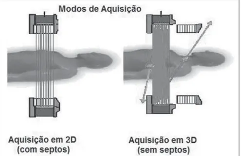
Positron emission tomography can be performed through two acquisition modes: 2D and 3D as shown on Figure 1.
The 2D acquisition mode utilizes tung-sten septa placed under the detectors. These septa act as antidiffusion grids utilized in
radiology. They cause a reduction in the photons incidence angle, resulting in a de-crease of up to 15% in the rate of false events produced by scattering photons. As a result, the images contrast resolution is improved although the equipment count rate performance decreases so the admin-istration of a higher radioactivity is re-quired for images acquisition(7,8).
In the 3D acquisition mode, the septa are retracted and an electronic collimation of the detected coincidences is utilized, resulting in up to six-fold higher count rates. As a result, there is an increase in the equipment sensitivity, allowing a reduction both in the radioactivity administered to the patient and in the acquisition time. On the other hand, as a disadvantage, the increase in the count rate receives the contribution of false coincidences (random and scatter-ing events), which cause a reduction in the images contrast resolution(7,8).
Centers where PET/CT scans are per-formed with the 2D acquisition mode re-place this protocol by the 3D mode as a strategy to reduce radiation doses for al-lowing the administration of lower radiop-harmaceutical activity.
In this context, studies aimed at demon-strating the appropriateness of 3D acquisi-tion mode to preserve the image quality with the administration of lower radiotracer ac-tivity are important for the clinical routine.
The present study is aimed at evaluat-ing whether images obtained with the 3D acquisition mode with two minutes (3D-2-min) and four minutes (3D-4-(3D-2-min) and lower FDG-18F activity, present the same image quality provided by the 2D acquisi-tion mode (2D-4-min), which is usually utilized as a standard in the department, and, based on this evaluation, validating a new protocol of 3D acquisition mode to replace the 2D mode, reducing the activity and, consequently, the radiation dose deliv-ered to the patients, without a loss in the image quality.
MATERIALS AND METHODS
The present study was developed with a Discovery ST PET/CT equipment (Gen-eral Electric Medical System; Milwaukee, USA) utilizing crystalline bismuth ger-manate (BGO)(9,10).
The experimental method employed image quality tests according to the 2001 version of the protocol of the National Electrical Manufacturers Association Stan-dards (NEMA) (NU 2-2001). A phantom (NEMA/IEC 2000; Biodex) was utilized to simulate a human thorax both in shape and size. This phantom includes six spheres with different diameters (1 cm, 1.3 cm, 1.7 cm, 2.2 cm, 2.8 cm, 3.7 cm) transversely positioned on a circumference (23 cm in diameter), and a cylinder (5 cm in diameter) positioned on the center(10).
In compliance with the protocol for test-ing image quality, the phantom and the four smaller spheres were filled with radioactive material at different concentrations, and the cylinder, with cotton to simulate the pulmo-nary parenchyma. With this arrangement, the authors could identify “hot” (four spheres) and “cold” (two spheres and the cylinder) images in relation to the back-ground (bg) radiation. The activities uti-lized in the phantom components were in compliance with the NEMA manual(10).
A NEMA phantom for scatter test is utilized to include information from scat-ter photons of other regions. This phantom is comprised of a cylinder with 70 cm in length and 20 cm in diameter, and a cen-tral canal through which passes a plastic tube filled with radioactive material. A whole-body study can be simulated with
this arrangement(10). The 3D acquisitions were performed two hours after the 2D acquisitions to take advantage from the 18F radioactive decay resulting in a reduction to about one-half of the initial activity. The images acquired were reconstructed on a Xeleris workstation (GEMS), by means of interactive ordered subset expectation maximization (OSEM), with 21 subsets and two iterations for the 2D mode, and Fourier rebinning-OSEM (FORE-OSEM) with 24 subsets and three iterations for the 3D mode.
Twenty-seven simulations were per-formed over a one-year period. In this pe-riod, the equipment was evaluated by means of daily, weekly and quarterly qual-ity control tests, according to the NEMA recommendations. The results of these tests are in compliance with the manufacturer standards.
The set of images resulting from each simulation were qualitatively analyzed on the workstation by three experienced radi-ologists who did not know the acquisition modes utilized. Each observer attributed a score 1 when at least one of the spheres was not identified or in cases where the image was dubious, or 2 for clearly visible spheres. The contrast value for the smallest sphere of each simulation was found through the image quality test of the NEMA protocol. It is defined as the relation between the activity calculated by the count of the de-signed region of interest (ROI) and the real activity. The contrast value in the “hot” spheres is calculated according to the fol-lowing formula(11):
where: CE = counts in the area correspond-ing to the smallest sphere; CB = count in the area corresponding to the bg; aE = activity concentration in the “hot” sphere; aB = ac-tivity concentration in the bg.
In the present study, only the contrast values for the smallest (1 cm) “hot” spheres were utilized.
Initially, all the data collected were de-scriptively analyzed. Subsequently, some summary measures, such as mean, standard deviation, among others, were calculated, and boxplots were constructed for quanti-tative variables(12).
The qualitative variables (observers’ scores) were analyzed through the calcula-tion of absolute and relative frequencies(12). The inferential analyses performed to confirm or contradict the evidences found by the descriptive analysis were: block vari-ances analysis, besides Bonferroni com-parison, as necessary(13); and estimation of generalized kappa coefficient of agree-ment(14).
A level of significance á corresponding to 5% was utilized to draw conclusions through the inferential analyses.
The data were appropriately stored in an Excel 2000 worksheet for Windows, and the software Statistical Package for Social Sciences (SPSS) version 11.0 for Windows was utilized for statistical analysis.
RESULTS
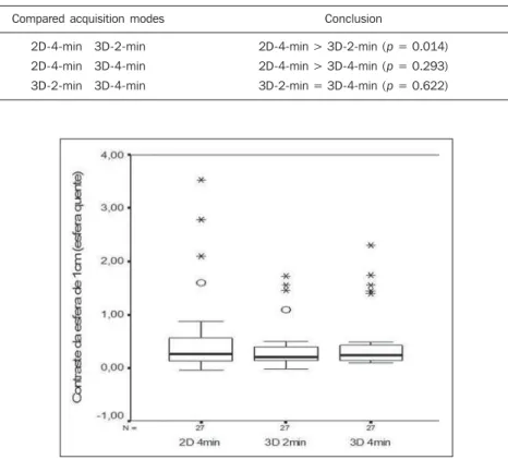
Data regarding contrast observed in the 1 cm sphere through the three acquisition modes are shown on Table 1 and Figure 2. The inferential results demonstrated that the mean 1 cm sphere contrast (“hot” sphere) is higher in the 2D acquisition mode (p = 0.014). Table 2 shows the comparison of mean values of the different acquisition modes for the 1 cm sphere contrast.
The evaluation of the interobserver agreement as regards the spheres visualiza-tion was also included in the present study. The rates of interobserver agreement in relation to the 2D-4-min, 3D-2-min and 3D-4-min acquisition modes were, respec-tively, 63.0%, 77.8% and 88.9%.
QE = × 100%
(CE/CB) – 1
(aE/aB) – 1
Table 2 Inferential results according to 1 cm sphere contrast (“hot” sphere).
Compared acquisition modes
2D-4-min 3D-2-min
2D-4-min 3D-4-min
3D-2-min 3D-4-min
Conclusion
2D-4-min > 3D-2-min (p = 0.014) 2D-4-min > 3D-4-min (p = 0.293) 3D-2-min = 3D-4-min (p = 0.622)
Table 1 Summary measures of 1 cm sphere contrast (“hot” sphere) according to the acquisition mode.
Summary measures
1 cm sphere contrast
Acquisition mode
2D-4-min
3D-2-min
3D-4-min
Mean
0.623
0.397
0.494 Median
0.270
0.200
0.240
Minimum
–0.040
–0.010
0.090
Maximum
3.530
1.720
2.290 SD
0.877
0.476
0.605
SD, standard deviation.
Generalized kappa coefficient was es-timated for quantifying the level of interob-server agreement. Table 3 with these esti-mates demonstrates a good interobserver agreement.
The images obtained were individually classified by each of the observers into vis-ible (score 2) or non-visvis-ible (score 1). This qualitative evaluation was performed for the different acquisition modes. The scores distribution among the acquisition modes is shown on Figure 3, where one can ob-serve that the percentage of times where all the spheres were identified was highest with the 3D-4-min acquisition mode. Ad-ditionally, the number of times where there was difficulty in visualizing the spheres with the 3D-4-min mode was three-fold
Figure 3. Scores distribution. The graphic demonstrates the scores distribu-tion for each acquisidistribu-tion mode according to the observers.
Modos de aquisição
Visível Não visível ou duvidosa
2D-4-min 3D-2-min 3D-4-min
100%
80%
60%
40%
20%
0%
52% 48%
63%
37%
85%
15%
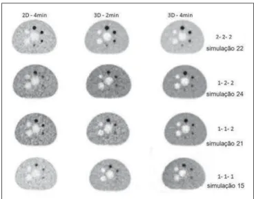
Figure 4. Images classification on different simulations. The figure presents the variation of image quality in different acquisition modes, with examples of the classification attributed to each simulation.
lower than with the 2D-4-min mode and two-fold lower than with the 3D-2-min mode.
Correlation between image quality and acquisition modes considering the category variables (visible or non-visible spheres) was determined by consensus among the observers based on a single score for the three acquisition modes on each simula-tion. The results of this distribution are shown on Table 4 and the image classifi-cation as well as their respective scores are shown on Figure 4.
These data were evaluated through the generalized kappa test whose results dem-onstrate a moderate correlation among the three acquisition protocols: 0.444 [0.227; 0.662].
DISCUSSION
Nuclear medicine services with BGO-based PET-CT scanners preferentially uti-lize the 2D acquisition mode which pro-vides better image quality. However, a higher FDG-18F activity is required in this acquisition mode and, consequently, the patient receives a higher radiation dose.
Studies comparing 2D and 3D acquisi-tion modes have been developed aiming at finding a balance in the utilization of these technological resources with dose optimi-zation. The 2001 version of the NEMA protocol for testing image quality was de-signed to simulate whole-body studies with both acquisition modes, and for this reason this protocol was utilized in the present study so that the patients were not unnec-essarily irradiated.
The analysis of the tests results demon-strated that the 2D acquisition mode pro-vides a better contrast resolution for “hot” spheres”. A similar result has been ob-served by Bettinardi et al.(16) and Mawlawi et al.(9), according to studies published in 2004, where these authors evaluated the performance of the same PET/CT system utilized in the present study, considering this equipment as efficient with both acqui-sition modes.
A relevant issue to be considered is that the patient bodily mass can influence the image quality. A study developed by El Fakhri et al.(17) has concluded that BGO-Table 3 Punctual and interval estimates* of generalized kappa coefficients of agreement.
*95% confidence interval, according to Fleiss(14). †Level of agreement according to Landis & Koch(15). Acquisition mode
2D-4-min
3D-2-min
3D-4-min
Coefficient of agreement
0.506 [0.288;0.724]
0.696 [0.478;0.914]
0.725 [0.507;0.942]
Level of agreement†
Moderate
Substantial
Substantial
Table 4 Distribution of spheres visualization demonstrating the correlation between acquisition modes.
2D-4-min
Visible
Non-visible
Non-visible
Non-visible
Visible
3D-2-min
Visible
Non-visible
Non-visible
Visible
Non-visible
3D-4-min
Visible
Non-visible
Visible
Visible
Visible
Total
13
4
5
4
1
%
48%
15%
19%
15%
based PET/CT scans produce similar re-sults in the detection of lesions with differ-ent sizes with both 2D and 3D acquisition modes, but the 3D mode presents a better performance in the detection of lesions in patients with body mass index < 33; on the other hand, the 2D mode is better for pa-tients with body mass index > 34.
A study published by Visvikis et al.(18) in 2005 also demonstrated that in studies performed with BGO-based equipment the images quality is similar with both the 2D and 3D acquisition modes. In this study, the author has observed a correlation among image quality, patient’s body weight and acquisition time, and has demonstrated that the increase in the patient weight leads to a progressive loss of image quality in both acquisition modes, but particularly with the 3D mode in patients weighting more than 70 kg.
Lodge et al. have compared 2D and 3D acquisition modes in a PET scanner with lutetium oxyorthosilicate crystal (LSO) and have observed that the 3D acquisition mode presented a better performance be-cause of the crystal characteristics, allow-ing a reduction of dose and acquisition time(19).
Although LSO-based equipment is more appropriate for 3D-acquisition, in-creased patient weight leads to a loss in the image quality, so an extended acquisition time is required to compensate this prob-lem(20).
Bettinardi et al. have evaluated the Dis-covery STE with a new configuration of BGO crystals, and have concluded that the greatest advantage of the 3D acquisition mode is the increase in the system’s sensi-tivity and the utilization of new reconstruc-tion algorithms allowing an improvement in the images quality, in the capability to detect lesions and in the accuracy of the method as compared with the 2D acquisi-tion(21).
The results of the present study demon-strated a moderate correlation among the 2D-4-min, 3D-2-min and 3D-4-min acqui-sition modes as regards the visualization of “hot” spheres, generalized kappa
coeffi-cient = 0.444 [0.227;0.662], highlighting the 3D-4-min acquisition mode, where all the spheres were visible for the highest number of times (85%). The rate of interob-server agreement was higher with the 3D-4-min than with the other acquisition modes (88.9%), with a generalized kappa coefficient = 0.725 [0.507;0.942]. These results are significant because they indicate that the 3D acquisition mode can be uti-lized with BGO-based PET/CT scanners for patients whose thorax dimensions are similar to the phantom’s. Based on the re-sults of the present study, the authors could establish protocols with lower FDG-18F activity, which will be very useful in the case of pediatric patients.
CONCLUSION
The experimental study has demon-strated that there is no loss in imaging qual-ity with the utilization of the 3D acquisi-tion mode with half the administered activ-ity The 3D-4-min acquisition mode has allowed the spheres visualization in the majority of tests, so this technique can be utilized as a strategy to reduce the FDG-18F activity delivered to patients with a biotype similar to the phantom.
The results of the present study suggest that the proposed 3D-4-min acquisition mode with BGO-based PET/CT scanners will allow a reduction in the radiation ac-tivity and, consequently, lower irradiation doses to the patient, affecting especially the application of PET in pediatrics.
REFERENCES
1. Lomeña F, Soler M. Clinical application of PET. Braz Arch Biol Technol. 2005;48(no. spe 2):179– 83.
2. Townsend DW, Beyer T, Blodgett TM. PET/CT scanners: a hardware approach to image fusion. Semin Nucl Med. 2003;33:193–204.
3. Hahn K, Pfluger T. Is PET/CT necessary in pae-diatric oncology? Against. Eur J Nucl Med Mol Imaging. 2006;33:966–8.
4. Stauss J, Franzius C, Pfluger T, et al. Guidelines for 18F-FDG PET and PET-CT imaging in paedi-atric oncology. Eur J Nucl Med Mol Imaging. 2008;35:1581–8.
5. Brix G, Lechel U, Glatting G, et al. Radiation ex-posure of patients undergoing whole-body
dual-modality 18F-FDG PET/CT examinations. J Nucl Med. 2005;46:608–13.
6. Cálculo da dose efetiva. [acessado em 30 de julho de 2008]. Disponível em: http://www.doseinfo--radar.com/RADARDoseRiskCalc.html
7. Townsend DW. Physical principles and technol-ogy of clinical PET imaging. Ann Acad Med Singapore. 2004;33:133–45.
8. Schöder H, Erdi YE, Larson SM, et al. PET/CT: a new imaging technology in nuclear medicine. Eur J Nucl Med Mol Imaging. 2003;30:1419–37. 9. Mawlawi O, Podoloff DA, Kohlmyer S, et al. Per-formance characteristics of a newly developed PET/CT scanner using NEMA standards in 2D and 3D modes. J Nucl Med. 2004;45:1734–42.
10. GE Healthcare. DST PET-CT NEMA test proce-dures, applicable to Discovery ST. Direction 5159176-100, Revision 2. Walkesha: General Electric Company; 2006.
11. National Electrical Manufacturers Association. Standards Publication NU-2-2001: performance measurements of positron emission tomography. Rosslyn: National Electrical Manufacturers As-sociation; 2001.
12. Bussab WO, Morettin PA. Estatística básica. 5ª ed. São Paulo: Saraiva; 2006.
13. Neter J, Kutner MH, Nachtsheim CJ, et al. Ap-plied linear statistical models. 4th ed. Boston: Irwin; 1996.
14. Fleiss JL. Statistical methods for rates and pro-portions. 2nd ed. New York: John Wiley; 1981.
15. Landis JR, Koch GG. The measurement of ob-server agreement for categorical data. Biometrics. 1977;33:159–74.
16. Bettinardi V, Danna M, Savi A, et al. Performance evaluation of the new whole-body PET/CT scan-ner: Discovery ST. Eur J Nucl Med Mol Imaging. 2004;31:867–81.
17. El Fakhri G, Holdsworth C, Badawi RD, et al. Impact of acquisition geometry and patient habi-tus on lesion detectability in whole body FDG-PET: a channelized hotelling observer study [ab-stract]. Nuclear Science Symposium Conference Record, 2002 IEEE. 2002;3:1402.
18. Visvikis D, Griffiths D, Costa DC, et al. Clinical evaluation of 2D versus 3D whole-body PET image quality using a dedicated BGO PET scan-ner. Eur J Nucl Med Mol Imaging. 2005;32: 1050–6.
19. Lodge MA, Badawi RD, Gilbert R, et al. Com-parison of 2-dimensional and 3-dimensional ac-quisition for 18F-FDG PET oncology studies per-formed on an LSO-based scanner. J Nucl Med. 2006;47:23–31.
20. Halpern BS, Dahlbom M, Quon A, et al. Impact of patient weight and emission scan duration on PET/CT image quality and lesion detectability. J Nucl Med. 2004;45:797–801.