Deconvoluting Protein (Un)folding Structural
Ensembles Using X-Ray Scattering, Nuclear
Magnetic Resonance Spectroscopy and
Molecular Dynamics Simulation
Alexandr Nasedkin1, Moreno Marcellini2, Tomasz L. Religa3, Stefan M. Freund4, Andreas Menzel5, Alan R. Fersht4, Per Jemth6, David van der Spoel2, Jan Davidsson1*
1Department of Chemistry-Ångström laboratory, Uppsala University, Box 523, SE-75110 Uppsala, Sweden,
2Uppsala Center for Computational Chemistry, Science for Life Laboratory, Department of Cell and Molecular Biology, Uppsala University, Box 596, SE-75124 Uppsala, Sweden,3Department of Physiology and Biophysics, Case Western Reserve University, Cleveland, Ohio 44106, United States,4Medical Research Council Laboratory of Molecular Biology, Cambridge CB2 0QH, United Kingdom,5Paul Scherrer Institut, 5232 Villigen-PSI, Switzerland,6Department of Medical Biochemistry and Microbiology, Uppsala University, BMC Box 582, SE-75123 Uppsala, Sweden
Abstract
The folding and unfolding of protein domains is an apparently cooperative process, but tran-sient intermediates have been detected in some cases. Such (un)folding intermediates are challenging to investigate structurally as they are typically not long-lived and their role in the (un)folding reaction has often been questioned. One of the most well studied (un)folding pathways is that ofDrosophila melanogasterEngrailed homeodomain (EnHD): this 61-resi-due protein forms a three helix bundle in the native state and folds via a helical intermediate. Here we used molecular dynamics simulations to derive sample conformations of EnHD in the native, intermediate, and unfolded states and selected the relevant structural clusters by comparing to small/wide angle X-ray scattering data at four different temperatures. The results are corroborated using residual dipolar couplings determined by NMR spectroscopy. Our results agree well with the previously proposed (un)folding pathway. However, they also suggest that the fully unfolded state is present at a low fraction throughout the investi-gated temperature interval, and that the (un)folding intermediate is highly populated at the thermal midpoint in line with the view that this intermediate can be regarded to be the dena-tured state under physiological conditions. Further, the combination of ensemble structural techniques with MD allows for determination of structures and populations of multiple inter-converting structures in solution.
a11111
OPEN ACCESS
Citation:Nasedkin A, Marcellini M, Religa TL, Freund SM, Menzel A, Fersht AR, et al. (2015) Deconvoluting Protein (Un)folding Structural Ensembles Using X-Ray Scattering, Nuclear Magnetic Resonance Spectroscopy and Molecular Dynamics Simulation. PLoS ONE 10(5): e0125662. doi:10.1371/journal.pone.0125662
Academic Editor:Sabato D'Auria, CNR, ITALY
Received:December 11, 2014
Accepted:March 11, 2015
Published:May 6, 2015
Copyright:© 2015 Nasedkin et al. This is an open access article distributed under the terms of the
Creative Commons Attribution License, which permits unrestricted use, distribution, and reproduction in any medium, provided the original author and source are credited.
Data Availability Statement:Data are available from
http://dx.doi.org/10.6084/m9.figshare.1356347.
Funding:Per Jemth and Jan Davidsson were funded by the Swedish Research Council, grants NT 2012-5096 (P.J.) and NT 2012-3908 (http://www.vr.se/). The funders had no role in study design, data collection and analysis, decision to publish, or preparation of the manuscript.
Introduction
The folding of proteins to their functional conformations has been studied extensively both ex-perimentally and through theoretical simulations. There has been great progress in under-standing (un)folding reactions, in particular for small fast folding (μs-ms) protein domains. Despite the apparent complexity, protein (un)folding reactions are usually fast (μs-s) and often occur without accumulation of intermediates, which can be illustrated using a smooth funneled energy landscape [1–3]. Nevertheless, there are several cases where intermediates accumulate in protein (un)folding reactions as on-pathway species [4–8]. There is however an ongoing de-bate about whether these intermediates are productive in the strict definition that they are obligatory species on the path to the native state.
Among fast-folding proteins,Drosophila melanogasterEngrailed homeodomain (EnHD) is one of the best studied systems. A combination of experimental and computational methods have demonstrated that EnHD folds by initial formation of secondary structure elements that subsequently dock to form the native state [9,10] in line with the diffusion-collision model [11]. Protein engineering in combination with nuclear magnetic resonance (NMR) showed that this (un)folding intermediate (or denatured state under physiological conditions, Dphys)
contains both native and non-native helices [12], and that the helix-turn-helix motif constitut-ing H2-H3 forms independently of H1 [13], and can thus be regarded the main structural unit of the intermediate.
Small- to wide-angle X-ray scattering (S/WAXS) in solution has emerged as a powerful structural probe of biomolecules that explicitly side-step the fundamental limitations of con-ventional X-ray diffraction methods that probe proteins in the crystalline phase. It is in general not feasible to directly extract 3D atomic structures from disordered systems. However, by fit-ting S/WAXS data against structures obtained from X-ray crystallography, NMR spectroscopy and/or from theoretical modeling it is possible to extract information about the structural en-semble in the sample [14]. WAXS extends the data present in the conventional SAXS regime (q*0.3 Å−1), where information about shape and size of macromolecules can be obtained, out to a regime where scattering fingerprints from internal protein structures are present and can thus be viewed as a high-resolution extension of SAXS which provides low-resolution structural information of proteins [15]. Structural intermediates have uniquely been identified for smaller molecules in several time-resolved WAXS experiments [16–18] demonstrating the achievable resolution of the technique. WAXS has recently been extended to also probe the re-arrangement of secondary structural elements within proteins [19]. In addition, a methodology based on refining the molecule of interest toward solution scattering data using MD simulation has very recently been developed and successfully applied on several molecular systems [20].
HN-N residual dipolar couplings (RDCs) are highly sensitive to the orientations of amide
bond vectors within the molecular frame [21,22]. Small changes in the relative bond vector ori-entation within that frame will result in a coupling different from that predicted from the refer-ence structure. A poor correlation between measured and predicted data suggests a change in secondary or tertiary structures. The agreement of the experimental data set to the structure is evaluated using either a simple correlation coefficient (R) or, more commonly, using the Cornilescu Q factor (Q = rms(Dcalc-Dobs)/rms(Dobs), where rms, Dcalc, and Dobsrepresent
root-mean-square deviation, predicted and observed RDCs, respectively) [23]. Solution structures corresponding to high resolution X-ray structures typically have Q<0.25 [24].
Kozaket al.[28] We demonstrate the presence of three distinct ensembles of species during (un)folding of EnHD. These ensembles correspond to the denatured state D, an (un)folding in-termediate I, corresponding to the denatured state under physiological conditions and the na-tive state N. In particular, data at temperatures ranging from 20°C to 55°C suggest that the intermediate state becomes populated near the midpoint (apparent midpoint for thermal un-folding is about 52°C), rather than a more extended denatured state. We also find that the en-sembles of substates within each population fit the S/WAXS data significantly better than for example the average NMR structure, thus capturing the flexible multi-state nature of proteins. The conclusions from S/WAXS and MD simulations are corroborated using residual dipolar couplings (RDCs) obtained from NMR experiments at different temperatures, showing the po-tential of the approach to detect and characterize protein (un)folding intermediates by X-ray scattering and MD simulations.
Materials and Methods
X-ray solution scattering
The S/WAXS experiments were carried out at the cSAXS beamline of the Swiss Light Source using a rapid-readout pixel detector, Pilatus [29]. The EnHD protein was expressed and puri-fied as previously described [30]. A solution of 1.1 mM EnHD in 50 mM HEPES, 100 mM NaCl at pH 8.0 was delivered into the monochromatic X-ray beam (12.44 keV,*1012 pho-tons/s in a 300 × 300μm spot) by being pumped through a 1 mm diameter (0.98 mm internal diameter) quartz capillary. Part of the tubing and the capillary was placed in-between two metal plates with a small hole for the X-ray to enter the capillary. The temperature was regulat-ed by a thermo coupler and a cooling system integratregulat-ed in the metal plates. About 30 cm of tubing on each side of the capillary was inserted in between the plates to ensure that the whole sample volume was heated to the desired temperature.
Pump triggering was integrated within the beam line control system, and for each acquisi-tion cycle the sample was pumped continuously (2μl/s) to ensure that a new sample volume was exposed to X-ray for each measurement. During the acquisition cycle a sequence of scatter-ing images were recorded usscatter-ing an integration time of 7 ms and a readout delay of 3 ms, equat-ing to a readout frequency of 100 Hz. Pixel maskequat-ing and radial integration of each Pilatus frame was computed at the beamline by in house software. A MATLAB script specifically writ-ten for this experiment was later utilized to filter out outliers (mainly caused by bubbles or ag-gregates in the solution), to normalize the intensity of the single azimuthally integrated frame to the incoming flux, for the statistical analysis, and to subtract the buffer signal from the protein solution.
In the scattering data there was some indications of aggregation (enhanced scattering at very lowq), especially at elevated temperatures, and therefore a lower limit ofq= 0.07 Å−1was used in the structural analysis (q= 4πsin(θ)/λ, where 2θis the scattering angle of incident X-ray beam andλis the wavelength of the X-ray photons). Traditional Guinier plots, atq span-ning from 0.01 to 0.1 Å−1for proteins of this size, give the radius of gyration, i.e. the average di-mension of the particles but not much information otherwise. Atq-values below 0.07 Å−1 particle scattering will thus completely dominate and this region will be of less importance in the structural analysis performed in this work.
Residual dipolar couplings
The HN-N residual dipolar couplings were measured as described previously [12] in 20 mM
data was analyzed using PALES software [32] using the 1ENH crystal structure as the template [33]. The conditions for the measurements are slightly different from those in the SAXS mea-surements in order to lower the stability of EnHD and ensure that the RDCs can be measured past the Tm of the protein on our Bruker DRX500 spectrometer equipped with a single axis gradient cryo-probe. Change in buffer condition causes decrease in Tm by about 10°C, yet since there are no histidine residues and all basic or acidic side chains are well solvated, there is no reason to assume the protonation state of the protein would change significantly.
Molecular dynamics simulations
MD simulations of the EnHD (PDB ID: 2JWT) were carried out based on the first NMR model of EnHD in the PDB file, i.e. the most optimized structure. Simulations were performed using the GROMACS package [34] and the AMBER99SB-ILDN force field [35] with the TIP3P water model [36]. The protein molecule was immersed in a periodic box containing 32759 water molecules. Additionally, 60 Na+ and 68 Cl- were added to reach a salt concentration of 50 mM corresponding to the experimental value and neutralizing the positive protein charge. The box size was*10 nm which prevented protein interactions through periodical bound-aries even in the unfolded state. A cutoff distance 1 nm has been used for calculations of Len-nard-Jones interactions and PME was used for the treatment of the long-range electrostatic interactions. An integration time step of 2 fs was used and the Berendsen algorithm for temper-ature and pressure control [37] was used with 0.1 and 1 ps coupling constants, respectively. The pressure was kept at 1 atm. In total eight trajectories of each 100 ns were generated. The temperature for each production run was 275, 300, 325, 350, 375, 400, 450 and 500 K. Before the production run, the system was minimized using steepest descent for 1000 steps after which the system was equilibrated in dynamic simulation 20 ps long with 1 fs time step at 275 K. Atomic coordinates from the last equilibration snapshot were then used as an input for the production runs at different temperature.
Cluster analysis
Protein structures for fitting to the experimental data were obtained after cluster analysis on each MD trajectory. Clustering has been accomplished by the algorithm due to Daura et al. [38] which is implemented in the GROMACS package [34]. Structures for the cluster analysis were sampled from MD-trajectory every 10 ps, for a total of 10000 structures from each trajec-tory. As the clustering criterion, the root mean square deviation (RMSD) of main-chain and C-beta atoms was used. Cut-off clustering distances of 1.0, 2.5 and 3.5 Å have been used. Only clusters consisting of ten or more structures have been taken into account for SAXS fitting. The representative structure for each cluster was taken to be the structure from the MD-trajectory most closely located to the center of the cluster in RMSD space.
Computations of X-ray scattering spectra and NMR restraints
Atomic distances have been calculated directly from the protein structure by g_disre pro-gram [40], which is a part of the GROMACS package. EnHD is in fast exchange in NMR chem-ical shift timescale, since it is an ultrafast folder (>100 000/s), those RDCs and NOEs could be
interpreted as being averaged over all the populated conformers. Ensemble-averaged NMR vio-lations have been calculated based on experimental distance restraints [12] and atomic dis-tances averaged over all conformationsiof the ensemble by applying power-averaging,d= (∑(di)−n)−1/n. The powernis usually taken between three and six, where the higher number gives better correlation between calculated and experimental distance restrains [41]. In the cur-rent workn= 3 was selected.
Optimization algorithm
The ensemble optimization approach by Bernado and co-workers [42] was utilized to obtain the ensemble of protein structures that reproduced the experimental scattering data using an it-erative genetic algorithm [43]. Ensembles were formed from an extensive pool of conformers generated by MD simulations. The fitting was performed to reproduce the logarithm of the ex-perimental scattering intensity, log I(q). The square difference between exex-perimental and en-semble-averaged X-ray scattering was used as the target function in the fitting. At each step of the algorithm several ensembles of structures, denominated chromosomes, were tested and sorted according to the value of the target function. The specified number of ensembles show-ing the lowest values of target function was selected to pass into the next iteration. The size of each ensemble had a maximum of 20 spectra. All spectra were of equal weight in the chromo-some, yet multiple repetitions of structures were allowed, which gives the possibility to change the partial weights of the structures that contributes to the ensemble. Optimization was per-formed for 10000 iterations and repeated 50 times. In total 20 ensembles were selected to pass through each iteration of the algorithm. The crossover operator was tuned to generate the same number of chromosomes as the initial population. The number of chromosomes generat-ed by the mutation operator excegenerat-edgenerat-ed the number of initial chromosomes by the factor of two. This last option was selected in order to achieve rapid convergence toward the optimal solution due to higher ensemble divergence created by the mutation operator. The code for the optimi-zation algorithm was implemented in a MATLAB package and is available from the authors upon request.
Results
Molecular dynamics simulations for sampling
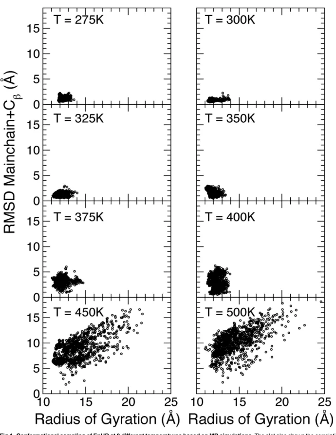
Fig 1. Conformational sampling of EnHD at 8 different temperatures based on MD simulations.The plot also shows the correlation between radius of gyration and root mean square deviation from the NMR structure. The RMSD have been calculated for the mainchain and Cβatoms of all 61 residues.
Modeling the structural ensembles from S/WAXS data
Conventional SAXS experiments give information mainly about the overall shape and size of proteins in solution. By extending the collected scattering angles, into the range between 0.4 and 0.7 Å−1where scattering more specifically related to secondary and tertiary protein struc-tures will be present, the conformational ensemble present in the sample can be characterized. In an optimization procedure [42], based on an iterative genetic algorithm [43], the ensemble of protein structures that best reproduced the experimental data was selected from a pool of MD and NMR structures as is further discussed in Materials and Methods.
X-ray scattering (0.01<q<0.7 Å−1) was collected from a 1.1 mM sample of EnHD at four
temperatures, 20, 30, 40 and 55°C (Fig 2a) in an attempt to structurally resolve conformational changes in the folding/unfolding process of the protein.
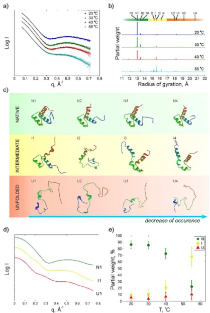
The scattering profiles (Fig 2a) show a pronounced broad peak at around 0.5 Å−1that fades away with temperature. This pattern provides the signature of a significant shift in the protein population towards less folded structures at the melting point.
Fig 2bshows the radius of gyration of the protein structure ensemble obtained from the fit-ting to the scattering data using the optimization algorithm (seeMaterials and Methods). Three groups of structures can be identified. The first group located around 13 to 14 Å (green) contains native-like structures which dominate at low temperatures. The second group (yellow) contains proteins with less organized structures with radii of gyration between 14.5 and 17 Å. The third group (red) at higher radii of gyration contains denatured structures where some of the helices are completely unfolded. InFig 2ca structural comparison of the four most abun-dant structures in each group is displayed. The numbering within each group is related to the relative weight of these structures in the fitting at most temperatures. The N1 structure, which is selected with high abundance in the native group, is the 12thstructure from the NMR
ensem-ble consisting of 25 structures (PDB ID: 2JWT [12]).Fig 2dshows the calculated scattering profiles for the most abundant structures in each group. A clear difference in scattering signa-tures between the three groups of strucsigna-tures is apparent, and it is obvious that the structure seen around 0.5 Å−1 in the experimental data at low temperature is related to a more folded structure while an unfolded structure gives a more straight scattering profile. InFig 2ethe rela-tive weight of the three groups at the four temperatures is shown. As expected, a transition from native like structures towards more unfolded structures with increasing temperature is observed.
Validation of the structural ensemble using RDCs
Prior comparison of the solution NMR parameters of EnHD to the X-ray data revealed an out-standing agreement (Cornilescu Q factor<0.25) of the EnHD HN-N RDCs when the protein
was folded [12]. For a fully unfolded protein, the HN-N RDCs in radially compressed
acrylam-ide gels will be slightly negative, and there will be no agreement between the measured RDCs and the crystal structure across all of the protein [44].
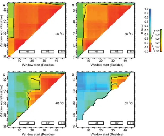
As we measured the RDCs at increasing temperatures for EnHD, we observed that while the agreement for the 10–55 residue range, which encompasses H1/H2/H3 was lost, the values
were still consistent with a preserved HTH motif at 40°C (Fig 3b). Even at 55°C, close to the ap-parent midpoint for thermal denaturation [9], the agreement of the HN-N RDCs to the crystal
structure for the 28–52 residue fragment was as good, suggesting a significant population of
Discussion
The ensemble optimization of conformations generated by MD simulations (Fig 1) against the S/WAXS data allows for deconvolution of structures and populations of multiple interconvert-ing structures in solution. This gives new insights into the structural complexity of protein (un) folding in Engrailed Homeodomain. The obtained S/WAXS structures at low temperatures are in good agreement with those obtained in previous NMR studies [12] while the structures at temperatures close to the thermal midpoint (Fig 2) are compatible with the HN-N residual
cou-plings (Fig 3).
Fig 2. X-ray scattering intensity as a function of the scattering vector q at four different temperatures.
b) histogram of radius of gyrations of proteins obtained from the optimization procedure at the different temperatures, c) the four most populated structures for native, intermediate and unfolded as determined by a cluster analysis, d) simulated scattering curves for the most populated native (N1), intermediate (I1) and unfolded conformations (U1) and e) population of native (N), intermediate (I) and unfolded (U) as a function of temperature. The error bar at e) indicates a standard deviation obtained in ensemble fitting. Scattering profiles at a) and d) and weight distribution at b) are shifted to increase visibility.
From the fitting of the X-ray scattering data we can resolve three groups of structures that seem to be present at all temperatures but with varying occupancy. Few structures are pre-sented in the optimized ensembles, which is not surprising due to high stability of the protein and fast convergence of the optimization algorithm. As expected, the occurrence of the native-like structures will strongly decrease at the thermal midpoint, from almost 90% to 20%. At 55°C an intermediate structure, I1, present already at low temperature, will increase in abun-dance while some more unfolded intermediate structures (I2 and I4) appear as well. It is partic-ularly interesting to note that at 55°C the population of intermediate and denatured states contains a high degree of secondary structure.
At the lowest temperatures one of the NMR structures [12], N1, was selected with a partial weight of 65% in the fitting, demonstrating that the S/WAXS data is consistent with the known experimental structure of EnHD and, of significance for the whole analysis, that the optimiza-tion algorithm works reliably. The other three structures in the native group are quite similar to the NMR structure where the main differences are located at the termini. In the intermediate group the helices are usually maintained with a few exceptions such as I2 for which two of the helices, H1 and H2, are more or less unfolded. Less than 6% of the total ensemble at 20°C con-sists of unfolded structures.
Fig 3. Agreement of HN-N RDCs to EnHD crystal structure at increasing temperatures.The Cornilescu
Q factors were calculated using a sliding window from‘Window start’to‘Window end’to estimate the agreement of the given residue range. Q = 0.25 was used as the cut-off for the agreement [24]. At 20°C (a) and 30°C (b) EnHD is fully folded, and any chosen residue window displays perfect agreement with the crystal structure. At 40°C (c) the agreement for the 10–55 fragment is not present, but the HTH motif remains structured. This indicates that H1 must be undocked from H2/H3. C. At 55°C (d) around the thermal midpoint of denaturation, some HN-N RDCs for H2/H3 remain in agreement with the crystal structure.
The averaged RMSD for S/WAXS-fitted ensembles is in the range of 1.1 to 3.5 Å, as shown inTable 1), which is comparable with structural ensembles defined by 2D NOESY NMR [12]. The number of violated restraints for 20, 30 and 40°C are below 1%. Similar number of violated restraints was obtained for NMR-refined and crystal structures [12]. Larger structural ensem-bles applied for calculation of NMR restrains violations slightly improve the number of violated restrains [45], yet fitting of SAXS data with such ensembles is computationally expensive. In addition, the use of only the NMR structures in the optimization against the X-ray data results in a much poorer fitting with about 50% largerχ-value, which again indicates the presence of
additional structures in the sample than those recovered from NMR structural ensemble [12]. It was previously shown that the H2-H3 helix-turn-helix motif of EnHD folds independent-ly of H1 and with a rate constant similar to the fast phase for folding of full-length EnHD [13]. This, and data on the destabilized L16A mutant of EnHD, which is molten-globule like under physiological conditions due to the lost tertiary contact at the 16thresidue [46], make a strong
case for the H2-H3 motif being present in the folding intermediate and/or the denatured states. The present data provide even more detail. Firstly, the proposed H2-H3 motif is present in some of the intermediate structures (I3 and I4,Fig 2e) but not in the other two. The RDC ma-trix at 40°C also suggests that the interaction between H1 and H2/H3 is weak. Note that the RDC plots present an ensemble average and hence the result at 40°C qualitatively agrees with structure analysis based on S/WAXS and MD. Secondly, the unfolded structures (Fig 2e) are relatively compact as well. Thirdly, the structures of the intermediates at 55°C contain a signifi-cant fraction of alpha helix, suggesting they could be characterized as native-like in circular di-chroism experiments for instance, and give an apparent higher population of native-like structures in such an analysis.
Protein folding is a complex process and sometimes intermediate states that may be de-tected using one technique cannot be observed using other means. Here we have shown that by combining experimental techniques with theoretical modeling it is possible to extract new in-formation from even a well-studied protein like EnHD. Simplified descriptions of protein fold-ing in terms of funnels [1,2] have been useful in formulating some general principles of folding. However, atomic-level descriptions of protein folding pathways are dominated by spe-cific interactions within proteins and between protein and solvent and require theoretical sim-ulations that use a realistic description of protein and solvent [47–53]. On the experimental side, local interactions require elucidation at the atomic and near atomic levels by such tech-niques as NMR [54,55] and byF-value analysis [56]. Where possible, multiple complementary experimental techniques should be used that give further local and global information on struc-ture. From the native state of a protein the folding road is uphill. Progress in studying protein folding under physiological conditions and in the context of, for instance, misfolding-induced disease depends on those methods and ideas [57]. Further, the methodology used here should
Table 1. The number of violated restraints according to Fletcher et al. [45] The total number of NOE re-straints is 735.The RMSD have been averaged over all structures in the S/WAXS refined ensemble.
Experimental temperature Violated restraints RMSD, Mainchain + Cβ,Å
20°C 3(<0.5%) 1.1
30°C 2(<0.5%) 1.1
40°C 3(<0.5%) 1.8
55°C 68(9%) 3.5
not only be restricted to folding but should, in principle, be applicable in any determination of protein structures and populations of multiple interconverting structures in solution.
Author Contributions
Conceived and designed the experiments: AN MM PJ JD. Performed the experiments: AN MM TLR AM JD. Analyzed the data: AN MM TLR PJ DvdS JD. Contributed reagents/materials/ analysis tools: PJ DvdS. Wrote the paper: AN MM TLR SMF AM ARF PJ DvdS JD.
References
1. Bryngelson JD, Wolynes PG. Spin glasses and the statistical mechanics of protein folding. P Natl Acad Sci USA. 1987; 84(21):7524–7528. Available from:http://www.pnas.org/content/84/21/7524.abstract. doi:10.1073/pnas.84.21.7524
2. Bryngelson JD, Onuchic JN, Socci ND, Wolynes PG. Funnels, pathways, and the energy landscape of protein folding: A synthesis. Proteins. 1995; 21(3):167–195. doi:10.1002/prot.340210302PMID: 7784423
3. Oliveberg M, Wolynes PG. The experimental survey of protein-folding energy landscapes. Q Rev Bio-phys. 2005 8; 38:245–288. doi:10.1017/S0033583506004185PMID:16780604
4. Jemth P, Gianni S, Day R, Li B, Johnson CM, Daggett V, et al. Demonstration of a low-energy on-path-way intermediate in a fast-folding protein by kinetics, protein engineering, and simulation. P Natl Acad Sci USA. 2004; 101(17):6450–6455. Available from:http://www.pnas.org/content/101/17/6450. abstract. doi:10.1073/pnas.0401732101
5. Ferguson N, Capaldi AP, James R, Kleanthous C, Radford SE. Rapid folding with and without populat-ed intermpopulat-ediates in the homologous four-helix proteins Im7 and Im9. J Mol Biol. 1999; 286(5):1597–
1608. Available from:http://www.sciencedirect.com/science/article/pii/S0022283698925487. doi:10. 1006/jmbi.1998.2548PMID:10064717
6. Khorasanizadeh S, Peters ID, Roder H. Evidence for a three-state model of protein folding from kinetic analysis of ubiquitin variants with altered core residues. Nat Struct Mol Biol. 1996 Feb; 3(2):193–205. doi:10.1038/nsb0296-193
7. Travaglini-Allocatelli C, Gianni S, Morea V, Tramontano A, Soulimane T, Brunori M. Exploring the Cyto-chrome c Folding Mechanism: CYTOCHROME c552 FROM THERMUS THERMOPHILUS FOLDS THROUGH AN ON-PATHWAY INTERMEDIATE. J Biol Chem. 2003; 278(42):41136–41140. Available from:http://www.jbc.org/content/278/42/41136.abstract. doi:10.1074/jbc.M303990200PMID: 12842869
8. Teilum K, Poulsen FM, Akke M. The inverted chevron plot measured by NMR relaxation reveals a na-tive-like unfolding intermediate in acyl-CoA binding protein. P Natl Acad Sci USA. 2006; 103(18):6877–
6882. Available from:http://www.pnas.org/content/103/18/6877.abstract. doi:10.1073/pnas. 0509100103
9. Mayor U, Johnson CM, Daggett V, Fersht AR. Protein folding and unfolding in microseconds to nano-seconds by experiment and simulation. P Natl Acad Sci USA. 2000; 97(25):13518–13522. Available from:http://www.pnas.org/content/97/25/13518.abstract. doi:10.1073/pnas.250473497
10. Mayor U, Guydosh NR, Johnson CM, Grossmann JG, Sato S, Jas GS, et al. The complete folding path-way of a protein from nanoseconds to microseconds. Nature. 2003 Feb; 421(6925):863–867. doi:10. 1038/nature01428PMID:12594518
11. Karplus M, Weaver DL. Protein folding dynamics: The diffusion-collision model and experimental data. Protein Sci. 1994; 3(4):650–668. doi:10.1002/pro.5560030413PMID:8003983
12. Religa TL. Comparison of multiple crystal structures with NMR data for engrailed home-odomain. J Bio-mol NMR. 2008; 40(3):189–202. doi:10.1007/s10858-008-9223-9PMID:18274703
13. Religa TL, Johnson CM, Vu DM, Brewer SH, Dyer RB, Fersht AR. The helixturnhelix motif as an ultra-fast independently folding domain: The pathway of folding of Engrailed homeodomain. P Natl Acad Sci USA. 2007; 104(22):9272–9277. Available from:http://www.pnas.org/content/104/22/9272.abstract. doi:10.1073/pnas.0703434104
15. Makowski L. Characterization of proteins with wide-angle X-ray solution scattering (WAXS). J Struct Functional Genomics. 2010; 11(1):9–19. doi:10.1007/s10969-009-9075-x
16. Neutze R, Wouts R, Techert S, Davidsson J, Kocsis M, Kirrander A, et al. Visualizing photochemical dy-namics in solution through picosecond X-ray scattering. Phys Rev Lett. 2001; 8719(19):–.
17. Davidsson J, Poulsen J, Cammarata M, Georgiou P, Wouts R, Katona G, et al. Structural determination of a transient isomer ofCH2I2by picosecond X-ray diffraction. Phys Rev Lett. 2005 Jun; 94:245503.
doi:10.1103/PhysRevLett.94.245503
18. Ihee H, Wulff M, Kim J, Adachi Si. Ultrafast X-ray scattering: structural dynamics from diatomic to pro-tein molecules. Int Rev Phys Chem. 2010; 29(3):453–520. doi:10.1080/0144235X.2010.498938 19. Westenhoff S, Nazarenko E, Malmerberg E, Davidsson J, Katona G, Neutze R. Time-resolved
structur-al studies of protein reaction dynamics: a smorgasbord of X-ray approaches. Acta Cryststructur-allogr A. 2010; 66(2):207–219. doi:10.1107/S0108767309054361PMID:20164644
20. Bjorling A, Niebling S, Marcellini M, van der Spoel D, Westenhoff S. Deciphering Solution Scattering Data with Experimentally Guided Molecular Dynamics Simulations. Journal of Chemical Theory and Computation. 2015; 11(2):780–787. doi:10.1021/ct5009735PMID:25688181
21. Bax A. Weak alignment offers new NMR opportunities to study protein structure and dynamics. Protein Sci. 2003; 12(1):1–16. doi:10.1110/ps.0233303PMID:12493823
22. Mohana-Borges R, Goto NK, Kroon GJA, Dyson HJ, Wright PE. Structural Characterization of Unfolded States of Apomyoglobin using Residual Dipolar Couplings. J Mol Biol. 2004; 340(5):1131–1142. Avail-able from:http://www.sciencedirect.com/science/article/pii/S0022283604005881. doi:10.1016/j.jmb. 2004.05.022PMID:15236972
23. Cornilescu G, Marquardt JL, Ottiger M, Bax A. Validation of Protein Structure from Anisotropic Carbonyl Chemical Shifts in a Dilute Liquid Crystalline Phase. J Am Chem Soc. 1998; 120(27):6836–6837. doi: 10.1021/ja9812610
24. Bax A, Grishaev A. Weak alignment NMR: a hawk-eyed view of biomolecular structure. Curr Opin Struc Biol. 2005; 15(5):563–570. Carbohydrates and glycoconjugates/Biophysical methods. Available from: http://www.sciencedirect.com/science/article/pii/S0959440X05001545. doi:10.1016/j.sbi.2005.08.006 25. Pelikan M, Hura GL, Hammel M. Structure and flexibility within proteins as identified through small
angle X-ray scattering. Gen Physiol Biophys. 2009; 28(0231–5882 (Linking)):174–189. Available from: http://www.ncbi.nlm.nih.gov/pubmed/19592714?itool=EntrezSystem2.PEntrez.Pubmed.Pubmed_ ResultsPanel.Pubmed_RVDocSum&ordinalpos=18. doi:10.4149/gpb_2009_02_174PMID: 19592714
26. Huang Jr, Warner LR, Sanchez C, Gabel F, Madl T, Mackereth CD, et al. Transient electrostatic interac-tions dominate the conformational equilibrium sampled by multidomain splicing factor U2AF65: a com-bined NMR and SAXS study. J Am Chem Soc. 2014; 136(19):7068–7076. doi:10.1021/ja502030n PMID:24734879
27. Jensen MR, Blackledge M. Testing the validity of ensemble descriptions of intrinsically disordered pro-teins. P Natl Acad Sci USA. 2014; 111(16):E1557–E1558. Available from:http://www.pnas. org/ content/111/16/E1557.short. doi:10.1073/pnas.1323876111
28. Kozak M, Lewandowska A, Ołdziej S, Rodziewicz-Motowidło S, Liwo A. Combination of SAXS and NMR Techniques as a Tool for the Determination of Peptide Structure in Solution. The Journal of Physi-cal Chemistry Letters. 2010; 1(20):3128–3131. doi:10.1021/jz101178t
29. Kraft P, Bergamaschi A, Bronnimann C, Dinapoli R, Eikenberry EF, Graafsma H, et al. Characterization and calibration of PILATUS detectors. IEEE T Nucl Sci. 2009 June; 56(3):758–764. doi:10.1109/TNS. 2008.2009448
30. Ades SE, Sauer RT. Differential DNA-binding specificity of the engrailed homeodomain: The role of res-idue 50. Biochemistry. 1994; 33(31):9187–9194. doi:10.1021/bi00197a022PMID:8049221
31. Cordier F, Grzesiek S. Direct Observation of Hydrogen Bonds in Proteins by Interresidue3hJ NCScalar
Couplings. J Am Chem Soc. 1999; 121(7):1601–1602. doi:10.1021/ja983945d
32. Zweckstetter M, Bax A. Prediction of sterically induced alignment in a dilute liquid crystalline phase: aid to protein structure determination by NMR. J Am Chem Soc. 2000; 122(15):3791–3792. doi:10.1021/ ja0000908
33. Clarke ND, Kissinger CR, Desjarlais J, Gilliland GL, Pabo CO. Structural studies of the engrailed home-odomain. Protein Sci. 1994; 3(10):1779–1787. doi:10.1002/pro.5560031018PMID:7849596
35. Lindorff-Larsen K, Piana S, Palmo K, Maragakis P, Klepeis JL, Dror RO, et al. Improved side-chain tor-sion potentials for the Amber ff99SB protein force field. Proteins. 2010; 78(8):1950–1958. doi:10.1002/ prot.22711PMID:20408171
36. Jorgensen WL, Chandrasekhar J, Madura JD, Impey RW, Klein ML. Comparison of simple potential functions for simulating liquid water. J Chem Phys. 1983; 79(2):926–935. doi:10.1063/1.445869 37. Berendsen HJC, Postma JPM, van Gunsteren WF, DiNola A, Haak JR. Molecular dynamics with
cou-pling to an external bath. J Chem Phys. 1984; 81(8):3684–3690. doi:10.1063/1.448118
38. Daura X, Jaun B, Seebach D, van Gunsteren WF, Mark AE. Reversible peptide folding in solution by molecular dynamics simulation. J Mol Biol. 1998; 280(5):925–932. doi:10.1006/jmbi.1998.1885PMID: 9671560
39. Svergun D, Barberato C, Koch MHJ. CRYSOL—a program to evaluate X-ray solution scattering of bio-logical macromolecules from atomic coordinates. J Appl Crystallogr. 1995 Dec; 28(6):768–773. doi:10. 1107/S0021889895007047
40. Seibert MM, Patriksson A, Hess B, van der Spoel D. Reproducible Polypeptide Folding and Structure Prediction using Molecular Dynamics Simulations. J Mol Biol. 2005; 354(1):173–183. Available from: http://www.sciencedirect.com/science/article/pii/S0022283605010958. doi:10.1016/j.jmb.2005.09.030 PMID:16236315
41. Tropp J. Dipolar relaxation and nuclear Overhauser effects in nonrigid molecules: The effect of fluctuat-ing internuclear distances. J Chem Phys. 1980; 72(11):6035–6043. doi:10.1063/1.439059
42. Bernado P, Mylonas E, Petoukhov MV, Blackledge M, Svergun DI. Structural characterization of flexi-ble proteins using small-angle X-ray scattering. J Am Chem Soc. 2007; 129(17):5656–5664. doi:10. 1021/ja069124nPMID:17411046
43. Jones G. In: Genetic and Evolutionary Algorithms. John Wiley & Sons, Ltd; 2002. Available from:http:// dx.doi.org/10.1002/0470845015.cga004.
44. Jensen MR, Ruigrok RW, Blackledge M. Describing intrinsically disordered proteins at atomic resolu-tion by NMR. Curr Opin Struc Biol. 2013; 23(3):426–435. New contructs and expressions of proteins / Sequences and topology. Available from:http://www.sciencedirect.com/science/article/pii/
S0959440X13000365. doi:10.1016/j.sbi.2013.02.007
45. Fletcher CM, Jones DM, Diamond R, Neuhaus D. Treatment of NOE constraints involving equivalent or nonstereoassigned protons in calculations of biomacromolecular structures. J Biomol NMR. 1996; 8 (3):292–310. doi:10.1007/BF00410328PMID:20686883
46. Religa TL, Markson JS, Mayor U, Freund SMV, Fersht AR. Solution structure of a protein denatured state and folding intermediate. Nature. 2005 Oct; 437(7061):1053–1056. doi:10.1038/nature04054 PMID:16222301
47. Krivov SV, Karplus M. Hidden complexity of free energy surfaces for peptide (protein) folding. P Natl Acad Sci USA. 2004; 101(41):14766–14770. Available from:http://www.pnas.org/content/101/41/ 14766.abstract. doi:10.1073/pnas.0406234101
48. Zhang H, Tan T, Hetnyi C, van der Spoel D. Quantification of Solvent Contribution to the Stability of Noncovalent Complexes. J Chem Theory and Computation. 2013; 9(10):4542–4551. doi:10.1021/ ct400404q
49. Caleman C, Hub JS, van Maaren PJ, van der Spoel D. Atomistic simulation of ion solvation in water ex-plains surface preference of halides. P Natl Acad Sci USA. 2011; 108(17):6838–6842. Available from: http://www.pnas.org/content/108/17/6838.abstract. doi:10.1073/pnas.1017903108
50. Rhee YM, Sorin EJ, Jayachandran G, Lindahl E, Pande VS. Simulations of the role of water in the pro-tein-folding mechanism. P Natl Acad Sci USA. 2004; 101(17):6456–6461. Available from:http://www. pnas.org/content/101/17/6456.abstract. doi:10.1073/pnas.0307898101
51. Zhang H, Tan T, Feng W, van der Spoel D. Molecular Recognition in Different Environments:β -Cyclo-dextrin Dimer Formation in Organic Solvents. J Phys Chem B. 2012; 116(42):12684–12693. doi:10. 1021/jp308416pPMID:23025718
52. Czaplewski C, Ripoll DR, Liwo A, Rodziewicz-Motowideo S, Wawak RJ, Scheraga HA. Can cooperativ-ity in hydrophobic association be reproduced correctly by implicit solvation models? Int J Quantum Chem. 2002; 88(1):41–55. doi:10.1002/qua.10077
53. Juraszek J, Bolhuis PG. Sampling the multiple folding mechanisms of Trp-cage in explicit solvent. P Natl Acad Sci USA. 2006; 103(43):15859–15864. Available from:http://www.pnas.org/content/103/43/ 15859.abstract. doi:10.1073/pnas.0606692103
55. Sekhar A, Vallurupalli P, Kay LE. Defining a length scale for millisecond-timescale protein conforma-tional exchange. P Natl Acad Sci USA. 2013; Available from:http://www.pnas.org/content/early/2013/ 06/25/1303273110.abstract. doi:10.1073/pnas.1303273110
56. Cho JH, O’Connell N, Raleigh DP, Palmer AG.Φ-value analysis for ultrafast folding proteins by NMR relaxation dispersion. J Am Chem Soc. 2010; 132(2):450–451. doi:10.1021/ja909052hPMID: 20028088
57. Teplow DB, Lazo ND, Bitan G, Bernstein S, Wyttenbach T, Bowers MT, et al. Elucidating Amyloid beta-Protein Folding and Assembly: A Multidisciplinary Approach. Accounts Chem Res. 2006; 39(9):635–
![Table 1. The number of violated restraints according to Fletcher et al. [45] The total number of NOE re- re-straints is 735](https://thumb-eu.123doks.com/thumbv2/123dok_br/16460554.198246/10.918.298.864.150.248/table-number-violated-restraints-according-fletcher-number-straints.webp)
