Effe ct o f me tabo lic co ntro l o n
parathyro id ho rmo ne se cre tio n
in diabe tic patie nts
Departamento de Clínica Médica, Faculdade de Medicina de Ribeirão Preto, Universidade de São Paulo, Ribeirão Preto, SP, Brasil
F.J.A. Paula, C.M.M. Lanna, T. Shuhama and M.C. Foss
Abstract
The metabolic derangement caused by diabetes mellitus may poten-tially affect bone mineral metabolism. In the present study we evalu-ated the effect of diabetes metabolic control on parathyroid hormone (PTH) secretion during stimulation with EDTA infusion. The study was conducted on 24 individuals, 8 of them normal subjects (group N: glycated hemoglobin - HbA1C = 4.2 ± 0.2%; range = 3.5-5.0%), 8 patients with good and regular metabolic control (group G-R: HbA1C = 7.3 ± 0.4%; range = 6.0-8.5%), and 8 patients with poor metabolic control (group P: HbA1C = 12.5 ± 1.0%; range: 10.0-18.8%). Blood samples were collected at 10-min intervals throughout the study (a basal period of 30 min and a 2-h period of EDTA infusion, 30 mg/kg body weight) and used for the determination of ionized calcium, magnesium, glucose and intact PTH. Basal ionized calcium levels were slightly lower in group P (1.19 ± 0.01 mmol/l) than in group N (1.21 ± 0.01 mmol/l) and group G-R (1.22 ± 0.01 mmol/l). After EDTA infusion, the three groups presented a significant fall in cal-cium, but with no significant difference among them at any time. Basal magnesium levels and levels determined during EDTA infusion were significantly lower (P<0.01) in group P than in group N. The induction of hypocalcemia caused an elevation in PTH which was similar in groups N and G-R but significantly higher than in group P throughout the infusion period (+110 min, N = 11.9 ± 2.1 vs G-R = 13.7 ± 1.6 vs P = 7.5 ± 0.7 pmol/l; P<0.05 for P vs N and G-R). The present results show that PTH secretion is impaired in patients with poorly controlled diabetes.
Co rre spo nde nce M.C. Foss
Departamento de Clínica Médica FMRP, USP
Av. Bandeirantes, 3900 14049-900 Ribeirão Preto, SP Brasil
E-mail: mcfoss@ fmrp.usp.br
Research supported by FAPESP, CNPq and FAEPA.
Received August 30, 2000 Accepted July 6, 2001
Ke y words
·Diabetes mellitus complications ·Mineral metabolism ·Parathormone secretion
Intro ductio n
Poor metabolic control of diabetes melli-tus can induce alterations in calcium homeo-stasis (1). As a consequence, an increase in parathyroid hormone (PTH) secretion may occur to correct for any possibility of a re-duction in calcium. However, the complete disturbance of mineral metabolism observed in diabetes mellitus also involves alterations
humerus and foot fractures among older women (2,3).
Seino and Ishida (1) reviewed several points concerning osteopenia and diabetes mellitus: a) urinary waste of calcium, phos-phorus and magnesium is correlated with the intensity of glycosuria (4-6) and McNair et al. (6) have shown that serum calcium and PTH are slightly lower in diabetics than in controls; b) with respect to alterations in vitamin D metabolism, Frazer et al. (7) re-ported that 1,25(OH)2D levels are lower in
patients with IDDM, whereas other studies have pointed out an increase in 1,25(OH)2D
clearance (8), and still others have suggested a reduction in the number of receptors for this hormone (9), and c) another line of investigation has related insulin deficiency to reduction in osteoblast activity as demon-strated by a reduction in serum osteocalcin levels in diabetic rats (10).
Since the classical Diabetes Control and Complications Trial in 1993 (11), and more recently the United Kingdom Prospective Diabetes Study (12), a factor to be always considered in the evaluation of diabetic com-plications is metabolic control and, more particularly, the hyperglycemic state. Thus, the major objective of the present study was to evaluate PTH secretion in diabetic pa-tients with different degrees of metabolic control, in view of the fact that a) PTH is a physiologically important hormone for the
maintenance of bone trophism (13), b) when secreted in excessive amounts its predomi-nant effect is osteolysis (14), and c) PTH is one of the major stimuli for 1,25(OH)2D
synthesis (15) and osteoblast activity (16). The model used was induction of hypocalce-mia by EDTA infusion.
Mate rial and Me thods
The study was conducted on 24 individu-als, 8 of them normal (group N, 5 males and 3 females), 8 with diabetes mellitus under good or regular metabolic control (group G-R, 4 males and 4 females), and 8 with diabetes mellitus under poor metabolic con-trol (group P, 4 males and 4 females). The three groups did not differ significantly in terms of age, body weight, height, or body mass index (Table 1).
The study was approved by the Ethics Committee of the University Hospital, Fac-ulty of Medicine of Ribeirão Preto, USP, and all subjects gave informed consent to partici-pate.
The three groups were defined on the basis of clinical history and fasting plasma glucose and glycated hemoglobin (HbA1c)
de-termination. The normal individuals had no personal or family history of diabetes melli-tus and presented fasting glycemia levels ranging from 3.9 to 5.3 mmol/l and HbA1c
ranging from 3.5 to 5.0%. The G-R group consisted of six patients with diabetes melli-tus type 1 and two patients with diabetes mellitus type 2, with fasting glycemia levels ranging from 3.9 to 9.0 mmol/l and HbA1c
ranging from 6.0 to 8.5%. Diabetic patients with poor metabolic control presented HbA1c
of 10 to 18.8% and fasting glycemia of 12.2 to 19.6 mmol/l. In this group, six individuals had diabetes mellitus type 1 and two diabe-tes mellitus type 2. Subjects with a history of tabacco smoking, alcoholism, use of drugs with known effects on mineral metabolism (estrogen, barbiturics, vitamin D, diuretics, corticosteroids, and thyroid hormones) and
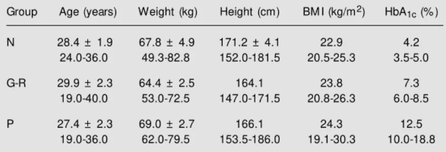
Table 1. Clinical characteristics of normal subjects (group N), and diabetic subjects w ith good or regular (group G-R) or poor (group P) metabolic control.
Group Age (years) Weight (kg) Height (cm) BM I (kg/m2) HbA 1c (% )
N 28.4 ± 1.9 67.8 ± 4.9 171.2 ± 4.1 22.9 4.2
24.0-36.0 49.3-82.8 152.0-181.5 20.5-25.3 3.5-5.0
G-R 29.9 ± 2.3 64.4 ± 2.5 164.1 23.8 7.3
19.0-40.0 53.0-72.5 147.0-171.5 20.8-26.3 6.0-8.5
P 27.4 ± 2.3 69.0 ± 2.7 166.1 24.3 12.5
19.0-36.0 62.0-79.5 153.5-186.0 19.1-30.3 10.0-18.8
The results are reported as means ± SEM and ranges. BM I = body mass index; HbA1c
diabetic nephropathy (microalbuminuria, creatinine and ureic nitrogen) were excluded from the study.
The experiments were conducted with the patients in the supine position in a meta-bolic study room at a constant temperature of 22oC. Cubital veins were cannulated in
each forearm with 20-gauge butterfly cath-eters for blood collections and solution infu-sion, respectively. Blood samples were col-lected at 10-min intervals during the basal period (30 min) and during a 2-h period of EDTA infusion. The infusion solution was prepared immediately before administration and contained 30 mg/kg weight EDTA, 2.7 mg/kg weight xylocaine, and 5% glucose for a total volume of 200 ml. The solution was infused with an infusion pump (Harvard ap-paratus) into the contralateral arm to that used for blood collection. The blood samples were used for the determination of plasma glucose by a glucose oxidase technique us-ing a Beckman glucose analyzer II (Beckman Instruments, Fullerton, CA, USA) and glycated hemoglobin was determined by an affinity chromatography method (17). Ion-ized calcium was measured in blood ob-tained with a 3-ml syringe containing cal-cium-titrated heparin (2560™ Radiometer, Copenhagen, Denmark) using an analyzer with a calcium-specific electrode (Radiom-eter ICA II) (18). The samples were col-lected anaerobically and the measurements were made immediately after collection. Blood samples for the determination of in-tact PTH were centrifuged in a refrigerated centrifuge and frozen at -70oC until the day
for determination. Serum PTH was analyzed in duplicate by an immunoradiometric method (Nichols Diagnostic Institutes, San Juan Capistrano, CA, USA). The intra- and interassay coefficients of variation were 4.9 and 11.0%, respectively. Serum magnesium was determined in duplicate by atom absorp-tion spectrophotometry using a Perkin-Elmer apparatus (Perkin Elmer, Norwalk, CT, USA).
Data are reported as means ± SEM and were analyzed using the Graphpad Prism 1999 software (Graphpad Software Inc., San Diego, CA, USA). The Friedman test, two-way analysis of variance by ranks, was used for paired parameters and the Kruskal-Wallis test was used for unpaired variables. Dunn’s post test was used in both analyses. The level of significance was set at P<0.05.
Re sults
The normal group presented a signifi-cantly lower fasting glycemia than group G-R (N = 4.4 ± 0.1 mmol/l vs G-R = 6.1 ± 0.6
mmol/l, P<0.05) and group P (P = 13.6 ± 1.7 mmol/l, P<0.005). The difference in glyce-mia levels between the two groups of dia-betic individuals was significant (P<0.01).
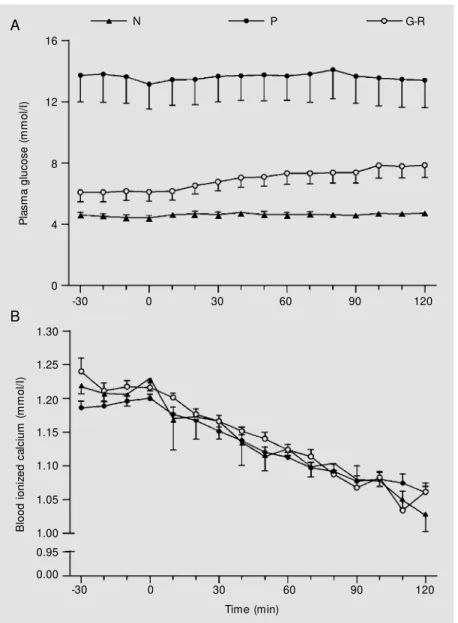
After the beginning of EDTA infusion, the normal group presented a slight variation in plasma glucose, with the highest values being reached at 120 min (4.7 ± 0.04 mmol/l). The diabetic groups also showed a small variation in glycemia during EDTA infu-sion, with the peak occurring at 120 min in the G-R group (7.9 ± 0.8 mmol/l) and at 80 min in the P group (14.1 ± 1.9 mmol/l). The plasma glucose levels of the P group were significantly higher than those for the G-R group (P<0.005) throughout the period of infusion and the latter group presented sig-nificantly higher plasma glucose levels than the N group throughout the study (P<0.05) (Figure 1A).
The P group tended to present lower calcium ion levels than the other two groups throughout the study. After the beginning of EDTA infusion, the three groups presented a significant reduction in circulating calcium ion levels corresponding to 18.6% (from 1.21 ± 0.01 to 1.02 ± 0.02 mmol/l) for group N, 18.4% (from 1.22 ± 0.01 to 1.03 ± 0.01 mmol/l) for group G-R, and 12.3% (from 1.19 ± 0.01 to 1.06 ± 0.01 mmol/l) for group P, respectively (Figure 1B).
were similar for groups N and G-R, while group P showed a slight tendency to lower serum PTH levels (N = 2.5 ± 0.6 vs G-R = 2.7
± 0.5 vs P = 2.2 ± 0.3 pmol/l). This behavior
became more marked during the stimulation tests, i.e., after the induction of hypocalce-mia serum PTH levels became significantly lower in P patients (P<0.05), whereas no difference in PTH levels was observed be-tween N and G-R patients. The maximum
PTH levels during the test were N = 11.9 ± 2.1 vs G-R = 13.7 ± 1.6 vs P = 7.5 ± 0.7 pmol/
l (Figure 2A).
Figure 2B shows that serum magnesium levels remained stable throughout the study in the three groups, ranging from 0.73 ± 0.03 to 0.76 ± 0.02 mmol/l in group N, from 0.69 ± 0.02 to 0.71 ± 0.03 mmol/l in group G-R, and from 0.64 ± 0.03 to 0.66 ± 0.02 mmol/l in group P. Magnesium levels were signifi-cantly lower in the P group than in the N group.
D iscussio n
It is well known that alterations in min-eral metabolism can induce disturbance in glucose metabolism (19-21) and glucose in-tolerance which also can interfere with min-eral metabolism (22). Previous studies have shown that poor diabetes mellitus control is associated with calcium, phosphorus and magnesium wastage (4,5). We evaluated PTH secretion in diabetic patients with different degrees of metabolic control and our results showed that patients with poor metabolic control present lower PTH secretion than well-controlled diabetic and normal subjects during acute stimulation.
Hyperglycemia that leads to osmotic di-uresis also causes magnesium depletion. It is possible that the reduced PTH secretion oc-curring in patients with poorly controlled diabetes is related to low serum magnesium (23). Magnesium interacts in a complex man-ner with PTH, interfering both with the se-cretion and action of this hormone. Acute hypomagnesemia stimulates PTH secretion, whereas chronic severe hypomagnesemia has a negative effect and is the exclusive cause of clinical hypoparathyroidism. Furthermore, hypomagnesemia is associated with a de-creased action of PTH on target tissues, pos-sibly by causing a functional impairment of the adenyl cyclase system (24). Magnesium is a predominantly intracellular ion and a normal serum magnesium level does not
P
la
s
m
a
g
lu
c
o
s
e
(
m
m
o
l/
I)
16
12
8
4
0
B
lo
o
d
i
o
n
iz
e
d
c
a
lc
iu
m
(
m
m
o
l/
I)
1.30
1.25
1.10
0.95
0.00 1.20
1.15
1.00 1.05
-30 0 30 60 90 120
-30 0 30 60 90 120
Time (min)
A
B
N P G-R
mean a normal content of body magnesium. Lima et al. (22) verified that only intracellu-lar magnesium levels correlate with meta-bolic control in diabetes mellitus. However, several studies have measured only circula-tory levels of magnesium as evidence of hypomagnesemia. Rude et al. (23) have shown that patients with low serum magne-sium presented undetectable basal PTH only when magnesium was below 0.35 mmol/l and that basal PTH was increased or normal in patients with 0.44 and 0.49 mmol/l serum magnesium levels, respectively. Our results are in accordance with those reported by Rude et al. (23), i.e., normal basal PTH levels in subjects with magnesium higher than 0.44 mmol/l. Furthermore, we observed that the parathyroid response to acute hypo-calcemia is impaired in diabetic patients with serum magnesium levels of 0.64 ± 0.03 mmol/l.
Calcium sensors in the parathyroid gland are especially sensitive to serum calcium variations and permit PTH to exert efficiently its most important function, i.e., to maintain calcium homeostasis (25). Another impor-tant physiological role of PTH is its partici-pation in the control of bone trophism (13). Whereas exaggerated and constant PTH se-cretion has a predominantly catabolic effect on bone, its periodic secretion is associated with the maintenance and even an increase in trabecular bone mass in humans (14,26) and with an increase in trabecular and corti-cal bone in rats (27). The action of PTH favoring the maintenance and/or increase in bone mass may be secondary to its metabolic actions of urine calcium retention and/or to increased intestinal calcium absorption sec-ondary to stimulation of 1,25(OH)2D
syn-thesis or may even be due to a direct activa-tion of osteoblasts, in view of the fact that previous studies have shown PTH receptors in these cells (28).
Our results clearly show that PTH secre-tion is impaired in patients with poorly con-trolled diabetes mellitus and suggest an
as-sociation between this occurrence and the low serum levels of magnesium observed in these patients. Previous studies have sug-gested that the osteopenia observed in dia-betic patients is due to the reduction in 1,25(OH)2D levels (7) and that insulin
defi-ciency could reduce osteoblast activity (12,13). However, other studies have
sug-Figure 2. Serum intact PTH (A) and serum magnesium (B) levels under basal conditions (-30 to 0 min) and during 2 h of EDTA infusion in normal subjects (group N), in diabetic patients w ith good or regular metabolic control (group G-R), and in diabetic patients w ith poor metabolic control (group P). A, P<0.05 group N vs group P (intervals +60 to +120 min). P<0.05 group G-R vs group P (intervals +50 to +120 min). B, P<0.01 group N vs group P (Kruskal-Wallis test).
S
e
ru
m
i
n
ta
c
t
P
T
H
(
p
m
o
l/
I)
18
120 15
12
9
6
3
0
0.9
0.8
0.7
0.6
0.5 0.4
0.0
S
e
ru
m
m
a
g
n
e
s
iu
m
(
m
m
o
l/
I)
-30 0 30 60 90 120
-30 0 30 60 90
Time (min)
N P G-R
A
gested that osteopenia is not a complication of diabetes mellitus (29). Probably, both hy-potheses can be considered true in view of the heterogeneous situation of diabetes mel-litus. Whereas patients with good metabolic control have normal calcium ion and PTH secretion, poorly controlled diabetics tend to present lower calcium ion levels, deficient PTH secretion and lower serum magnesium levels. Thus, on the basis of these alter-ations, it is possible that osteopenia is a nonclassical complication of diabetes which
is likely to be related to hyperglycemia. It is also possible that decreased PTH secretion contributes to a reduction of 1,25(OH)2D
levels and of osteoblast activity in diabetics.
Ackno wle dgm e nts
The authors gratefully acknowledge S.L. Brandão Filho for expert technical help, C. Guimarães and E.F. Verceze for secretarial assistance, and E. Greene for revising the English text.
Re fe re nce s
1. Seino Y & Ishida H (1995). Diabetic osteo-penia. Pathophysiology and clinical as-pects. Diabet es and M et abolism Re-view s, 11: 21-35.
2. Forsen L, M eyer HE, M idthjell K & Edna TH (1999). Diabetes mellitus and the inci-dence of hip fracture: results from the Nord-Trondelag health survey. Diabetolo-gia, 42: 920-925.
3. Schw artz AV, Sellmeyer DE, Ensrud KE, Cauley JA, Tabor HK, Schreiner PJ, Jamal SA, Black DM & Cummings SR (2001). Older w omen w ith diabetes have an in-creased risk of fracture: a prospective study. Journal of Clinical Endocrinology and M etabolism, 86: 32-38.
4. M onnier L, Colette C, Aguirre L, Sony C & M irouze J (1978). Intestinal and renal han-dling of calcium in human diabetes melli-tus: influence of acute oral glycose load-ing and diabetic control. European Journal of Clinical Investigation, 8: 225-231. 5. Paolisso G, Scheen A, D’Onofrio F &
Lefèbvre P (1990). M agnesium and glu-cose homeostasis. Diabetologia, 33: 511-514.
6. M cNair P, M adsbad S, Christiansen C, Christensen M S & Transbal I (1982). Hy-ponatremia and hyperkalemia in relation to hyperglycemia in insulin treated dia-betic out-patients. Clinica Chimica Acta, 120: 243-250.
7. Frazer TE, White NH, Hough S, Santiago JV, M cGee BR, Bryle O, M allon J & Avioli LV (1981). Alterations in circulating vita-min D metabolites in the young insulin-dependent diabetic. Journal of Clinical Endocrinology and M etabolism, 53: 1154-1159.
8. Verhaeghe J, Suiken AM H, Van Bree R,
Vanlterck E, Jans I, Visser WJ, Thomasset M , Allew aert K & Bouillon R (1993). In-creased clearance of 1,25(OH)2D3 and
tis-sue-specific responsiveness to 1,25(OH)2D3
in diabetic rats. American Journal of Phys-iology, 265: E215-E223.
9. Ishida H, Cunningham NS, Henry HL & Norman AW (1988). The number of 1,25-dihydroxyvitam in D3 receptors is
de-creased in both intestine and kidney of genetically diabetic ob/db mice. Endocri-nology, 122: 2436-2443.
10. Ishida H, Seino Y, Takeshita N, Kurose T, Tsuji K, Okamoto Y, Someya Y, Hara K, Akiyama Y, Imura H & Nozaw a M (1992). Effect of pancreas transplantation on de-creased levels of circulat ing bone g-carboxyglutamic acid containing protein and osteopenia in rats w ith streptozoto-cin-induced diabetes. Acta Endocrinolo-gica, 127: 81-85.
11. Diabetes Control and Complications Trial Research Group (1993). The effect of in-tensive treatment of diabetes on the de-velopment and progression of long term complications in insulin-dependent diabe-tes mellitus. New England Journal of M edicine, 329: 977-986.
12. UK Prospective Diabetes Study (UKPDS) Group (1998). Intensive blood-glucose control w ith sulfonylureas or insulin com-pared w ith conventional treatment and risk of complications in patients w ith type 2 diabetes (UKPDS 33). Lancet, 352: 837-853.
13. Rosen CJ & Donahue LR (1996). Parathy-roid hormone and osteoporosis. Current Opinion in Endocrinology and Diabetes, 3: 532-539.
14. Garton M , M artin J, Stew art A, Krukow ski
Z, M atheson N, Robins S, Low redge N & Reid D (1995). Changes in bone mass and metabolism after surgery for primary hy-perparathyroidism. Clinical Endocrinology, 42: 493-500.
15. Hughs-Daw son B (1994). Regulation of parathyroid hormone by dietary calcium and vitamin D. In: Bilezakian JP, M arcus R & Levine NA (Editors), The Parathyroid. Basic and Clinical Concepts. 1st edn. Raven Press, New York.
16. Canalis E, Hock JM & Raisz LG (1994). Anabolic and catabolic effects of parathy-roid hormone on bone and interactions w ith grow th factors. In: Bilezakian JP, M arcus R & Levine NA (Editors), The Para-thyroid. Basic and Clinical Concepts. 1st edn. Raven Press, New York.
17. Klenk DC, Hermanson GT, Krohn RI, Fuji-moto EJ, M allia AK, Smith PK, England JD, Wiedmeyer H, Little RR & Goldstein DE (1982). Determination of glycosylated hemoglobin by affinity chromatography: comparison w ith colorimetric and ion-ex-change method and effects of common interferences. Clinical Chem istry, 28: 2088-2094.
18. Bow ers Jr GN, Brasard C & Sena SF (1986). M easurements of ionized calcium in serum w ith ion-selective electrodes: a mature technology that can meet the daily service needs. Clinical Chemistry, 32: 1437-1447.
19. Paula FJA, Plens AECM & Foss M C (1998). Effects of hypophosphatemia on glucose tolerance and insulin secretion. Hormone and M etabolic Research, 30: 281-284.
Bourton AJM (1994). Impaired glucose tolerance and insulin sensitivity in primary hyperparathyroidism. Clinical Endocrinol-ogy, 40: 47-53.
21. Rosolova H, M ayer O & Reaven G (1997). Effect of variation in plasma magnesium concentration on resistance to insulin-mediated glucose disposal in nondiabetic subjects. Journal of Clinical Endocrinol-ogy and M etabolism, 82: 3783-3785. 22. Lima M L, Rodrigues LE, Cruz T, Barbosa
K, Pousada CP & Canguçu V (1998). The effect of magnesium supplementation in increasing doses on the control of type 2 diabetes. Diabetes Care, 21: 682-686. 23. Rude RK, Oldhan SB, Sharp CF & Singer
FR (1978). Parathyroid hormone secretion
in magnesium deficiency. Journal of Clini-cal Endocrinology and M etabolism, 47: 800-806.
24. Rude RK (1996). M agnesium depletion and hypermagnesemia. In: Favas M J (Edi-tor), Primer on the M etabolic Bone Dis-eases and Disorders of M ineral M etabo-lism. 3rd edn. Lippincot-Raven, Philadel-phia.
25. Brow n EM & Pollak Hebert SC (1998). The extracellular calcium-sensing recep-tor: its role in health and disease. Annual Review of M edicine, 49: 15-29. 26. Hodsman AB, Steer BM , Fraher LJ &
Drost DJ (1991). Bone densitometric and histomorphometric response to sequen-tial human PTH (1-38) and salmon
calcito-nin in osteoporotic patients. Bone M in-eral, 14: 67-83.
27. Guiness-Hey M & Hock JM (1984). In-creased trabecular bone mass in rats treated w ith human synthetic parathyroid hormone. M etabolic Bone Disease and Related Research, 5: 177-181.
28. M cSheehy PHJ & Chambers TJ (1986). Osteoblastic cells mediate osteoclastic responsiveness to PTH. Endocrinology, 118: 824-828.