© 2012 Sociedade Brasileira de Hemodinâmica e Cardiologia Intervencionista. Published by Elsevier Editora Ltda. All rights reserved.
RFRIST Study (Fractional Flow Reserve in Functional
Quantification of Renal Allograft Artery Stenosis):
Rationale and Study Design
Alexandre Vidal Bonim
1, Claudia Maria Rodrigues Alves
2, Adriano Henrique Pereira Barbosa
3,
José Osmar Medina Pestana
4, Marcelo Costa Batista
5, Antônio Carlos Carvalho
6ABSTRACT
Background: Renal allograft artery stenosis is the most
preva-lent vascular complication after renal transplantation. The diagnostic limitations of noninvasive tests are well deined and angiography remains the gold standard for diagnosis and therapeutic deinition. The use of fractional low reserve for a better stratiication of native renal artery stenosis may be useful for an adequate selection of patients for percutaneous treatment, however this method has not yet been validated in patients undergoing transplantation. The objective of this study is to describe and standardize the fractional low re-serve protocol in patients with renal allograft artery stenosis in a group of patients selected for percutaneous renal inter-vention, correlating the method with angiography Methods:
Cross-sectional, single center pilot study (Hospital São Paulo/ Universidade Federal de São Paulo, São Paulo, SP, Brazil), including 10 patients with a clinical picture compatible with renal allograft artery stenosis, with angiography showing graft stenosis > 60% and admitted for percutaneous renal interven-tion. Graft dysfunction assessment will include biomarkers of renal function. Conclusions: In this study, a fractional low reserve protocol for the functional assessment of renal allograft artery stenosis will be standardized, evaluating an alternative method capable of identifying patients most likely to beneit from percutaneous renal intervention.
DESCRIPTORS: Renal artery obstruction. Kidney transplantation. Renal insuficiency, chronic.
1 Postgraduate Physician at Hemodynamics and Interventionist Cardiology Sector of Escola Paulista de Medicina da Universidade Federal de São Paulo. São Paulo, SP, Brazil.
2 In-charged Physician for Hemodynamics and Cardiology Sector of Escola Paulista de Medicina da Universidade Federal de São Paulo. São Paulo, SP, Brazil.
3 Physician at Hemodynamics and Interventionist Cardiology Sector of Escola Paulista de Medicina da Universidade Federal de São Paulo. São Paulo, SP, Brazil.
4 Principal Professor of Nefrology at Escola Paulista de Medicina da Universidade Federal de São Paulo. São Paulo, SP, Brazil.
5 Principal Professor of Nefrology at Escola Paulista de Medicina da Universidade Federal de São Paulo. São Paulo, SP, Brazil.
6 Principal Professor of Cardiology at Escola Paulista de Medicina da Universidade Federal de São Paulo. São Paulo, SP, Brazil.
Correspondence to: Alexandre Vidal Bonim. Rua Botucatu, 740 – Vila Clementino – São Paulo, SP, Brazil – CEP 04023-900
E-mail: dr_alexbonfa@yahoo.com.br
Received on: 7/24/2012 • Accepted on: 11/16/2012 RESUMO
Estudo RFRIST (Reserva de Fluxo Fracionada na Quantificação Funcional das Estenoses em Artérias de
Rins Transplantados): Racional e Desenho do Estudo
Introdução: A estenose da artéria do enxerto renal é a
com-plicação vascular mais prevalente após transplante renal. As limitações dos exames não-invasivos para seu diagnóstico são bem deinidas e a angiograia permanece como padrão de referência para diagnóstico e deinição terapêutica. A utilização da reserva de luxo fracionada renal para melhor estratiicação das estenoses de artéria renal nativa pode ajudar na adequada seleção de pacientes para tratamento percutâneo, porém tal método ainda não está padronizado em pacientes submetidos a transplante. O objetivo deste estudo será des-crever e padronizar o protocolo de reserva de luxo fracionada em pacientes com estenose da artéria do enxerto renal em um grupo de pacientes selecionados para intervenção renal percutânea, correlacionando o método com a angiograia.
Métodos: Estudo piloto, prospectivo, transversal, unicêntrico
(Hospital São Paulo/Universidade Federal de São Paulo, São Paulo, SP, Brasil), em que serão selecionados 10 pacientes com quadro clínico compatível com estenose da artéria do enxerto renal, com angiograia do enxerto evidenciando estenose
≥ 60%, admitidos para intervenção renal percutânea. Como
avaliação da disfunção do enxerto serão realizadas dosagens de biomarcadores da função renal. Conclusões: No presente estudo, um protocolo original de reserva de luxo fracionada para avaliação funcional de estenose da artéria do enxerto renal será padronizado, avaliando um método auxiliar na investigação funcional que possa identiicar pacientes que realmente se beneiciem com a intervenção renal percutânea.
DESCRITORES: Obstrução da artéria renal. Transplante de rim.
Insuiciência renal crônica.
R
enal transplantation has been performed for the treatment of patients with chronic renal failure since 1954. Nevertheless, renal transplantation can give rise to urologic, vascular, and even clinical com-plications due to the use of immunosuppressive drugs. Vascular complications are infrequent in kidney transplantations; however, they are one of the major causes of morbidity and mortality, occasionally result-ing in renal graft dysfunction, graft removal, or death.1,2Among the vascular complications are renal graft artery stenosis, arteriovenous istula, and renal vein throm-bosis. Renal graft artery stenosis is the most common and occurs in 1% to 15% of transplanted patients.3-5
According to the United States Renal Data System registry, 41,867 renal transplantations were performed in the period from 2000 to 2005, of which 823 de-veloped renal graft artery stenosis, corresponding to a rate of 8.3 cases per 1,000 patient-years.6 According to
data from the Brazilian Transplantation Registry, 19,101 procedures were performed in that same period, but no data are available regarding the incidence of renal graft artery stenosis.7 In the present institution, Lopes
et al.8 reported a renal graft artery stenosis rate of
ap-proximately 1.6%.
Although no speciic study has conirmed the associa-tion between vascular complicaassocia-tions and graft posiassocia-tion, the most often used technique is graft implantation in the common iliac or external artery, using termino-lateral anastomosis due to its speed and simplicity. The main causes of renal graft artery stenosis are inappropriate suture due to technical failure, intimal lesion during graft perfusion or when performing the implant, and excessive length of the renal artery causing torsion or tension at the suture site.1,4-9 Atherosclerosis and ibrosis
at the anastomosis site are also causes of renal artery stenosis, and generally occur at a later stage after transplantation.9
In general, clinical indings that suggest renal graft artery stenosis are severe arterial hypertension refractory to drug treatment, and audible murmur at the site of the graft associated with unexplained progressive graft dysfunction. The presence of hypertension in renal transplantation recipients is a clinical inding in 50% to 60% of cases in the long-term.9,10 In the study by
Mendes et al.,9 in all cases of haemodynamically
sig-niicant renal graft artery stenosis (deined as stenosis
> 60%), there was progressive loss of graft function and
dificult-to-control hypertension (with the use of three or more antihypertensive drugs).
Colour Doppler ultrasonography is an excellent diagnostic method for initial assessment of renal graft artery stenosis, with a sensitivity of 94% and a speciicity of 87% when compared to conventional angiography.10
Computed tomography angiography (CTA) has a sensiti-vity and a speciicity of 100% and 96%, respectively; its disadvantages are the use of ionising radiation and
iodinated contrast.11 Magnetic resonance angiography
(MRA) using gadolinium has been shown to be a good method for the detection of renal artery stenosis. Korst et al.12 found a sensitivity and a speciicity of 100%
and 85%, respectively. It is an excellent technique for patients with impaired renal function and allergy to iodinated contrast medium, in whom digital angiography cannot be performed.13
In general, all patients submitted to renal trans-plantation at this institution who have high clinical suspicion of graft stenosis undergo Doppler ultrasound assessment, and when the results are altered (peak systolic velocity > 180 cm/s), a renal angiography is
indicated. Angiography, an examination considered to be the gold standard for the deinitive diagnosis of renal artery stenosis, is used to conirm the injury indicated by Doppler ultrasonography and/or CTA, and guides the therapeutic approach. However, angiography is based on indings of visual estimation or by quantita-tive renal angiography (QRA), thereby determining the severity of a stenosis while quantifying t. Stenoses are considered severe when there is > 60% of vessel
lu-men obstruction.7,11
Once the diagnosis is attained, treatment options include percutaneous treatment using a conven-tional percutaneous intervention with a balloon or, preferably, stenting (percutaneous renal intervention [PRI]) and revascularisation surgery. The PRI results in resolution or improvement in 76% of cases, with the discontinuation of at least two antihypertensive medications, and in some patients already on dialy-sis, it is possible to achieve graft function recovery.7
Success rates are high, and restenosis with stenting is below 10%, mainly due to the high luminal diameter of the vessel and the immunosuppressive therapy used after transplantation.7,9
In contrast, some patients submitted to renal graft angiography have moderate stenosis (> 30% and < 60%),
in addition to cases in which the visual estimation/ QRA cannot deine the actual severity of the stenosis. This usually occurs in stenosis with ostial location or even as a result of the limitations of luminography. In this scenario, the idea of using a method that would help in the functional stratiication of renal graft artery stenosis has been proposed. Developed and validated for the functional stratiication of coronary stenosis, the fractional low reserve for native renal artery stenosis has been studied by Leesar et al.14 and Kapoor et al.15
determined, respectively, for maximum systolic gradient
> 21 mmHg, and 79%, 82%, and 88%, respectively,
for a Pd/Pa ratio < 0.9.
Regarding renal graft artery stenosis, there have been no studies that used fractional low reserve for the functional analysis of these stenoses. Thus, the usefulness of this method in the functional evaluation of these patients and the possibility of predicting the therapeutic effectiveness of the PRI has not yet been explored.
METHODS
Fractional flow reserve: definitions and principles
Renal fractional low reserve is deined as the ratio between the maximum blood low to the renal paren-chyma in the presence of a determined stenosis (or stenoses) and this same low if there were no stenoses. This ratio represents the fraction of normal maximum renal low that can be achieved in the presence of stenosis. The fractional low reserve can be readily determined by dividing the mean pressure distal to the renal lesion by the mean pressure in the aorta during maximal vasodilation induced by administering an intra-arterial bolus dose of 30 mg papaverine in the renal graft. A fractional low reserve of 0.6 indicates
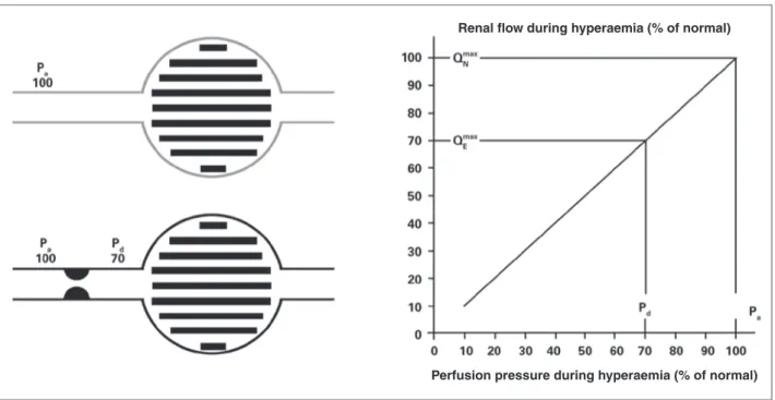
that the maximum amount of blood (and oxygen) that irrigates that particular area of renal tissue is only 60% of what it would be if the culprit artery was completely normal. If, after a PRI, the fractional low reserve in that case increases to 0.90, it means the maximum achievable low into the parenchymal area supplied by that artery (and hence, the oxygen supply) increased by 50% and is now 90% of the attainable value if the artery was completely normal. Figures 1 and 2 show, schematically, how to determine the fractional low reserve and how the low measurement can be inferred by dividing pressures.
Protocol description
Study goals
1. To describe and standardise the study protocol of fractional low reserve in patients with severe ste-nosis of the renal graft artery who are selected for PRI, observing whether the cutoff used for native renal artery stenosis (maximum systolic gradient >
21 mm or Pd/Pa < 0.9) is reproducible for renal
graft artery stenosis.
2. To compare the results obtained through direct measurements of pressure gradients (pre- and post-stenosis) at rest and after hyperaemic stimulation performed with a conventional 4F catheter, as well as the pressure measured by the pressure wire.
Figure 1 – Deinition of renal fractional low reserve. If there is no stenosis (gray lines), the perfusion pressure determines the maximum normal low rate (100%) of the renal parenchyma (Pa = Pd). In case of stenosis (black lines), responsible for a gradient of 30 mmHg during maximum hyperaemia, the distal perfusion pressure will decrease to 70 mmHg. As the ratio between renal low and pressure is linear during hyperaemia (chart), renal low will reach only 70% of its normal value. Pa, perfusion pressure, Pd, distal pressure.
Renal flow during hyperaemia (% of normal)
3. To evaluate the association between fractional low reserve values before and after the procedure and procedural success through ultra-sensitive markers of renal function.
Study population
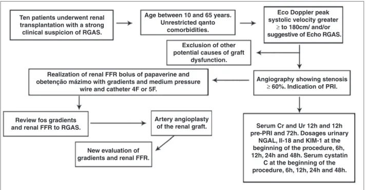
A total of ten 15 to 65 year-old patients will be initially assessed, after an informed consent is signed. The patients will have been referred to the Interven-tional Cardiology Service of the Hospital São Paulo (Escola Paulista de Medicina, Universidade Federal de São Paulo (UNIFESP/EPM), São Paulo, SP, Brazil), with a clinical picture of renal graft artery stenosis, associated with Doppler ultrasound and/or CTA suggestive of renal graft artery stenosis to undergo renal angiography and, when the presence of this stenosis is confirmed, will have an indication for PRI (renal graft artery stenosis ≥ 60% at visual
esti-mation). The project was approved by the research ethics committee, protocol No. 51,518 – Plataforma Brasil – Brazilian Ministry of Health. Figure 3 sum-marises the study design. Patients will be admitted 12 hours before the procedure, and intravenous hydration will be carried out with 0.9% saline solu-tion at 1 mL/kg/hour, 12 hours before and after the test. Low-osmolarity contrast will be used to minimise the risk of contrast-induced nephropathy. Dual anti-platelet therapy will be administered (with 300 mg acetylsalicylic acid and 300 mg clopidogrel, 12 hours prior to the procedure). A dose of 100 mg acetylsali-cylic acid will be maintained indefinitely, as well as 75 mg clopidogrel for 30 days.
The angiograms of all patients selected for the study will be analysed by an experienced interventional physician. Then, artery lesions considered to be severe (visual estimates showing stenosis ≥ 60%), and thus with
an indication for PRI, will be submitted to the proto-col for the analysis of the mean systolic translesional
pressure gradients and fractional low reserve pre- and post-treatment under hyperaemia with papaverine.
Materials
6F or 7F therapeutic guide-catheters without lat-eral holes will be used for selective catheterisation of the artery to be approached. The measurement of the translesional pressure gradient will be mandatorily performed with 4F guide catheters, as larger-calibre catheters interfere with the assessment of stenosis du-ring their retreat to perform the gradient measurement. The guidewire to be used for measurement of fractional low reserve uses a speciic sensor located 3 cm from its tip, has a diameter of 0.014 inch (0.36 mm), and has a soft tip, similar in calibre to the guide wires routinely used in PRI procedures. PressureWire Certus Hydrophilic guidewires (Radi Medical Systems AB Inc., Uppsala, Sweden) will also be used. The guide wire is connected to a monitor, which, by converting the pressure signal to an electrical signal, presents the results directly on the screen, allowing the data to be viewed and saved in a few seconds.
The angiograms will be performed with Philips Allure FD10 equipment (lat panel), which has software with appropriate settings for acquisition and processing of images in rotational angiography of the abdomen.
Hyperaemia
It is essential to induce maximal vasodilation in the two compartments of renal circulation, in the vessel itself and in the microcirculation, to calculate the renal fractional low reserve. Administration of 30 mg papaverine by an intra-arterial route in bolus is the available option. The drug’s action lasts up to 50 seconds; it has no side effects and can be used in catheter withdrawal manoeuvres. The systolic and mean translesional gradients are calculated simultaneously during the analysis of the renal fractional low reserve.
Systematisation for the initial angiographies, angiographic definition of lesion severity, measurement of renal fractional flow reserve, and PRI with a stent and new measurement of fractional flow reserve.
A total of ten patients who have a clinical picture of renal artery graft stenosis and angiography showing graft stenosis ≥ 60% and are admitted to undergo PRI
will be selected. For the evaluation of graft dysfunc-tion, serum measurements of urea and creatinine will be performed 12 hours before and 12 and 72 hours after the procedure. Measurement of urinary neutrophil gelatinase-associated lipocalin (NGAL), interleukin-18 (IL-18), kidney injury molecule-1 (KIM-1), and serum cystatin C will be performed at the beginning and 6 hours, 12 hours, 24 hours, and 48 hours after the procedure.
Figure 2 – Renal fractional low reserve is calculated by dividing the distal pressure in the renal artery by the pressure in the aorta during maximum hyperaemia. FFR, fractional low reserve, QStenosis, low in renal graft artery with stenosis; QNormal, low in the renal graft artery in the absence of stenosis; Pd, distal pressure; PV, venous pressure; PA, pressure in the aorta; RMIO, renal resistance.
Hydraulic equation Pressure (∆P) = Flow (Q) × Resistance (R)
Q = ∆P/R
QStenosis
QNormal
The following steps will be performed during the procedure:
1. Local anaesthesia of the arterial puncture site with 2% lidocaine (10 – 15 mL).
2. Insertion of a 6F or 7F arterial sheath.
3. Introduction of a 4F Judkins right (JR) catheter after the stenosis and maximum hyperaemia induced with 30 mg papaverine in bolus followed by evaluation of translesional gradients.
4. Selective catheterisation of the renal graft artery with an RDC guide catheter or a JR 4 of PRI, 6F, or 7F without lateral holes.
5. Administration of 100 UI/kg IV heparin. 6. Calibration of guide-catheter and pressure wire. 7. Connection of the pressure wire to the analyser,
equating the pressures of the guide-catheter and wire pressure when the sensor of the latter is close to the tip of the guide-catheter.
8. Placement of the pressure wire in the distal bed of the renal graft artery to be analysed.
9. Evaluation of graft stenosis by measuring the frac-tional low reserve during maximum hyperaemia induced by an intra-arterial injection of 30 mg papaverine in bolus in the renal graft.
10. Performing PRI with stent implantation.
11. New gradient assessment through maximum hy-peraemia with papaverine using the pressure wire and a 4F catheter.
Procedural success analysis
Angiographic success
Procedural angiographic success will be considered when the stenosis is resolved after the PRI (residual
≤ 30%)7 with no major complications.
Functional success
Following the principles established by the hydraulic equation to calculate the renal fractional low reserve as shown in Figure 2, after normalising the low with the resolution of the previously existing stenosis, there should be no pressure difference between the pressure distal to the treated site and the proximal pressure (Pd = Pa), which means that the Pd/Pa ratio = 1. Likewise, the resolution of the systolic and mean translesional pressure gradients can be estimated.
To analyse renal function improvement, kidney injury markers, which have been developed and validated to predict the degree of renal dysfunction, will be quan-tiied. The most commonly used laboratory parameter for the clinical diagnosis of acute renal failure (ARF)
Figure 3 – Overview of the study protocol. RGAS, renal graft artery stenosis; Echo, echocardiography; CTA, computed tomography angiography; FFR, fractional low reserve; PRI, percutaneous renal intervention; Ur, urea; Cr, creatinine; NGAL, neutrophil gelatinase-associated lipocalin; IL-18, interleukin-18; KIM-1, kidney injury molecule-1.
Ten patients underwent renal transplantation with a strong clinical suspicion of RGAS.
Realization of renal FFR bolus of papaverine and obetenção mázimo with gradients and medium pressure
wire and catheter 4F or 5F.
Age between 10 and 65 years. Unrestricted qanto
comorbidities.
Exclusion of other potential causes of graft
dysfunction.
Angiography showing stenosis
≥ 60%. Indication of PRI.
Review fos gradients and renal FFR to RGAS.
Artery angioplasty of the renal graft.
New evaluation of gradients and renal FFR.
Serum Cr and Ur 12h and 12h pre-PRI and 72h. Dosages urinary
NGAL, Il-18 and KIM-1 at the beginning of the procedure, 6h, 12h, 24h and 48h. Serum cystatin
C at the beginning of the procedure, 6h, 12h, 24h and 48h.
Eco Doppler peak systolic velocity greater
is the elevation in plasma creatinine. However, it is necessary that the glomerular iltration rate is reduced by 50% in order to increase the creatinine. The ideal biomarker should be quick and easy to measure, ob-tainable through minimally invasive procedures, and capable of detecting early tubular injury and relect-ing changes of renal function. Currently, KIM-1, IL-18, NGAL, and cystatin C are considered as the urinary molecules with the greatest potential for early detection of acute kidney injury for clinical use.16,17
Parikh et al.18 evaluated the urinary measurement
of NGAL associated with lipocalin in 63 patients sub-mitted to renal transplantation and found a sensitivity and speciicity of 90% and 83%, respectively, to predict ARF (cutoff of 53 pg/mL) measured two hours after transplantation. The same authors also evaluated the measurement of IL-18 with excellent results in predict-ing ARF in post-renal transplantation.
Therefore, the measurements of the renal injury markers (IL-18, NGAL, urinary KIM-1, and serum cys-tatin C) will be performed shortly before the procedure (time zero), 6, 12, 24, and 48 hours after the PRI, and their values will be compared with the plasma creatinine measured 12 hours before and 12 and 72 hours after PRI. Consequently, it will be possible to assess the effects of the procedure (handling of arte-rial stenosis, contrast-induced nephropathy) associated with improved perfusion of the renal parenchyma after stenosis resolution.
Statistical analysis
Due to the small number of patients in this pilot study, it is possible that much of the data will not be comparable. This project will serve to establish the submission method for a supplemental project to the funding agency and sample expansion. The most important data collected here will be the difference between fractional low reserve before and after treat-ment, assuming that the same value found in studies of native artery stenosis is reproduced here (fractional low reserve of 0.9).
Continuous variables will be expressed as the means and standard deviations and compared by Stu-dent’s t-test. Categorical variables will be described as absolute frequencies and compared by the chi-squared test. Spearman’s test will be used for the correlation between the haemodynamic parameters of the study and the percentage of renal graft artery stenosis at visual estimation. The Wilcoxon test will be used to compare the measurements of kidney injury biomarkers before and after stent implantation at the times described. P-values < 0.05 will be considered indicative of statistical
signiicance. All analyses will be performed using the SPSS 11.5 software package (SPSS – Chicago, USA). It is noteworthy that, due to the small sample size, there will not be suficient statistical power to evaluate the
eficiency of PRI in the context of treatment for renal graft artery stenosis.
CONCLUSIONS
The use of the fractional low reserve for better stratiication of renal graft artery stenosis may improve the selection of patients for percutaneous treatment, thus reducing risks and preserving resources. Since the study will be conducted with patients with conirmed diagnosis of severe renal graft artery stenosis (> 60%),
and since the patients will be treated regardless of the values found for fractional low reserve, it will not be possible to conclude that the fractional low reserve is an additive method for better stratiication of steno-ses, but only that the protocol was described in this sample. However, the results may be of great value for the development of future studies, expanding the use of methods and reining information obtained by other diagnostic tests.
Thus, a future study designed is being that can validate the fractional low reserve as a method to stratify patients who can greatly beneit from PRI in the clinical scenario described, using a more robust sample of patients.
CONFLICT OF INTEREST
The authors declare no conlicts of interest.
REFERENCES
1. Del Valle Sanz Y, Lorente Ramos RM, Berrocal Frutos T, Pietro Arellano C, Rodríguez Lemos R, García-Messeguer MC. Complicaciones vasculares en el transplante renal pediátrico: diagnóstico ecográico. An Esp Pediatr. 1999;50(3):263-8. 2. Droupy S, Eschwège P, Blanchet P, Hammoudi Y, Bensadoun H,
Bellamy J, et al. Recipient arterial repairs during renal trans-plantation. Transplant Proc. 2002;34(3):814.
3. Höhnke C, Abendroth D, Schleibner S, Land W. Vascular complications in 1,200 kidney transplantations. Transplant Proc. 1987;19(5):3691-2.
4. Sutherland RS, Spees EK, Jones JW, Fink DW. Renal artery stenosis after renal transplantation: the impact of the hypo-gastric artery anastomosis. J Urol. 1993;149(5):980-5. 5. De Bruney B, Monoharan G, Pijls NH, Verhamme K, Madaric J,
Bartunek J, et al. Assessment of renal artery stenosis severity by pressure gradient measurements. J Am Coll Cardiol. 2006; 48(9):1851-5.
6. Hurst FP, Abbot KC, Neff RT, Elster EA, Falta EM, Lentine KL, et al. Incidence, predictors and outcomes of transplant renal artery stenosis after kidney transplantation: analysis of USRDS. Am J Nephrol. 2009;30(5):459-67.
7. Associação Brasileira de Transplante de Órgãos (ABTO). RBT Registro Brasileiro de Transplante. 2010cited 15 Feb 2011;16(4). [cited 15 Feb. 2011]. Available from: http://www.abto.org.br/ abtov02/portugues/populacao/rbt/anoXVI_n4_completo/index. aspx
9. Mendes WDS, Silva LF, Espinosa G, Fernandes AL, Furtado R, Gonçalves R. Estenose arterial nos transplantes renais. Rev Bras Col Cir. 2005;32(5):237-43.
10. De Cobelli F, Venturini M, Vanzulli A, Sironi S, Salvioni M, Angeli E, et al. Renal arterial stenosis: prospective comparison of color Doppler US and breath-hold, three-dimensional, dynamic, gado-linium-enhanced MR angiography. Radiology. 2000;214(2):373-80. 11. Hofmann LV, Smith PA, Kuszyk BS, Kraus E, Fishman EK.
Three-dimensional helical CT angiography in renal transplant recipients: a new problem-solving tool. AJR Am J Roentgenol. 1999;173(4):1085-9.
12. Korst MB, Joosten FB, Postma CT, Jager GJ, Krabbe JK, Barentsz JO. Accuracy of normal-dose contrast-enhanced MR angiography in assessing renal artery stenosis and accessory renal arteries. AJR Am J Roentgenol. 2000;174(3):629-34. 13. Dong Q, Schoenberg SO, Carlos RC, Neimatallah M, Cho KJ,
Williams DM, et al. Diagnosis of renal vascular disease with MR angiography. Radiographics. 1999;19(6):1535-54.
14. Leesar MA, Varma J, Shapira A, Fahsah I, Raza ST, Elghoul Z, et al. Prediction of hypertension improvement after stenting of renal artery stenosis: comparative accuracy of translesional pressure gradients, intravascular ultrasound, and angiography. J Am Coll Cardiol. 2009;53(25):2363-71.
15. Kapoor N, Fahsah I, Karim R, Jevans AJ, Leesar AM. Physio-logical assessment of renal artery stenosis: comparisons of resting with hyperemic renal pressure measurements. Catheter Cardiovasc Interv. 2010;76(5):726-32.
16. Mehta RL, Chertow GM. Acute renal failure definitions and classiication: time for change?. J Am Soc Nephrol. 2003;14(8):2178-87.
17. Star RA. Treatment of acute renal failure. Kidney Int. 1998;54(6): 1817-31.