Submitted28 June 2016
Accepted 25 August 2016
Published20 September 2016
Corresponding author
Yann Quilichini, [email protected]
Academic editor
Tokuko Haraguchi
Additional Information and Declarations can be found on page 12
DOI10.7717/peerj.2488 Copyright
2016 Quilichini et al.
Distributed under
Creative Commons CC-BY 4.0
OPEN ACCESS
Spermatozoon ultrastructure in two
monorchiid digeneans
Yann Quilichini1, Abdoulaye J.S. Bakhoum1,2, Jean-Lou Justine3,
Rodney A. Bray4, Cheikh T. Bâ2and Bernard Marchand1
1GEM - Service d’Etude et de Recherche en Microscopie Electronique (SERME), UMR 6134 –SPE, CNRS –
University of Corsica, Corte, Corsica, France
2Laboratory of Evolutionary Biology, Ecology and Management of Ecosystems, University Cheikh Anta Diop
of Dakar, Dakar, Senegal
3ISYEB, Institut de Systématique, Évolution, Biodiversité (UMR7205 CNRS, EPHE, MNHN, UPMC), Muséum
National d’Histoire Naturelle de Paris, Sorbonne Universités, Paris, France
4Department of Life Sciences, National History Museum of London, London, United Kingdom
ABSTRACT
Spermatological characteristics of species from two monorchiid genera, Opisthomonorchis and Paramonorcheides, have been investigated, for the first time, by means of transmission electron microscopy. The ultrastructural study reveals that the mature spermatozoon of Opisthomonorchis dinema and Paramonorcheides selarisshare several characters such as the presence of two axonemes of different lengths showing the 9+‘‘1’’ pattern of the Trepaxonemata, a nucleus, two mitochondria, two bundles of parallel cortical microtubules, external ornamentation of the plasma membrane, spine-like bodies, granules of glycogen and similar morphologies of the anterior and posterior extremities. The slight differences between the male gamete of O. dinema andP. selaris are the length of the first axoneme and the position of the second mitochondrion. This study also elucidates the general morphology of the spermatozoon in all monorchiid species described so far, which corresponds to a unique spermatozoon type. Other interesting finds concern the spermatological similarities between monorchiid spermatozoa and the mature spermatozoon reported in the apocreadiidNeoapocreadium chabaudi. These similarities allow us to suggest a close phylogenetical relationship between the Monorchiidae and the Apocreadiidae, although more studies are needed, especially in the unexplored taxa.
SubjectsCell Biology, Parasitology, Zoology
Keywords Sperm, Ultrastructure, Digenea, Monorchiidae,Opisthomonorchis,Paramonorcheides, TEM
INTRODUCTION
Helicometroides atlanticus(Levron, Ternengo & Marchand, 2004a;Diagne et al., 2015) have been explored for spermatological characteristics. Moreover, the mature spermatozoon of M. parvusandH. atlanticusexhibit some differences especially in their respective anterior extremity.
The aim of this work was to bring more spermatological descriptions from two additional genera, Opisthomonorchisand Paramonorcheides, in order to elucidate the general morphology of the spermatozoon in the Monorchiidae. In addition, the mature spermatozoon of the monorchiids was compared to those of the other digeneans with a brief comment on its phylogenetic relevance.
MATERIALS AND METHODS
Fish were bought dead, but very fresh, at the fishmarket in Nouméa, New Caledonia. Specimens of Opisthomonorchis dinemaBray & Justine, 2013 were collected from the digestive tract of Carangoides dinema Bleeker (Perciformes, Carangidae) caught off Nouméa, New Caledonia, from the same fish specimen (MNHN JNC3224) as the paratypes of the species (Bray & Justine, 2013). Specimens ofParamonorcheides selaris Lakshmi & Madhavi, 2009 were collected from the digestive tract ofSelar crumenophthalmus(Bloch, 1793) (Perciformes, Carangidae) caught off Nouméa, New Caledonia, on 10 Sept. 2009 (fish specimen MNHN JNC 3043).
Digenean specimens were fixed in cold (4◦C) 2.5% glutaraldehyde in 0.1 M sodium
cacodylate buffer at pH 7.2, rinsed in 0.1 M sodium cacodylate buffer at pH 7.2, post-fixed in cold (4◦C) 1% osmium tetroxide in the same buffer for 1 h, dehydrated in ethanol
and propylene oxide series, embedded in Spurr resin and polymerized at 60◦C for 24 h.
Ultrathin sections (60–90 nm) of the seminal vesicle were obtained on an ultramicrotome (Power tome PC, RMC BoeckelerR). The sections were placed on 300 and 200 mesh copper grids and stained with uranyl acetate and lead citrate according toReynolds (1963) methodology. To locate glycogen granules, the Thiéry technique (1967) was also used in several sections placed on gold grids.
All grids were examined on a Hitachi H-7650 transmission electron microscope, operating at an accelerating voltage of 80 kV, in the ‘‘Service d’Étude et de Recherche en Microscopie Électronique’’ of the University of Corsica (Corte, France).
RESULTS
The organization of the mature spermatozoon of Opisthomonorchis dinema and Paramonorcheides selarisis described after observations of cross- and longitudinal sections (Figs. 1–4). Thus, in both monorchiid species four regions, with distinctive ultrastructural characteristics, are evident, from the anterior to the posterior spermatozoon extremities. The granules of glycogen present inO. dinemaandP. selarisspem cells were detected by the cytochemical test of Thiéry (Fig. 3EforP. salaris).
Ax1
Aae2
A B C
D E F
G H I
J K L
Aae2 Eo
Cm
Eo Eo
Sb Aae1
Cm
Eo
M1
M1
Cm
Cm Cm
M1
Cm
Cm Pae1
S
N M2
N
N
N
Cm
S
Cm
Figure 1 Cross-sections of the mature spermatozoon ofOpisthomonorchis dinemaobserved in TEM.
Ax1
Aae2 Ax1
Sdm Ase
Ax1
A B C
D
E F G
H I J
Eo
Eo
M1
Cm Ax1
Sb Eo
Eo
Cm
Cm
M1 Cm
Cm
Cm M2
M2
Cm
N
Cm M2 Cm
Pae1 Ax1
Ax2
Ase
Eo Aae2
Aae2
B
A C
D E
N
N N
N
N Cm
Cm M2
M2
D
G
N
Eo
Figure 3 Cross-section of regions III and IV of the mature spermatozoon ofParamonorcheides selaris
observed in TEM. (A) Posterior part of the region III characterized by the nucleus, the second axoneme, the second mitochondrion and cortical microtubules; (B and C) region IV characterized by the nucleus, the second axoneme which disorganizes progressively, and the posterior extremity of the last cortical mi-crotubules; (D) posterior part of region IV containing only the nucleus; (E) positive test of Thiéry for la-belling of glycogen. Scale bars: 0.2µm (A–D), 0.5µm (E). Cm, cortical microtubules; D, doublets of crotubule; Eo, external ornamentation of the plasma membrane; G, granules of glycogen; M2, second mi-tochondrion; N, nucleus.
membrane. InO. dinemathe anterior spermatozoon tip exhibits both anterior extremities of the axonemes, surrounded by a layer of cortical microtubules (about 26) and external ornamentation (Figs. 1A,1Band4I). InP. selarislongitudinal and cross-sections of the anterior spermatozoon tip show that it tapers to a point (Fig. 2A), and that it contains the anterior extremities of both axonemes and the cortical microtubules (about 26) (Fig. 2C) as well as exhibiting external ornamentation of the plasma membrane (Figs. 2B,2CandFig. 4I). In the middle part of the region I, when both axonemes are formed, spine-like bodies appear interrupting the external ornamentation of the spermatozoa ofO. dinemaandP. selaris(Figs. 1C,2Dand4I). The posterior part of the region I is characterized, for each species, by the presence of external ornamentation of the plasma membrane associated with cortical microtubules and the first mitochondrion (Figs. 1D,1E,2Eand4I). No glycogen granule have been highlighted by the Thiéry’s method in this region.
N
II
Ax1 Ax2
Pse
Ase
I
III
IV Pm
M2
N Pae1
Pae2 Sb
M1
Aae2 Cm
Pae1
Sb
a
Aae2
Az
M1
Pae2
Pae2 N
N
N M2
Eo
Cm
b
III
Aae1
Eo
Ax1
M2 M2
G G
ornamentation and the presence of the axonemes and the first mitochondrion (Figs. 1F,2FandFig. 4II). In addition, for each monorchiid species, the posterior part of the region II contains a reduced number of cortical microtubules: about 5 inO. dinema (Fig. 1G) and 13 inP. selaris(Fig. 2G). Only few granules of glycogen are observed in this region.
Region III exhibits several ultrastructural characteristics that distinguish the mature spermatozoa ofO. dinema(Figs. 3Aand4IIIA) from those ofP. selaris(Figs. 3B,4IIIB): – In the anterior part of the region III, the spermatozoon is characterized by the
disorganization of the first axoneme inO. dinema(Figs. 1H,4IIIA), whereas inP. selaris the two axonemes and the second mitochondrion are observed (Figs. 2H,4IIIB). – The middle part of the region III is characterized inO. dinemaby the presence of only
one axoneme, the nucleus, few cortical microtubules and the second mitochondrion (Figs. 1Iand4IIIA). In contrast, inP. selaris, the second mitochondrion, the nucleus and few cortical microtubules were accompanied by the two axonemes (Figs. 2I,4IIIB). Note that this part corresponds to the disorganization of the first axoneme inP. selaris (Figs. 2Jand4IIIB).
– In the posterior part of the region III, the second mitochondrion, accompanied by one axoneme, the nucleus and cortical microtubules is observed in bothO. dinema(Figs. 1I and4III) and inP. selaris(Fig. 3A,4III).
This region is characterized, for the two species, by a high number of glycogen granules. Region IV represents the posterior spermatozoon extremity. When the second mitochondrion disappears in bothO. dinemaandP. selarismature spermatozoa, cross-sections exhibit only one axoneme, the nucleus and very few cortical microtubules (Figs. 1J,3B and4IV). Moreover, for each species, the second axoneme disorganizes exhibiting doublets and singlets of microtubules (Figs. 1K,3C). Consequently, the posterior spermatozoon tips inO. dinemaandP. selarisshow only the nucleus (Figs. 1L,3Dand4IV). For the two species, only few glycogen granules have been observed in the anterior part of this region.
DISCUSSION
The mature spermatozoon described inOpisthomonorchis dinemaandParamonorcheides selarispossesses two axonemes of the 9+‘‘1’’ trepaxonematan pattern, two mitochondria, a nucleus, two bundles of parallel cortical microtubules, an external ornamentation of the plasma membrane, spine-like bodies and granules of glycogen. In addition to these ultrastructural characters, the male gamete of O. dinemaandP. selarisexhibits similar anterior and posterior spermatozoon morphologies.
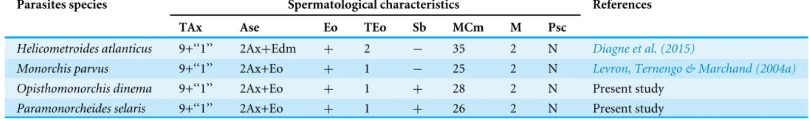
Table 1 Comparative ultrastructural characteristics of the spermatozoon in the Monorchiidae.Ase, anterior spermatozoon extremity; Ax, ax-oneme; Cm, cortical microtubules; Edm, electron-dense material; Eo, external ornamentation of the plasma membrane; M, number of mitochon-dria; MCm, maximum number of cortical microtubules; N, nucleus; TAx, type of axoneme; TEo, type of external ornamentation according to Quili-chini et al., (2011a); Psc, posterior spermatozoon character; Sb, spine-like bodies;+/−, 2013, presence/absence of considered character.
Parasites species Spermatological characteristics References
TAx Ase Eo TEo Sb MCm M Psc
Helicometroides atlanticus 9+‘‘1’’ 2Ax+Edm + 2 − 35 2 N Diagne et al. (2015)
Monorchis parvus 9+‘‘1’’ 2Ax+Eo + 1 − 25 2 N Levron, Ternengo & Marchand (2004a)
Opisthomonorchis dinema 9+‘‘1’’ 2Ax+Eo + 1 + 28 2 N Present study
Paramonorcheides selaris 9+‘‘1’’ 2Ax+Eo + 1 + 26 2 N Present study
General organisation of the mature spermatozoon in the Monorchiidae
From the anterior part to the posterior part of the gametes, six characteristics are presented here to describe the architecture of the mature spermatozoa in Monorchiidae. Moreover, monorchiid spermatozoa are compared to those reported from the other digenean species.
Type of axoneme
The structure of the axoneme with 9+‘‘1’’ pattern of Trepaxonemata (Ehlers, 1984) has been observed inO. dinemaandP. selarisas described previously inM. parvusandH. atlanticus (Table 1). A similar morphology of the axoneme has been reported in most digenean species (Quilichini et al., 2011a;Foata et al., 2012;Miquel et al., 2013;Ndiaye et al., 2012; Ndiaye et al., 2015a) with the exception of schistosomes in which the 9+‘‘1’’ special pattern was described and some didymozoid species showing the 9+0 pattern (Justine & Mattei, 1983;Justine, Jamieson & Southgate, 1993;Yang, Dong & Jiang, 2003).
Their different length is another aspect related to axonemes. In both O. dinemaand P. selaris, the posterior extremity of the axoneme 2 is longer than that of the first one. Moreover, the spermatozoon ofO. dinemais distinguished by the first axoneme that does not reach the nuclear region, while inP. selaristhe first axoneme is observed in the nuclear area like inM. parvusandH. atlanticus(Levron, Ternengo & Marchand, 2004a;Diagne et al., 2015).
Anterior spermatozoon morphology
The anterior spermatozoon extremity ofO. dinemaandP. selariscontains two axonemes, cortical microtubules and external ornamentation of the plasma membrane. Within the Monorchiidae, an anterior spermatozoon extremity showing two axonemes, cortical microtubules and external ornamentation has been reported inMonorchis parvus(Levron, Ternengo & Marchand, 2004a). Whereas in the other monorchiid species studied so far, namelyH. atlanticus(Diagne et al., 2015), only electron-dense material and two axonemes have been observed in the anterior spermatozoon extremity.
Ndiaye et al., 2012) and the mesometridsCentroderma spinosissimaandWardula capitellata (Bakhoum et al., 2012b;Bakhoum et al., 2013). In the two latter families, the presence of a lateral expansion is described in the anterior spermatozoon extremity of the studied species.
Other types of spermatozoa showing two axonemes in their anterior extremities have been reported in several digenean species such as the deropristidDeropristis inflata(Foata, Quilichini & Marchand, 2007), the omphalometridRubenstrema exasperatum(Bakhoum et al., 2011) or the pleurogenidsPleurogenes claviger,Pleurogenoides mediansandProsotocus confusus(Miquel et al., 2013).
External ornamentation and its location
In all monorchiid species studied, the presence of external ornamentation associated with cortical microtubules has been observed in the anterior region of the spermatozoon as in most digenean species (Justine & Mattei, 1982;Miquel et al., 2006;Quilichini et al., 2007; Bâ et al., 2011;Ndiaye et al., 2012;Ndiaye et al., 2015a;Ndiaye et al., 2015b;Bakhoum et al., 2015a;Bakhoum et al., 2015b). However, the location of the external ornamentation distinguishes the spermatozoa ofO. dinema andP. selarisfrom those of other digenean species. In fact, based on the diagram of the localization of the external ornamentation established byQuilichini et al. (2011a), the mature spermatozoon ofO. dinemaandP. selaris presents the type 1 of external ornamentation, i.e., located in proximal part of the anterior spermatozoon region. It is also interesting to note that in bothO. dinemaandP. selaris, the external ornamentation is extended from centriole level to the area containing the first mitochondrion. Such disposition of the external ornamentation is also reported in the monorchiidM. parvusbut not inH. atlanticus(Table 1). This latter species seems to exhibit type 2 external ornamentation according to the diagram ofQuilichini et al. (2011a).
Spine-like bodies
Since their first description by Miquel, Nourrisson & Marchand (2000) the spine-like bodies have frequently been reported in the mature spermatozoon especially in its anterior part. In the male gamete of O. dinemaandP. selaris, spine-like bodies are present in the ornamented area associated with cortical microtubules. Moreover, in both monorchiid species the morphology of the spine-like bodies follows that reported in most digenean species i.e., appearance of a small vesicle interrupting the external ornamentation of the plasma membrane (Miquel et al., 2006;Quilichini et al., 2011b;Bakhoum et al., 2015b; Ndiaye et al., 2015a;Ndiaye et al., 2015b).
Within the Monorchiidae, the absence of spine-like bodies was mentioned in the male gamete ofM. parvusandH. atlanticus(Levron, Ternengo & Marchand, 2004a;Diagne et al., 2015) (Table 1) hence the spine-like bodies are described here, for the first time, in the mature spermatozoon of monorchiid species.
Taking into account all these aspects, the presence or absence of spine-like bodies in the Monorchiidae needs more ultrastructural investigations.
Number of mitochondria
Eucestoda, the absence of mitochondria has been highlighted as a synapomorphy (Justine, 1991a). Besides its interest in phylogenetic relationships, the other criterion related to mitochondria is their number.
Based on many cross- and longitudinal sections, two mitochondria are evident in the mature spermatozoon ofO. dinemaandP. selaris. The presence of two mitochondria have also been reported inM. parvus,H. atlanticus(Table 1) and in several species belonging, for instance, to the families Acanthocolpidae (Bakhoum et al., 2015a), Apocreadiidae (Kacem et al., 2010), Deropristidae (Foata, Quilichini & Marchand, 2007) or Notocotylidae (Ndiaye et al., 2003;Ndiaye et al., 2015b). Other digenean species possess one mitochondrion [e.g., Carmyerius endopapillus(Seck, Marchand & Bâ, 2008),Wardula capitellata; (Bakhoum et al., 2012b)] or three mitochondria [(e.g.,Anisocoelium capitellatum(Ternengo et al., 2009), Euryhelmis squamula(Bakhoum et al., 2009).
In the present study, the position of the second mitochondrion is an ultrastructural characteristic that distinguishes the mature spermatozoa of O. dinemafrom those of P. selaris, H. atlanticusand M. parvus. The second mitochondrion appears after the disorganization of the first axoneme inO. dinema, whereas in the remaining monorchiid the appearance of the second mitochondrion is noted before the posterior extremity of the first axoneme.
Posterior spermatozoon characters
In Digenea the posterior spermatozoon extremity is morphologically variable and is, therefore, valuable in the establishment of spermatozoon models (Quilichini et al., 2010; Ndiaye et al., 2015b;Bakhoum et al., 2012a; Bakhoum et al., 2015a). In all monorchiid species described so far the posterior spermatozoon extremity contains a nucleus as inO. dinemaandP. selaris(Table 1). This morphology of the posterior spermatozoon extremity is frequently reported in digenean (Kacem et al., 2010;Kacem et al., 2015a;Kacem et al., 2015b;Zhukova, Mordvinov & Kiseleva, 2014;Quilichini et al., 2015) and corresponds to the fasciolidean type or type 2 according to the diagram ofQuilichini et al. (2010).
Other types of posterior spermatozoon extremities have been reported in digenean species. This is the case of mature spermatozoa exhibiting one axoneme as described in most microphalloid species (Bakhoum et al., 2012a;Miquel et al., 2013;Bruňanská et al., 2014) and opisthorchioid species (Quilichini et al., 2009;Foata et al., 2012;Zhukova, Mordvinov & Kiseleva, 2014). In the other hand, in the families Opecoelidae and Opistholebetidae (Miquel, Nourrisson & Marchand, 2000;Levron, Ternengo & Marchand, 2003; Levron, Ternengo & Marchand, 2004b; Quilichini et al., 2010; Quilichini et al., 2011b), there are mature spermatozoa showing only cortical microtubules in their posterior extremities.
The phylogenetic relationships in the Monorchiidae and its systematic placement within the Digenea have been controversial. The Monorchiidae has been considered polyphyletic because of the great diversity exhibited in the morphology of its members (Madhavi, 2008). The most recent classification based on DNA sequences includes the Monorchiidae and Lissorchiidae in the superfamily Monorchioidea (Bray, 2008), which was included in the new suborder Monorchiata (Olson et al., 2003).
Here, the understanding of the relationships and phylogenetic affinities in Monorchiidae are attempted with ultrastructural characteristics of the spermatozoa, although there still remain some unexplored groups. The morphology of the spermatozoa described in O. dinema,P. selaris and other monorchiid species is similar in several points to that reported in the apocreadiidNeoapocreadium chabaudi(Kacem et al., 2010). The main similarities concern (1) the type of axoneme, (2) the morphology of both anterior and posterior spermatozoon extremities, (3) the presence of type 1 external ornamentation according toQuilichini et al. (2011a), (4) the two bundles of parallel cortical microtubules and (5) the presence of spine-like bodies.
Besides these similarities, it is interesting to note that the systematic position of the Apocreadiidae is unresolved. Some authors have grouped this family with the Haploporoidea and Monorchioidea (Cribb et al., 2001). Whereas other researchers suggested the removal of the Apocreadiidae from the Lepocreadioidea (seeBray & Cribb, 2012). Thus, to our knowledge no robust data are available to validate the phylogenetic position of the Apocreadiidae.
The present spermatological findings suggest, for the first time, a close relationship between the Monorchiidae and the Apocreadiidae based on ultrastructural characteristics of their mature spermatozoa. However, further studies are needed in order to support our hypothesis. Moreover, ultrastructural studies in the Lissorchiidae (probably a sister group) are also needed to test their close relationships with the Monorchiidae.
Conclusion
The mature spermatozoa ofOpisthomonorchis dinemaandParamonorcheides selarisshare several ultrastructural features with those reported inMonorchis parvusandHelicometroides atlanticus(Table 1). These similarities allow us to define the type of spermatozoon in the Monorchiidae, which exhibits the following features:
– Presence of an anterior extremity showing two axonemes accompanied by cortical microtubules and external ornamentation,
– Presence of the association ‘‘external ornamentation+cortical microtubules’’,
– Location of the external ornamentation corresponding to the type 1 accordingQuilichini et al. (2011a),
– Presence of two bundles of parallel cortical microtubules and two mitochondria, – Posterior spermatozoon extremity containing only the nucleus according toQuilichini
et al. (2010).
the Monorchiidae and Apocreadiidae, although additional spermatological evidences are needed especially, from the unexplored taxa belonging to the families mentioned above and the Lissorchiidae.
The ultrastructural characteristics described in digenean and in particular, the Monorchiidae could be used as phylogenetic tools and when establishing spermatozoon models, given that they may allow the distinguishing of genera, families or superfamilies within the Digenea.
ADDITIONAL INFORMATION AND DECLARATIONS
Funding
A.J.S. Bakhoum has a post-doctoral fellowship (No. CE/01/2014) from the ‘‘Collectivité Territoriale de Corse –Direction de l’Enseignement Supérieur et de la Recherche’’. The funders had no role in study design, data collection and analysis, decision to publish, or preparation of the manuscript.
Grant Disclosures
The following grant information was disclosed by the authors:
Collectivité Territoriale de Corse –Direction de l’Enseignement Supérieur et de la Recherche: CE/01/2014.
Competing Interests
Pr. Jean-Lou Justine is an Academic Editor for PeerJ.
Author Contributions
• Yann Quilichini and Abdoulaye J.S. Bakhoum conceived and designed the experiments, performed the experiments, analyzed the data, contributed reagents/materials/analysis tools, wrote the paper, prepared figures and/or tables, reviewed drafts of the paper.
• Jean-Lou Justine and Rodney A. Bray contributed reagents/materials/analysis tools, reviewed drafts of the paper.
• Cheikh T. Bâ reviewed drafts of the paper.
• Bernard Marchand conceived and designed the experiments, analyzed the data, contributed reagents/materials/analysis tools, prepared figures and/or tables, reviewed drafts of the paper.
Data Availability
The following information was supplied regarding data availability:
REFERENCES
Bâ CT, Ndiaye PI, Dione A, Quilichini Y, Marchand B. 2011.Ultrastructure of the
spermatozoon ofHolorchis micracanthum(Digenea: Lepocreadiidae), an intestinal parasite ofPlectorhinchus mediterraneus(Pisces, Teleostei) in Senegal.Parasitology Research109:1099–1106DOI 10.1007/s00436-011-2352-1.
Bakhoum AJS, Bâ CT, Fournier-Chambrillon C, Torres J, Fournier P, Miquel J. 2009. Spermatozoon ultrastructure ofEuryhelmis squamula(Rudolphi, 1819) (Digenea, Opisthorchioidea, Heterophyidae), an intestinal parasite ofMustela vison(Carnivora, Mustelidae).Revista Ibero-latinoamericana de Parasitología1:37–45.
Bakhoum AJS, Bâ CT, Shimalov CV, Torres J, Miquel J. 2011.
Spermatologi-cal characters of the digeneanRubenstrema exasperatum(Rudolphi, 1819) (Plagiorchioidea, Omphalometridae).Parasitology Research108:1283–1293 DOI 10.1007/s00436-010-2178-2.
Bakhoum AJS, Feliu C, Bâ CT, Miquel J. 2012a.Spermiogenesis and spermatozoon of
the liver flukeMediogonimus jourdanei(Microphalloidea: Prosthogonimidae), a parasite ofMyodes glareolus(Rodentia: Cricetidae).Folia Parasitologica59:32–42 DOI 10.14411/fp.2012.006.
Bakhoum AJS, Kacem H, Neifar L, Miquel J. 2013.Ultrastructure of the spermatozoon
ofCentroderma spinosissima(Stossich, 1886) (Digenea: Mesometridae) and its phylogenetic potential.Tissue and Cell45:428–433 DOI 10.1016/j.tice.2013.07.006.
Bakhoum AJS, Ndiaye PI, Sène A, Bâ CT, Miquel J. 2012b.Spermiogenesis and
ultrastructure of the spermatozoon ofWardula capitellata(Digenea, Mesometridae), an intestinal parasite of the sparid teleostSarpa salpain Senegal.Acta Parasitologica 57:34–45.
Bakhoum AJS, Quilichini Y, Justine J-L, Bray RA, Bâ CT, Marchand B. 2015a.
Ultra-structural study of sperm cells in Acanthocolpidae: the case ofStephanostomum murielaeandStephanostomoides tenuis(Digenea).PeerJ3:e744
DOI 10.7717/peerj.744.
Bakhoum AJS, Quilichini Y, Justine J-L, Bray RA, Bâ CT, Marchand B. 2015b.
Neo-multitestis aspidogastriformisBray and Cribb, 2003 (Digenea, Lepocreadiidae): mature spermatozoon and sperm morphologies in the Lepocreadioidea.Cell Biology International39:799–807 DOI 10.1002/cbin.10449.
Bakhoum AJS, Quilichini Y, Miquel J, Feliu C, Bâ CT, Marchand B. 2014.Collyricloides
massanae(Digenea, Collyriclidae): spermatozoon ultrastructure and phylogenetic importance.Parasite21: Article 59DOI 10.1051/parasite/2014061.
Bray RA. 2008. Superfamily Monorchioidea Odhner, 1911. In: Bray RA, Gibson DI,
Jones A, eds.Keys to the Trematoda. vol. 3. London: CAB International and The Natural History Museum, 143–144.
Bray RA, Cribb TH. 2012.Reorganisation of the superfamily Lepocreadioidea Odhner,
Bray RA, Justine J-L. 2013.Three species of opisthomonorchiine monorchiids (Digenea) inCarangoidesspp. (Perciformes: Carangidae) from off New Caledonia, with a description ofOpisthomonorchis dineman. sp.Systematic Parasitology85:147–156 DOI 10.1007/s11230-013-9415-x.
Bruňanská M, Brázová T, Zhokhov AE, Poddubnaya LG. 2014.Ultrastructural features
of the spermatozoon and its differentiation inBrandesia turgida(Brandes, 1888) (Digenea, Microphalloidea, Pleurogenidae).Parasitology Research113:2483–2491 DOI 10.1007/s00436-014-3897-6.
Cribb TH, Bray RA, Littlewood DTJ, Pichelin SP, Herniou EA. 2001. The Digenea. In:
Littlewood DTJ, Bray RA, eds.Interrelationships of the Platyhelminthes. London: Taylor and Francis, 168–185.
Diagne PM, Quilichini Y, Bâ CT, Ndiaye PI, Dione A, Marchand B. 2015.Ultrastructure
of the spermatozoon ofHelicometroides atlanticus(Digenea, Monorchiidae), an intestinal parasite ofParapristipoma octolineatum(Pisces, Teleostei) in Senegal. Tissue and Cell 47:198–204DOI 10.1016/j.tice.2014.12.006.
Ehlers U. 1984.Phylogenetisches System der Plathelminthes.Verhandlungen des
Naturwissenschaftlichen Vereins Hamburg, NF27:291–294.
Foata J, Quilichini Y, Greani S, Marchand B. 2012.Sperm ultrastructure of the digenean
Aphallus tubarium(Rudolphi, 1819) Poche, 1926 (Platyhelminthes, Cryptogonimi-dae) intestinal parasite ofDentex dentex(Pisces, Teleostei).Tissue and Cell 44:15–21 DOI 10.1016/j.tice.2011.10.001.
Foata J, Quilichini Y, Marchand B. 2007.Spermiogenesis and sperm ultrastructure
ofDeropristis inflataMolin, 1859 (Digenea, Deropristidae), a parasite ofAnguilla anguilla.Parasitology Research101:843–852.
Justine J-L. 1991a.Phylogeny of parasitic Platyhelminthes—a critical study of
synapo-morphies proposed on the basis of the ultrastructure of spermiogenesis and sperma-tozoa.Canadian Journal of Zoology69:1421–1440DOI 10.1139/z91-203.
Justine J-L. 1991b.Cladistic study in the Monogenea (Platyhelminthes), based upon a
parsimony analysis of spermiogenetic and spermatozoal ultrastructural characters. International Journal for Parasitology21:821–838
DOI 10.1016/0020-7519(91)90151-V.
Justine J-L. 1995.Spermatozoal ultrastructure and phylogeny of the parasitic
Platy-helminthes.Mémoires du Muséum National d’Histoire Naturelle166:37–54.
Justine J-L. 1998.Spermatozoa as phylogenetic characters for the Eucestoda.Journal of
Parasitology 84:385–408DOI 10.2307/3284502.
Justine J-L, Jamieson BGM, Southgate VR. 1993.Homogeneity of sperm structure in
six species of Schistosomes (Digenea, Platyhelminthes).Annales de Parasitologie Humaine et Comparée68:185–187DOI 10.1051/parasite/1993684185.
Justine J-L, Mattei X. 1982.Réinvestigation de l’ultrastructure du spermatozoïde
Justine J-L, Mattei X. 1983.A spermatozoon with two 9+0 axonemes in a parasitic flatworm,Didymozoon(Digenea: Didymozoidae).Journal of Submicroscopic Cytology and Pathology15:1101–1105.
Kacem H, Bakhoum AJS, Neifar L, Miquel J. 2010.Spermiogenesis and spermatozoon
ultrastructure of the digeneanNeoapocreadium chabaudi(Apocreadiidae), a parasite ofBalistes capriscus(Pisces, Teleostei).Parasitology International 59:358–366 DOI 10.1016/j.parint.2010.04.008.
Kacem H, Ndiaye PI, Neifar L, Torres J, Miquel J. 2015a.Ultrastructure of the
sperma-tozoon of the digeneanTergestia acanthocephala(Stossich, 1887) (Gymnophalloidea: Fellodistomidae): an intestinal parasite ofBelone belone gracilis(Pisces: Teleostei). Tissue and Cell 47:235–241DOI 10.1016/j.tice.2015.01.008.
Kacem H, Ndiaye PI, Neifar L, Torres J, Miquel J. 2015b.Spermatological characters
of the digeneanLecithostaphylus retroflexus(Molin, 1859) (Microphalloidea: Zoogonidae), a parasite of the teleost fishBelone belone gracilis.Tissue and Cell 47:431-437.
Levron C, Miquel J, Oros M, Scholz T. 2010.Spermatozoa of tapeworms
(Platy-helminthes, Eucestoda): advances in ultrastructural and phylogenetic studies. Biological Reviews85:523–543.
Levron C, Ternengo S, Marchand B. 2003.Ultrastructure of spermiogenesis and the
spermatozoon ofHelicometra fasciata(Digenea, Opecoelidae), a parasite ofLabrus merula(Pisces, Teleostei).Acta Parasitologica48:255–264.
Levron C, Ternengo S, Marchand B. 2004a.Ultrastructure of spermiogenesis and
the spermatozoon ofMonorchis parvusLooss, 1902 (Digenea, Monorchiidae), a parasite ofDiplodus annularis(Pisces, Teleostei).Parasitology Research93:102–110 DOI 10.1007/s00436-004-1115-7.
Levron C, Ternengo S, Marchand B. 2004b.Spermiogenesis and sperm ultrastructure
ofPoracanthium furcatum(Digenea, Opecoelidae), a parasite ofMullus surmuletus (Pisces, Teleostei).Acta Parasitologica49:190–200.
Madhavi R. 2008. Family Monorchiidae Odhner, 1911. In: Bray RA, Gibson DI, Jones
A, eds.Keys to the Trematoda, vol. 3. London: CAB International and The Natural History Museum, 145–175.
Miquel J, Fournier-Chambrillon C, Fournier P, Torres J. 2006.Spermiogenesis and
spermatozoon ultrastructure of the cranial digeneanTroglotrema acutum(Leuckart, 1842).Journal of Parasitology 92:441–453DOI 10.1645/GE-743R.1.
Miquel J, Nourrisson C, Marchand B. 2000.Ultrastructure of spermiogenesis and
the spermatozoon ofOpecoeloides furcatus(Trematoda, Digenea, Opecoelidae), a parasite ofMullus barbatus(Pisces, Teleostei).Parasitology Research86:301–310 DOI 10.1007/s004360050047.
Miquel J, Vilavella D, Świderski Z, Shimalov VV, Torres J. 2013.Spermatological
Ndiaye PI, Bakhoum AJS, Sène A, Diagne PM, Miquel J. 2015a.The ultrastructural char-acters of the mature spermatozoon ofOpechona bacillaris(Molin, 1859) (Digenea, Lepocreadiidae) a parasite ofScomber coliasGmelin, 1789 (Scombridae) off the coast of Dakar (Senegal).Acta Zoologica (Stockholm)96:91–98DOI 10.1111/azo.12054.
Ndiaye PI, Miquel J, Feliu C, Marchand B. 2003.Ultrastructure of spermiogenesis and
spermatozoa ofNotocotylus neyraiGonzález Castro, 1945 (Digenea, Notocotylidae), intestinal parasite ofMicrotus agrestis(Rodentia: Arvicolidae) in Spain.Invertebrate Reproduction and Development 43:105–115DOI 10.1080/07924259.2003.9652529.
Ndiaye PI, Quilichini Y, Sène A, Bâ CT, Marchand B. 2011.Ultrastructure of the
spermatozoon of the digeneanCricocephalus albus(Kuhl & van Hasselt, 1822) Looss, 1899 (Platyhelminthes, Pronocephaloidea, Pronocephalidae), parasite of ‘‘the hawksbill sea turtle’’Eretmochelys imbricata(Linnaeus, 1766) in Senegal.Zoologischer Anzeiger250:215–222 DOI 10.1016/j.jcz.2011.04.002.
Ndiaye PI, Quilichini Y, Sène A, Tkach VV, Bâ CT, Marchand B. 2012.Ultrastructural
study of the male gamete ofPleurogonius truncatusPrudhoe, 1944 (Platyhelminthes, Digenea, Pronocephalidae) parasite ofEretmochelys imbricata(Linnaeus, 1766). Comptes Rendus Biologies335:239–246DOI 10.1016/j.crvi.2012.02.004.
Ndiaye PI, Torres J, Eira C, Shimalov VV, Miquel J. 2015b.Ultrastructure of the
spermatozoon of the trematodeNotocotylus noyeri(Digenea: Notocotylidae), a parasite ofMicrotus arvalis(Rodentia: Cricetidae).Folia Parasitologica62: Article 001.
Olson PD, Cribb TH, Tkach VV, Bray RA, Littlewood DTJ. 2003.Phylogeny and
classification of the Digenea (Platyhelminthes: Trematoda).International Journal for Parasitology 33:733–755DOI 10.1016/S0020-7519(03)00049-3.
Quilichini Y, Foata J, Justine J-L, Bray RA, Marchand B. 2009.Sperm ultrastructure of
the digeneanSiphoderina elongata(Platyhelminthes, Cryptogonimidae) intestinal parasite ofNemipterus furcosus(Pisces, Teleostei).Parasitology Research105:87–95 DOI 10.1007/s00436-009-1366-4.
Quilichini Y, Foata J, Justine J-L, Bray RA, Marchand B. 2010.Ultrastructural study of
the spermatozoon ofHeterolebes maculosus(Digenea, Opistholebetidae), a parasite of the porcupinefishDiodon hystrix(Pisces, Teleostei).Parasitology International 59:427–434DOI 10.1016/j.parint.2010.06.002.
Quilichini Y, Foata J, Justine J-L, Bray RA, Marchand. 2011a.Spermatozoon
ultrastruc-ture ofGyliauchensp. (Digenea: Gyliauchenidae), an intestinal parasite ofSiganus fuscescens(Pisces: Teleostei).Biological Bulletin221:197–205.
Quilichini Y, Foata J, Justine J-L, Bray RA, Marchand B. 2011b.Sperm ultrastructure
ofHelicometra epinepheli(Platyhelminthes, Digenea, Opecoelidae), parasite of Epinephelus fasciatus(Pisces, Teleostei).Histology and Histopathology26:1019–1028.
Quilichini Y, Foata J, Marchand B. 2007.Ultrastructural study of the
Quilichini Y, Foata J, Orsini A, Marchand B. 2007.Spermiogenesis and spermatozoon ultrastructure ofNicolla wisniewskii(Digenea: Opecoelidae), an intestinal parasite of brown troutSalmo trutta(Pisces: Teleostei).Journal of Parasitology93:469–478 DOI 10.1645/GE-1085R.1.
Quilichini Y, Ndiaye PI, Sène A, Justine JL, Bray RA, Tkach VV, Bâ CT, Marchand
B. 2015.Ultrastructural characters of the spermatozoa in Digeneans of the genus
BianiumStunkard, 1930 (Digenea, Lepocreadiidae) parasites of fishes: a com-parative study ofBianium plicitumandBianium arabicum.Parasitology Research 114:3747–3757.
Reynolds ES. 1963.The use of lead citrate at high pH as an electron-opaque stain in
electron microscopy.Journal of Cell Biology17:208–212DOI 10.1083/jcb.17.1.208.
Seck MT, Marchand B, Bâ CT. 2008.Spermiogenesis and sperm ultrastructure of
Carmyerius endopapillatus(Digenea, Gastrothylacidae), a parasite ofBos taurusin Senegal.Acta Parasitologica53:9–18.
Ternengo S, Quilichini Y, Katharios P, Marchand B. 2009.Sperm ultrastructure of the
gall bladder flukeAnisocoelium capitellatum(Digenea: Cryptogonimidae), a parasite ofUranoscopus scaber(Pisces: Uranoscopidae).Parasitology Research104:801–807 DOI 10.1007/s00436-008-1259-y.
Thiéry JP. 1967.Mise en évidence des polysaccharides sur coupes fines en microscopie
électronique.Journal of Microscopy 6:987–1018.
Yang M-Y, Dong H-F, Jiang M-S. 2003.Ultrastructural observation of spermatozoa and
fertilization inSchistoma japonicum.Acta Tropica85:63–70.
Zhukova MV, Mordvinov VA, Kiseleva E. 2014.Ultrastructure of spermatozoa in the