1
Different manifestations of Class II Division 2 incisor retroclination – Morphologic Study
ABSTRACT
Introduction: The aim of the present study was to investigate whether there is a
different transverse morphologic pattern of dental arches among the different manifestations of Class II Division 2 incisor retroclination and evaluate to what extent the pattern of smaller-than-average teeth in Class II Division 2 malocclusion is common to all groups studied. This information may clarify whether different Class II Division 2 phenotypes represent a single etiology or multiple etiologies. Methods: The sample comprised 108 Class II Division 2 malocclusions which were divided into two groups according to the type of incisor retroclination: Group I composed of 43 Class II Division 2 with retroclination exclusively of the maxillary central incisors; Group II composed of 65 Class II Division 2 with retroclination of the four maxillary incisors. Maxillary and mandibular intercanine and intermolar widths as well as the mesiodistal crown dimension of the four maxillary and mandibular incisors were determined using the initial study models of patients. Mean values of all variables were compared between the two groups by gender using ANOVA. Results: From the comparison between the two groups analyzed, no statistically significant differences were found for all transverse measurements (p>0.05). For all mesiodistal measurements analyzed, statistically significant differences between the groups were only found for the mean value of both maxillary lateral incisors mesiodistal dimension in both sexes (p<0.05). Conclusions: It is not possible to attribute a characteristic pattern of dental arch width and of incisor mesiodistal dimension to the different manifestations of incisor retroclination in Class II Division 2 malocclusion.
INTRODUCTION
The morphologic characterization has proved to be an important aid in determining the way certain genetic factors are being expressed, and also the extent to which malocclusion phenotype can be influenced by environmental factors. The similarity of morphologic features is often used as the main criterion for classification and grouping of malocclusion and, consequently, decisive for diagnosing and addressing orthodontic treatment.
Class II Division 2 malocclusion has been described as displaying a phenotype resulting from multiple morphologic features, which are not always present or express themselves in
2
variable degree. Maxillary incisors retroclination is clearly the most peculiar feature and the main distinctive sign of this singular malocclusion, which, however, does not always manifest itself in the same way, existing different forms of maxillary incisor retroclination described in the literature1-3. The diverse morphologic characteristics attributed to Class II Division 2 malocclusion have been interpreted as different manifestations of the same clinical entity, without any related studies to support this view. When we consider the diversity of characteristics associated with Class II Division 2, some of them not always present or, when they are, occurring in different levels, particularly the different forms of incisor retroclination, it is fair to speculate whether we are in the presence of different clinical entities or whether we are facing different degrees or even different manifestations of the same clinical entity.
A reduced dental pattern in the mesiodistal direction has been consistently attributed to Class II Division 2 malocclusion, an evidence first revealed by Beresford4, who, when determining the maxillary central incisor mesiodistal dimension in the different Angle classes, only found statistically significant differences in Class II Division 2, where he observed narrower incisors. Besides the maxillary central incisors, Milicic et al 5 also analyzed the mesiodistal dimension of the maxillary laterals and of the four mandibular incisors, and, by comparing a Class II Division 2 malocclusion group and a Class II Division 1 malocclusion group with a control group in normocclusion, concluded that Angle’s Division 2 exhibited smaller incisors in the mesiodistal direction. Also Peck et al 6 observed a pattern of smaller-than-average anterior teeth associated with Class II Division 2 malocclusion, when comparing 23 severe manifestations of this malocclusion with a reference group. A reduced dental pattern has also been observed in the labiopalatal direction, associated with Class II Division 2 malocclusion7,8.
Whereas there is significant scientific evidence regarding a pattern of smaller-than-average teeth associated with Class II Division 2 malocclusion4-6, little consensus can be found in the literature concerning the existence of a transverse morphologic pattern of arch width, characteristic of this peculiar malocclusion9-15. These studies on tooth morphology, however, fail to mention the constitution of samples regarding the different manifestations of incisor retroclination, neither do they investigate the different Class II Division 2 phenotypes separately.
Some authors explain the different manifestations of maxillary incisor retroclination in Class II Division 2 malocclusion as being the consequence of the maxillary arch space conditions, i.e., they result from the space availability in the anterior region during the incisor eruption2,3. This view might presuppose the existence of distinctive transverse dental arch
3
features as well as different anterior dental dimension patterns associated with the various incisor retroclination manifestations.
The aim of this study was to investigate whether there is a distinctive transverse morphologic pattern of the arches among the different incisor retroclination manifestations. It also evaluated whether the pattern of smaller-than-average teeth in Class II Division 2 malocclusion is common to all groups studied. This information may aid in understanding the different clinical presentations of Class II Division 2 patients as a single clinical entity or etiologically diverse entities.
This research may open the way for future studies which can contribute for a better understanding of the ethiopathogenic mechanisms involved in the different Class II Division 2 phenotypes in terms of incisor retroclination. A deep knowledge of malocclusion ethiology will be paramount for the prevention and treatment of orthodontic disorders. Without a clear understanding of the ethiologic factors responsible for this peculiar malocclusion, we run the risk of using empirical or exclusively symptomatic therapies.
MATERIAL AND METHODS
This retrospective study was approved by the Ethics Commission of the Faculty of Dental Medicine of the University of Porto, Portugal. The sample was collected from the private practice of the first two authors of this paper. From the consecutive analysis of the initial orthodontic records of 4364 patients seeking orthodontic treatment between 2002 and 2010, 215 Class II Division 2 malocclusions were diagnosed. These 215 patients, all non-syndromic of Caucasian origin, were distributed into two groups on the basis of the type of maxillary incisor retroclination, after having been applied the following inclusion criteria: (1) molar distocclusion, at least unilateral in centric occlusion; (2) presence of all four maxillary incisors; (3) no history of orthodontic treatment, maxillofacial or plastic surgery and trauma in the maxillary anterior teeth; (4) absence of prosthetic crowns or extensive restorations in the six maxillary anterior teeth; (5) the angle between the maxillary incisor long axis and the palatal plane less than or equal to 100º; (6) overbite equal to or greater than 50% and (7) previous eruption of the maxillary and mandibular second permanent molars. Patients with retroclination involving three incisors were excluded. The total sample was thus made up of 108 subjects (66 females, 42 males) with a mean age of 22.6 years (SD, 9.1; range, 12-50 years) distributed into the two groups as follows:
Group I composed of 43 Class II Division 2 malocclusions (27 females, 16 males) with retroclination exclusively of both maxillary central incisors, with a mean age of 22.3 years (SD, 9.3; range, 12-50 years);
4
Group II composed of 65 Class II Division 2 (39 females, 26 males) with retroclination of all four maxillary incisors, with a mean age of 22.9 years (SD, 9.1; range 12-43 years).
For this morphologic study, the initial orthodontic study models were used. Of the 108 cases studied, there were 75 plaster models available in good condition and the remaining 33 presented digital models obtained from the initial plaster models using the Bibliocast system (Bibliocast Ibérica, Lda., Porto, Portugal) (Table I).
Four variables representative of the dental arch widths were evaluated and the mesiodistal dimension of the mandibular and maxillary four anterior teeth was determined. The criteria defined for each variable were the following:
Maxillary Intercanine Width (MaxIC) – Linear measurement between the tip of the right maxillary canine cusp and the tip of the left maxillary canine cusp or the center of the wear facet, in cases where attrition of the cusp tip was evident.
Mandibular Intercanine Width (MandIC) – Linear measurement between the tip of the right mandibular canine cusp and the tip of the left mandibular canine cusp or the center of the wear facet, in cases where attrition of the cusp tip was evident.
Maxillary Intermolar Width (MaxIM) – Linear measurement between the central fossae of the right and left maxillary permanent first molars.
Mandibular Intermolar Width (MandIM) – Linear measurement between the tip of the mandibular right permanent first molar centrobuccal cusp and the tip of the mandibular left permanent first molar centrobuccal cusp. Whenever the cusp tip exhibited attrition, the center of the wear facet was used.
Maximum mesiodistal crown diameter of the right maxillary lateral incisor (MD12).
Maximum mesiodistal crown diameter of the right maxillary central incisor (MD11).
Maximum mesiodistal crown diameter of the left maxillary central incisor (MD21).
Maximum mesiodistal crown diameter of the left maxillary lateral incisor (MD22).
Maximum mesiodistal crown diameter of the right mandibular lateral incisor (MD42).
Maximum mesiodistal crown diameter of the right mandibular central incisor (MD41).
Maximum mesiodistal crown diameter of the left mandibular central incisor (MD31).
Maximum mesiodistal crown diameter of the left mandibular lateral incisor (MD32). All measurements were rounded to the nearest 0.1 mm and taken by using a digital odontometric caliper (Mestra, Bilbao, Spain) on the plaster models and with the Cecile 3 tool (Bibliocast SARL, Paris, France) on the digital models. All variables were determined by the same examiner.
5
The focus of this study was to compare groups with regard to dental arch width and dental mesiodistal dimension. There is scientific evidence of sexual dimorphism in the human tooth size16,17 as well as in the maxillary arch widths18 (i.e. on average males exhibit wider teeth and wider arches than females). Knowing that there exists sexual dimorphism for the variables considered, the comparison between groups formed on the basis of a sample comprising a significantly higher number of female subjects than males had to take into consideration the gender.
Evaluation of measurement error
Estimates of measurement error were determined for four variables using the double determination method16. For the variables MaxIC, MandIM, MD12 and MD11 a second measurement was made by the same examiner on 20 subjects randomly selected, 30 days after the first measurement. After checking the assumption of normality (K-S test with values p >0.05), a paired sample t-test was performed, which did not reveal any statistically significant differences (p >0.05) in the mean values obtained through double determination for each one of the variables studied. The low method error found reveals a high consistency and reproducibility of the measurement technique and the references used (Table II).
Statistical analysis
The statistical analysis was performed using IBM SPSS Statistics 20.0. Since the measurements were evaluated in a quantitative scale the most suitable procedures for comparison of these measures involved comparison of mean values in terms of groups. Because there were two fixed factors (sex and group), each one with two levels, the most suitable procedure was the ANOVA test, which allowed comparison of mean values between the two levels of factors. The decision rule consists of detecting statistically significant evidence for probability values (value of the proof test) less than 0.05.
RESULTS
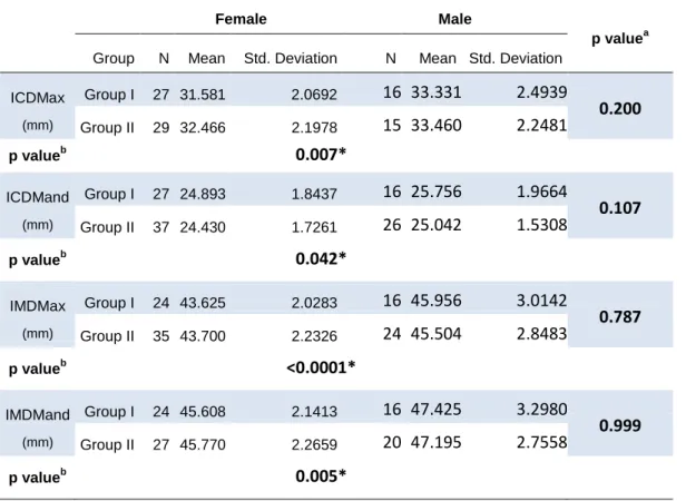
Missing or impacted teeth essential to determine the transverse variables studied did not allow the evaluation of all four arch width measurements in the 108 cases. Table III shows the descriptive statistics for the four transverse measurements analyzed. Having been confirmed that the transverse measurements result from a normal distribution (p>0.05), considering the assumption of independence between groups and the homogeneity of variance having been verified, the ANOVA procedure revealed the absence of statistically
6
significant differences for all transverse measurements by comparing the two groups studied. Arch width mean values proved to be significantly higher in males than in females (Table III).
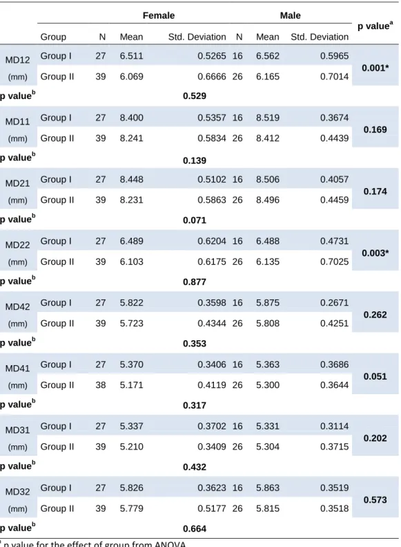
Table IV contains the descriptive statistics for the eight variables studied, representative of the anterior dental mesiodistal dimensions in the two groups formed. Once the assumptions of normality of the dental measurements, independence between groups and homogeneity of variance were confirmed, the ANOVA test revealed statistically significant differences (p<0.05) for MD12 and for MD22 between groups and for both sexes. These differences reveal mean values significantly higher in Group I than in Group II for MD12 and MD22. No significant differences in the mesiodistal mean values were found between genders (Table IV).
DISCUSSION
In the last years there has been some discussion about the validity and reliability of the digital models used for scientific studies and orthodontic diagnosis versus the traditional plaster models. Available studies about the Bibliocast system, which compare measurements performed on digital models and on plaster models, conclude that the method can be a tool with high levels of reliability, validity and reproducibility19-21. Diop Ba et al21 compared linear measurements on plaster models and on digital models obtained with the Bibliocast system in 57 patients. Using a digital caliper and the Bibliocast Cecile 3 software the overjet, the overbite, the mesiodistal and buccolingual diameters of the 12 maxillary and mandibular teeth, and the anterior and overall ratios were determined. For all variables studied no statistically significant differences were observed between the two measurement methods. Although the above-mentioned studies support the use of digital models to determine linear measurements, plaster models and digital models should be proportionately distributed in all the groups and no significant differences should exist for the mean values obtained through both measurement methods in order to validate their incorporation in this study with greater consistency. The comparison between the two methods with the Student’s t-test for independent samples did not reveal any statistically significant differences (p > 0.05) for the mean values of all variables obtained through the two measurement methods.
There is lack of consensus between the studies that compare interdental and alveolar widths of Class II Division 2 malocclusion carriers with control groups or with other types of malocclusion9-15, and also an obvious shortage of studies characterizing transversely the maxillary basal bone of this malocclusion. The diversity of results found in the literature will, certainly, have multiple causes such as the genetic variation of the populations, the origin and formation of control groups, the references used in the variables studied and the formation of
7
the study samples, particularly regarding the different manifestations of maxillary incisor retroclination.
Some authors have explained the different manifestations of maxillary incisor retroclination as resulting from the space conditions of the maxillary anterior arch. According to these authors, the effect of a high lower lip line on the incisor position when space in the anterior region is lacking, promotes the retroclination exclusively of the maxillary centrals. Where there is lack of space, the central incisors, when erupting, suffer retroclination under the influence of a high lip line, whereas when the lateral incisors erupt they are prevented from retroclining due to the lack of space in the maxillary anterior region. When there is enough space available, however, a higher number of teeth tend to retrocline during eruption2,3. These views might imply different mean arch widths depending on the various manifestations of maxillary incisor retroclination. The results achieved in this study for the four transverse variables analyzed reveal no statistically significant differences between the two groups formed. The absence of significant differences in the transverse measurements between the various types of Class II Division 2 studied do not seem to provide support for those who attribute the different manifestations of the maxillary incisor retroclination to the space conditions of the maxillary arch.
Although there is significant scientific evidence regarding the pattern of smaller-than-average teeth associated with Class II Division 2 malocclusion4-6, and just like for the dental arch width, there are no known studies that compare the dental mesiodistal dimension between Class II Division 2 malocclusion groups differing in their phenotype expressiveness. The comparison between the two groups formed did not reveal any statistically significant differences for all dental mesiodistal measurements studied except for the mesiodistal dimension of both maxillary lateral incisors. The statistically significant differences observed for the maxillary lateral incisor result from the fact that this tooth displays a lower mean mesiodistal dimension in the group with retroclination of the four maxillary anterior teeth than in the group with retroclination exclusively of both maxillary centrals. These results appear to be a consequence of the high number of congenital microdontic lateral incisors found in Group II by contrast with Group I, 16 and 2 respectively. The term microdontic applies to peg-shaped laterals that exhibit a conical shape with mesiodistal width greatest at the cervical margin and to small incisors when the maximum mesiodistal width is equal to or smaller than that of its mandibular counterpart. This observation and the absence of significant differences in the maxillary central incisor mesiodistal dimension between the different manifestations of incisor retroclination provide poor support for those theories that advocate the maxillary anterior
8
incisor positioning in Class II Division 2 malocclusion to result from the space availability during incisor eruption.
Results from arch width studies of Class II Division 2 malocclusion patients, when gender is taken into consideration, have not been uniform. Isik et al10 did not find any significant inter-gender differences for the transverse arch measures studied, whereas Huth et al9 observed sexual dimorphism for the maxillary and mandibular intermolar width but not for
intercanine widths. Sexual dimorphism was found for all transverse measurements studied in this research in both Class II Division 2 groups, in the sense that dental arch width was larger in males than in females. No sexual dimorphism was found for any mesiodistal measurement in both Class II Division 2 groups. These results contrast with studies conducted in samples of the general populationwhich have consistently demonstrated a bigger dental pattern in the male gender than in the female 16,17.
The integration of the findings obtained in this study did not demonstrate the existence of significant differences for the morphologic features evaluated between the various manifestations of the maxillary incisor retroclination in Class II Division 2 malocclusion. However, an interpretation of these findings does not exclude the distinct etiologic origin between the different incisor retroclination manifestations. An analysis of the morphologic findings demonstrates that there is poor support for the theories attributing the various forms of this peculiar incisor feature to the availability of space in the anterior region of the maxillary jawduring incisor eruption. A phenotype of retroclination exclusively of both central incisors or a phenotype of retroclination of all maxillary incisors could be under the influence of a distinct genetic basis affecting pre-and/or post-eruptive incisor pathway. In one study carried out by Milne and Cleall22, it is suggested that the maxillary incisors follow the same eruptive axis before and after emerging into the oral cavity, with the eruptive pathway depending on the axial inclination of tooth buds. Markovic’s study 23 on twins and triplets is important, as the author not only registered a concordance for the Class II Division 2 malocclusion between monozygotic twins but also a similarity with regard to the incisor position. A deep knowledge of the processes involved in the pre-and post-eruptive tooth pathway could be decisive for a better understanding of the incisor retroclination mechanism in Class II Division 2 malocclusion.
CONCLUSIONS
9
1. No significant differences were found for the mean maxillary and mandibular dental arch width between groups of Class II Division 2 differing in terms of the maxillary incisor retroclination manifestation.
2. No significant differences were found for the anterior dental mesiodistal dimension, except for the maxillary lateral incisor, between the distinct maxillary incisor manifestations in Class II Division 2 malocclusion. The reduced mesiodistal mean dimension found for the maxillary lateral incisor in the group where retroclination involved all the maxillary anterior incisors can be justified by the high prevalence of congenital microdontic lateral incisor found in this group.
3. The sexual dimorphism observed for the maxillary and mandibular arch width in both Class II Division 2 groups was not found in any of the mesiodistal tooth measurements evaluated.
4. The absence of significant differences in the dental arch width and in the anterior dental mesiodistal dimension do not support those theories advocating that the different manifestations of upper incisor retroclination result from the space available in the maxillary anterior region at the time of eruption.
REFERENCES
1. Canut JAB. Ortodoncia Clinica. Barcelona: Ediciones Científicas y Técnicas, S.A.; 1992.
2. van der Linden FPGM. Development of Dentition. Chicago: Quintessence Publishing Co.; 1983.
3. Korkhaus G, Bruhn C, Hofrath H. La Escuela Odontologica Alemana. Rio de Janeiro: Editorial Labor; 1939.
4. Beresford JS. Tooth size and class distinction. Dent Pract Dent Rec 1969;20:113-20.
5. Milicic A, Slaj M, Kovacic J. Dimensions of deciduous and permanent incisors in cases with Class II division 1 and 2 malocclusions. Bilt Udruz Ortodonata Jugosl 1990;23:7-14.
6. Peck S, Peck L, Kataja M. Class II Division 2 malocclusion: A heritable pattern of small teeth in well-developed jaws. Angle Orthod 1998;68:9-20.
7. McIntyre GT, Millett DT. Crown-root shape of the permanent maxillary central incisor. Angle Orthod 2003;73:710-5.
8. Robertson NR, Hilton R. Feature of the Upper Central Incisors in Class Ii, Division 2. Angle Orthod 1965;35:51-3.
9. Huth J, Staley RN, Jacobs R, Bigelow H, Jakobsen J. Arch widths in class II-2 adults compared to adults with class II-1 and normal occlusion. Angle Orthod 2007;77:837-44.
10. Isik F, Nalbantgil D, Sayinsu K, Arun T. A comparative study of cephalometric and arch width characteristics of Class II division 1 and division 2 malocclusions. Eur J Orthod 2006;28:179-83.
11. Xu JP, Shen PM. [Study on the dental arch width in Class II malocclusion]. Shanghai Kou Qiang Yi Xue 2005;14:597-600.
12. Uysal T, Memili B, Usumez S, Sari Z. Dental and alveolar arch widths in normal occlusion, class II division 1 and class II division 2. Angle Orthod 2005;75:941-7.
10
13. Walkow TM, Peck S. Dental arch width in Class II Division 2 deep-bite malocclusion. Am J Orthod Dentofacial Orthop 2002;122:608-13.
14. Buschang PH, Stroud J, Alexander RG. Differences in dental arch morphology among adult females with untreated Class I and Class II malocclusion. Eur J Orthod 1994;16:47-52.
15. Moorrees CF, Gron AM, Lebret LM, Yen PK, Frohlich FJ. Growth studies of the dentition: a review. Am J Orthod 1969;55:600-16.
16. Kieser JA. Human adult odontometrics. Cambridge: Cambridge University Press; 1990. 17. Garn SM, Lewis AB, Kerewsky RS. Sex Difference in Tooth Size. J Dent Res 1964;43:306. 18. Lee RT. Arch width and form: a review. Am J Orthod Dentofacial Orthop 1999;115:305-13. 19. Watanabe-Kanno GA, Abrão J, Miasiro Junior H, Sánchez-Ayala A, Lagravère MO. La détermination de la dysharmonie dento-dentaire et des rapports de Bolton a l’aide de modèles numériques Cécile3 de bibliocast. International Orthodontics 2010;8:215-26.
20. Watanabe-Kanno GA, Abrão J, Miasiro Junior H, Sánchez-Ayala A, Lagravère MO. Reproducibility, reliability and validity of measurements obtained from Cecile3 digital models. Braz Oral Res 2009;23:288-95.
21. Diop ba K, Diagne F, Diouf JS, Ndiaye R, Diop F. Données odontométriques au sein d’une population sénégalaise : comparaison entre la méthode manuelle et l’analyse numérique. International Orthodontics 2008;6:285-99.
22. Milne IM, Cleall JF. Cinefluorographic study of functional adaptation of the oropharyngeal structures. Angle Orthod 1970;40:267-83.
23. Markovic MD. At the crossroads of oral facial genetics. Eur J Orthod 1992;14:469-81.
Table I. Distribution per group according to the type of model.
Group Digital models Plaster models Total Digital proportion Plaster proportion I 13 30 43 30.2% 69.8% II 20 45 65 30.7% 69.3%
Table II. Estimate of measurement error.
Mean difference Std. Deviation p valuea
ICDMax-1 – ICDMax-2 (mm) 0.03000 0.28303 0.641
IMDMand-1 – IMDMand-2 (mm) -0.31000 1.36532 0.323
MD12-1 – MD12-2 (mm) 0.02000 0.16092 0.585
MD11-1 – MD11-2 (mm) -0.02500 0.12085 0.367
a
11
Table III. Summary statistics of the transverse measurements according to the Group for
female and male.
Female Male
p valuea
Group N Mean Std. Deviation N Mean Std. Deviation
ICDMax (mm) Group I 27 31.581 2.0692 16 33.331 2.4939 0.200 Group II 29 32.466 2.1978 15 33.460 2.2481 p valueb 0.007* ICDMand (mm) Group I 27 24.893 1.8437 16 25.756 1.9664 0.107 Group II 37 24.430 1.7261 26 25.042 1.5308 p valueb 0.042* IMDMax (mm) Group I 24 43.625 2.0283 16 45.956 3.0142 0.787 Group II 35 43.700 2.2326 24 45.504 2.8483 p valueb <0.0001* IMDMand (mm) Group I 24 45.608 2.1413 16 47.425 3.2980 0.999 Group II 27 45.770 2.2659 20 47.195 2.7558 p valueb 0.005*
a p value for the effect of group from ANOVA b
p value for the effect of sex from ANOVA *significant at 0.05 level
12
Table IV. Summary statistics of the dental mesiodistal dimension according to the Group for
female and male.
Female Male
p valuea
Group N Mean Std. Deviation N Mean Std. Deviation
MD12 (mm) Group I 27 6.511 0.5265 16 6.562 0.5965 0.001* Group II 39 6.069 0.6666 26 6.165 0.7014 p valueb 0.529 MD11 (mm) Group I 27 8.400 0.5357 16 8.519 0.3674 0.169 Group II 39 8.241 0.5834 26 8.412 0.4439 p valueb 0.139 MD21 (mm) Group I 27 8.448 0.5102 16 8.506 0.4057 0.174 Group II 39 8.231 0.5863 26 8.496 0.4459 p valueb 0.071 MD22 (mm) Group I 27 6.489 0.6204 16 6.488 0.4731 0.003* Group II 39 6.103 0.6175 26 6.135 0.7025 p valueb 0.877 MD42 (mm) Group I 27 5.822 0.3598 16 5.875 0.2671 0.262 Group II 39 5.723 0.4344 26 5.808 0.4251 p valueb 0.353 MD41 (mm) Group I 27 5.370 0.3406 16 5.363 0.3686 0.051 Group II 38 5.171 0.4119 26 5.300 0.3644 p valueb 0.317 MD31 (mm) Group I 27 5.337 0.3702 16 5.331 0.3114 0.202 Group II 39 5.210 0.3409 26 5.304 0.3715 p valueb 0.432 MD32 (mm) Group I 27 5.826 0.3623 16 5.863 0.3519 0.573 Group II 39 5.779 0.5177 26 5.815 0.3518 p valueb 0.664 a
p value for the effect of group from ANOVA b p value for the effect of sex from ANOVA *significant at 0.05 level


