Angle Class II, division 2 malocclusion with severe
overbite and pronounced discrepancy*
Daniela Kimaid Schroeder**
This article reports the treatment of a young patient at 13.8 years of age who presented with an Angle Class II, division 2 malocclusion, prolonged retention of deciduous teeth, dental crossbite and severe overbite, among other abnormalities. At first, the approach involved rapid maxillary expansion followed by the use of Kloehn headgear and fixed orthodontic appliance. Treatment results demonstrate the importance of careful diagnosis and planning as well as the need for patient compliance during treatment. This case was presented to the Brazilian Board of Orthodontics and Facial Orthopedics (BBO). It is representative of the free category and fulfills part of the requirements for obtaining the BBO Diploma.
Abstract
Keywords: Class II, division 2. Crossbite. Severe overbite. Prolonged retention of deciduous teeth.
** MSc in Orthodontics, Federal University of Rio de Janeiro (UFRJ). Diplomate of the Brazilian Board of Orthodontics. * Case report, free category - approved by the Brazilian Board of Orthodontics.
HISTORY AND ETIOLOGY
The patient sought orthodontic treatment at 13.8 years of age. Her main complaint was the fact that her teeth took too long to fall and she was ashamed to smile. No significant informa-tion was found in her past medical and dental records. Her malocclusion, mainly presented lack of space for the alignment of certain teeth, which compromised her facial aesthetics signifi-cantly (Fig 1), and had as major etiological fac-tor the prolonged retention of deciduous teeth. Her menarche had occurred at age 12.
DIAGNOSIS
Her dental pattern (Fig 1, 2) was an Angle Class II, division 2, right subdivision, excessive-ly upright upper and lower incisors, severe deep bite (100%), upper and lower midlines shifted 3 mm to the right, lack of space for eruption of tooth 13 and alignment of other teeth, dental crossbites and atretic arches.
FIGURE 1 - Initial facial and intraoral photographs.
A B
A B
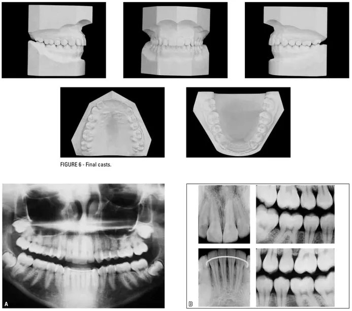
an interincisal angle of 157°, IMPA of 75°, 1-NA of 7º and 2.5 mm, and 1-NB of 12º and 4 mm. These features can be seen in figure 4 and table 1.
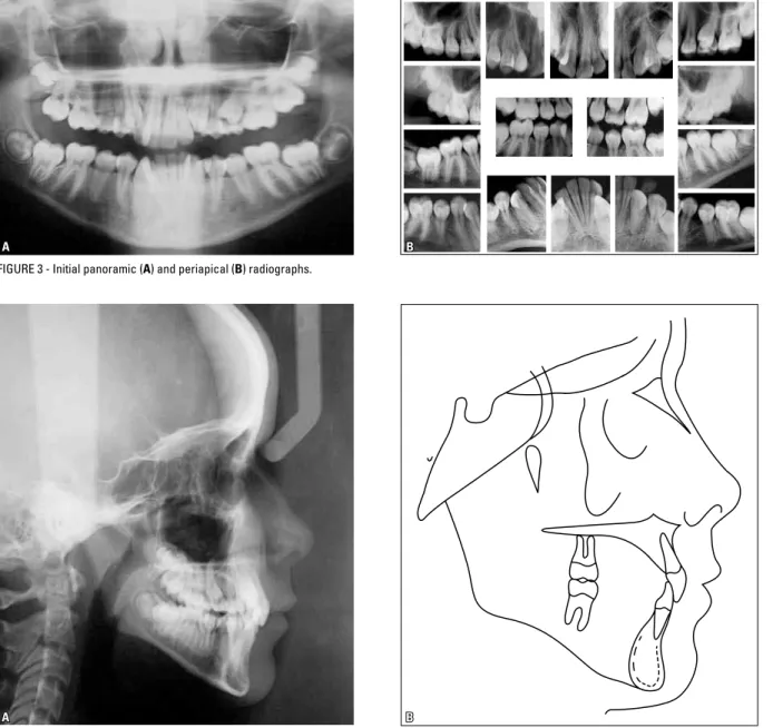
An analysis of the periapical and panoramic radiographs (Fig 3) reassured that the patient did not present with any condition that might compromise her orthodontic treatment.
The patient had a slightly convex profile and an unpleasant smile due to crowding and incor-rect tooth inclinations (Figs 1 and 4).
TREATMENT GOALS
In the anteroposterior direction, the aim was to establish an Angle Class I relationship and improve upper and lower incisor incli-nation. In the vertical direction, it would be necessary to reduce the severe overbite by leveling the upper and lower arches. In the transverse direction, upper and lower arch expansion was performed to increase interca-nine width.
FIGURE 3 - Initial panoramic (A) and periapical (B) radiographs.
With this, it was expected that crossbites would be eliminated, and adequate overbite and upper and lower midline correction would be achieved, significantly improving smile aesthetics.
TREATMENT PLAN
A treatment plan was established, starting with palatal expansion to increase the trans-verse maxillary dimension and make room for tooth alignment.
After removing the expansion appliance, an asymmetric Kloehn headgear (AKHG) would be used with the purpose of correcting the molar relationship and creating space. Con-currently with the AKHG, upper orthodontic appliance would be installed, alignment and leveling started in this arch, and only when the amount of overbite permited, the lower orthodontic appliance would be bonded. To improve the form of the lower arch and make room for alignment and leveling of the lower teeth, the plan was to use archwires featuring greater intercanine width, since the canine lin-gual inclination and an atretic arch would al-low such expansion.
To assist in opening space for tooth 13 and thus correct the upper midline, a compressed open spring would be placed between teeth 12 and 14, starting from the 0.018-in archwire.
To finish the case, the use of upper and low-er 0.019 x 0.025-in archwires would be coor-dinated, with first and third order bends, and individualized intermaxillary elastic mechan-ics would be applied, according to the needs of this particular case.
After the active treatment phase, an upper wraparound-type retention plate would be used and, in the lower arch, a 0.028-in intercanine arch.
TREATMENT PROGRESS
To expand the palate a modified Haas appli-ance was employed with activation of 2/4 turn of the screw once a day. The same appliance was
used as a retainer for 6 months. The maxilla was expanded, which enhanced the form of the up-per arch and consequently of the lower arch.
After removing the expansion screw, the asymmetric AKHG was adjusted by keeping its external right arm longer and open, with a force of 350g, to be worn for approximately 14 hours/ day. This corrected the molar relationship and helped to make space for upper tooth alignment.
Slot 0.022 x 0.028-in standard edgewise metal brackets with no torques or angulations were used. The orthodontic appliance was ini-tially installed on the upper arch. It was only af-ter adequate space and height had been achieved that the lower arch appliance was bonded.
On the upper arch, 0.014-in to 0.020-in archwires were used for alignment and leveling and from the moment that 0.018-in archwires began to be used, an open spring was com-pressed between teeth 12 and 14 to help create space for positioning tooth 13 and subsequent midline correction. After alignment and level-ing of all teeth, individualized 0.019 x 0.025-in sta0.025-inless steel archwires were 0.025-inserted on the upper arch to finish the case.
The same alignment and leveling procedures used for the upper arch were also performed on the lower, although the archwires were con-toured in order to expand the lower arch by up-righting the canines and premolars and allowing protrusion of the incisors, which were retro-clined before treatment. This enabled a correct alignment, leveling and midline correction. To finish the case, a 0.019 x 0.025-in stainless steel archwire with custom-made bends was used.
The patient had her upper and lower third mo-lars extracted.
TREATMENT RESULTS
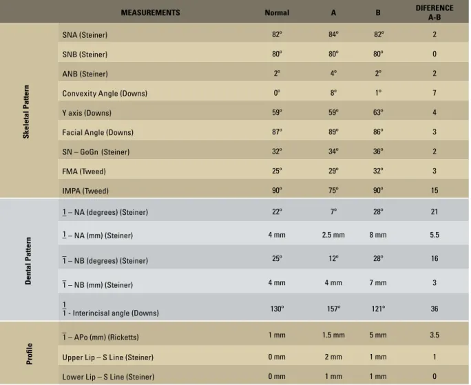
In reviewing the patient’s final records, it be-came clear that the major goals set at the begin-ning of treatment were attained (Figs 5, 6, 8). In the maxilla, ANB was reduced by 2º and the position of the maxilla relative to the overall pro-file improved considerably, reducing the angle of convexity from 8º to 1º. In addition, there was adequate vertical control and considerable en-hancement of the upper arch form (Figs 5, 6, 8).
The teeth exhibited adequate alignment and im-proved incisor inclination. The overbite was also corrected and intercanine width increased by 11 mm, as initially planned, while the intermolar width was maintained.
In the mandible, a clockwise rotation occurred as the FMA angle (Tweed) increased from 28º to 32º (Figs 8, 9 and Table 1) due to the use of the headgear as well as leveling. From a dental standpoint, adequate alignment was achieved, the curve of Spee was leveled and the incisors were protruded with an increase in the IMPA angle (Tweed) from 75º to 90º (Figs 8, 9 and Table 1).
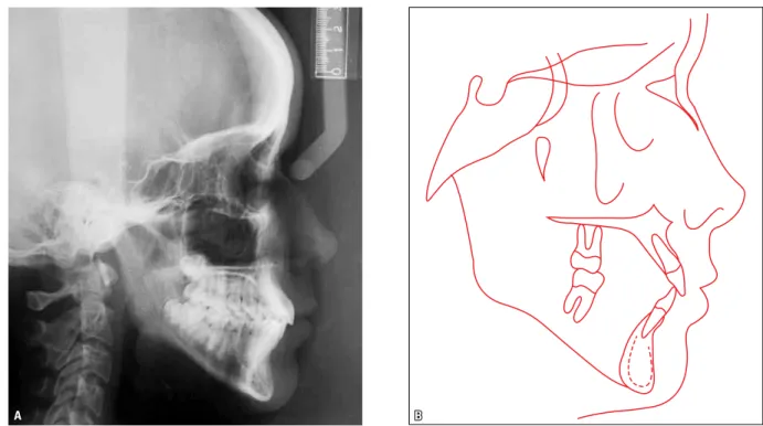
A B FIGURE 6 - Final casts.
FIGURE 7 - Final panoramic (A) and interproximal periapical (B) radiographs.
Regarding occlusion, the dental midlines were coincident to the facial midline, the molars and canines came into normal occlusion, vertical overbite became appropriate and disocclusion guides satisfactory.
The panoramic radiograph (Fig 7A) revealed adequate root parallelism. The gentle rounding of the apices of the upper incisor roots observed
in the Figure 7B is compatible with the amount of movement produced. The profile cephalomet-ric radiograph (Fig 8A) shows improved overbite and interlabial relationship.
A
A
B
B FIGURE 8 - Final profile cephalometric radiograph (A) and cephalometric tracing (B).
TABLE 1 - Summary of cephalometric measurements.
MEASUREMENTS Normal A B DIFERENCE
A-B
Skeletal Pattern
SNA(Steiner) 82º 84º 82º 2
SNB(Steiner) 80º 80º 80º 0
ANB(Steiner) 2º 4º 2º 2
Convexity Angle(Downs) 0º 8º 1º 7
Y axis(Downs) 59º 59º 63º 4
Facial Angle(Downs) 87º 89º 86º 3
SN – GoGn (Steiner) 32º 34º 36º 2
FMA(Tweed) 25º 29º 32º 3
IMPA(Tweed) 90º 75º 90º 15
Dental Pattern
–1 – NA (degrees)(Steiner) 22º 7º 28º 21
–1 – NA (mm)(Steiner) 4 mm 2.5 mm 8 mm 5.5
–
1 – NB (degrees)(Steiner) 25º 12º 28º 16
–
1 – NB (mm)(Steiner) 4 mm 4 mm 7 mm 3
–1
1 - Interincisal angle (Downs) 130º 157º 121º 36
Proile
–
1 – APo (mm)(Ricketts) 1 mm 1.5 mm 5 mm 3.5
Upper Lip – S Line(Steiner) 0 mm 2 mm 1 mm 1
Lower Lip – S Line(Steiner) 0 mm 1 mm 1 mm 0
FINAL CONSIDERATIONS
At first, the possibility of treating this case with tooth extractions was raised due to an ap-parent lack of space for the upper and lower teeth. However, the lack of space was the result of altered axial inclinations, tooth migration and atresia of the dental arches. The patient’s age allowed these problems to be corrected using orthodontic resources, whereby space was cre-ated without compromising periodontal sup-port, esthetics and function.2,3,4,5 Stability is yet
another factor that should be taken into account when protruding teeth and expanding dental arches. It is believed that because intercanine distances were widened by correcting upper and lower canine position and not by bringing the teeth out of their bone bases, it is highly likely that stability will be maintained after correc-tion.1 Even so, retention was carefully planned and half-yearly follow-up visits scheduled.
1. Giannely A. Evidence-based therapy: an orthodontic dilemma. Am J Orthod Dentofacial Orthop. 2006 May;129(5):596-8. 2. Haas AJ. Palatal expansion: just the beginning of dentofacial
orthopedics. Am J Orthod. 1970 Mar;57(3):219-55.
3. Haas AJ. Long-term post-treatment evaluation of rapid palatal expansion. Angle Orthod. 1980 Jul;50(3):189-217.
REFERENCES
4. Hershey H, Houghton CW, Burstone CJ. Unilateral face-bows: a theoretical and laboratory analysis. Am J Orthod. 1981 Mar;79(3):229-49.
5. Turpin DL. Correcting the Class II subdivision malocclusion. Am J Orthod Dentofacial Orthop. 2005 Nov;128(5):555-6.
Contact address Daniela Kimaid Schroeder
Rua Visconde de Pirajá, 444, sala 205 – Ipanema CEP: 22.410-002 – Rio de Janeiro/RJ, Brazil E-mail: [email protected]
to another town for two years, for educational purposes. During this period, she missed too many appointments, significantly increasing treatment time to 48 months.
The patient’s compliance in wearing the headgear was unstable, alternating moments
of total collaboration with others of sheer negligence, despite our constant reminders and encouragement. As can be seen in the fi-nal records, the overall result was considered adequate in terms of occlusion and facial and dental aesthetics.