1. Division of Rheumatology, Faculdade de Medicina da Universidade de São Paulo, São Paulo, Brazil
High levels of serum hyaluronic acid
in adults with dermatomyositis
ACTA REUMATOL PORT. 2015;40:150-155
AbstrAct
Background/objectives: Hyaluronic acid (HA) is rarely
described in dermatomyositis (DM). Thus, we deter-mined any clinical association of serum levels of hyaluronic acid (HA) in patients with dermatomyosi-tis (DM).
Materials and Methods: This cross-sectional
singlecenter analysis 75 DM and 75 healthy individuals, du -ring the period from January 2012 to July 2013. An anti-HA antibody assay was performed using specific ELISA/EIA kits, according to the manufacturer’s proto-col.
Results: The patients with DM and control subjects
had comparable demographic distributions (p>0.05). The median time duration between disease diagnosis and initial symptoms was 6.0 [3.0-12.0] months, with a median DM disease duration of 4.0 [1.0-7.0] years. The median level of serum HA was significantly in-creased in patients with DM compared to the control group [329.0 (80.0-958.0) versus 133.0 (30.0-262.0) ng/mL, respectively; p<0.001]. Additional analysis in-volving patients with DM showed that the serum level of HA did not correlate with age, duration between di -sease diagnosis and initial symptoms, di-sease duration, disease status, serum muscle enzyme levels or cumu-lative prednisolone dose (p>0.05). Serum HA also did not correlate with gender, ethnicity, auto-antibodies or drug use (p>0.05), but did correlate with cutaneous features, such as photosensitivity (p=0.001), “shawl” sign (p=0.018), “V-neck” sign (p=0.005) and cuticular hypertrophy (p=0.014).
Conclusions: A high level of serum AH was observed
in DM compared to healthy individuals. In DM, HA did not correlate to demographic, autoantibodies and the
-Victorino AA1, Silva MG1, Shinjo SK1
rapy parameters. However, HA correlated specifically with some cutaneous features, suggesting that this gly-cosaminoglycan could be involved in modulating cu-taneous inflammation in this population. More studies are necessary to understand the correlation between AH and patients with DM.
Keywords: Cutaneous features; Dermatomyositis;
Gly-cosaminoglycan; Hyaluronic acid; Idiopathic inflam-matory myopathies.
IntroductIon
Dermatomyositis (DM) is a rare systemic idiopathic in-flammatory myopathy associated with high morbidity and functional disabilities. The annual incidence of DM is 0.5 to 8 cases per million inhabitants, with a two to one female to male ratio1. The disease has a bimodal
age distribution, affecting individuals from 5 to 15 and from 45 to 55 years of age, although the average age of diagnosis occurs at approximately 40 years of age, and the disease can affect individuals of any age1.
In addition to progressive symmetrical muscle weak-ness, DM is characterized by classic skin lesions, such as heliotrope rash and/or Gottron’s papules1,2.
More-over, other cutaneous manifestations can be present, including periungual telangiectasia, mechanic’s hand, skin ulcers, vasculitis, “V-neck” sign, “shawl” sign, cal-cinosis cutis and Raynaud’s phenomenon1,3.
Hyaluronic acid (HA) is a glycosaminoglycan that comprises the extracellular connective tissue matrix. HA is distributed in various tissues, such as synovial fluid, ophthalmological vitreous humor, umbilical cord, connective tissues and cartilage4,5. HA is produced
mainly by fibroblasts, is transported by the lymph and is rapidly metabolized by the liver4,5. Moreover, there is
response by stimulating the expression of inflamma-tory genes in various immune cells at the site of injury. HA is active in regulating the inflammatory res ponse through cell recruitment, cytokine release and cell mi-gration5. HA also stimulates the release of
inflamma-tory factors, such as TNF-alpha and IL-1b, and other cytokines by fibroblasts, which assist in the inflam-matory response4,5.
An increased level of serum HA has been reported in systemic autoimmune diseases, including rheuma-toid arthritis, systemic sclerosis, psoriatic arthritis and systemic lupus erythematosus6-14. Patients with
rheumatoid arthritis, for instance, have elevated con-centrations of glycosaminoglycans, including HA in the blood and synovial fluid9, which positively
corre-late with articular destruction9. Yoshizaki et al.11obser
-ved a high level of serum HA in patients with systemic sclerosis compared to healthy individuals, and HA cor-related with disease severity and immunological abnor malities in this population. In psoriatic arthritis, there is also positive correlation between the increased le vels of serum HA and cutaneous manifestations, but not with articular involvement12.
Because HA is rarely described in DM17,18, we aim to
evaluated the serum level of this glycosaminoglycan in a large sample of adult patients with DM in compari-son to healthy individuals.
MAterIAls And Methods
The present cross-sectional study was performed at a single center and included 75 patients with DM (Bo-han and Peter criteria)2followed at a myopathy unit,
from January 2012 to July 2013. Patients with clini-cally amyopathic DM, cancer associated myositis, and acute and/or chronic infections (viral, bacterial or fun-gal) were excluded. As a control group, 75 age- and gender-matched adult healthy volunteers were recruited during the same period. The study was appro -ved by the local Ethics Committee.
All of the patients underwent a standardized inter-view and defined protocol to collect the following in-formation:
a) Demographic data: current age, gender, and ethni -city;
b) Clinical features: duration between initial symptoms and disease diagnosis; constitutional symptoms; articular involvement, pulmonary involvement cha racterized by abnormalities on pulmonary compu
-ter tomography (in-terstitial lung disease); and skin changes (heliotrope, Gottron’s papules, photosensi-tivity, calcinosis cutis, skin ulcers, vasculitis, “V neck” sign, “shawl” sign, periungual telangiectasia, and Raynaud’s phenomenon). These cumulative pa-rameters were determined as “yes” or “no”. In addi-tion, questionnaires were used to assess the current status of disease activity: (A) manual muscle strength testing - MMT-819,20; (B) physician global
assessment of disease activity (VAS)21,22; (C) patient
VAS21,22; and (D) Health Assessment Questionnaire
(HAQ)21,23,24;
c) Laboratory data: sample sera were collected and centrifuged immediately at 3,000 rpm for 15 minu -tes, at 4 ºC, and stored at - 70 ºC. From these samples, the following laboratory parameters were ana -lyzed: creatine phosphokinase (CPK: reference va lue: 24 - 173 U/L) and aldolase (1.0 - 7.5 U/L) using automatized kinetics; antinuclear antibody (ANA) using a HEp-2 cells; anti-Mi-2 antibody using a commercially available line blot test kit (Myositis Profile Euroline Blot test kit, Euroimmun, Lübeck, Germany) according to a previously des -cribed method25. The anti-HA antibody assay was
performed using specific ELISA/EIA kits, according to the manufacturer’s protocol.
Statistical analysis. The Kolmogorov-Smirnov test was used to evaluate the distribution of each parameter. The demographic and clinical features are expressed as the means and standard deviations (SD) for continu-ous variables or as the frequencies and percentages for categorical variables. The median (25 - 75th percentile) was calculated for continuous varia bles that were not normally distributed. Comparisons between the tients’ and controls’ categorical parame ters and pa-tients’ categorical parameters and the interquartile dis-tribution of serum HA serum level were made using Pearson’s chi-squared test or Fisher’s exact test. For continuous variables, the Mann-Whitney or Student’s t-test were used. For correlations between serum AH and continuous variables, the Spearman correlation was used. Values of p < 0.05 were conside red to be sig-nificant. All of the analyses were performed with the SPSS 15.0 statistics software (Chicago, USA).
results
The present study included 75 adult DM patients and 75 adult healthy controls. The DM patients and
con-trol patients had a comparable mean age (44.2 ± 14.6 vs. 44.5 ± 12.0 years, respectively) and gender and ethni city distributions (p > 0.05) (Table I). The medi-an duration between disease diagnosis medi-and initial symptoms was 6.0 [3.0 - 12.0] months, with a me dian DM disease duration of 4.0 [1.0 - 7.0] years.
The serum CPK level was similar in the DM and control groups, whereas the serum aldolase level was higher in the DM patients.
The median serum HA level was significantly in-creased in the DM patients compared to the control group [329.0 (80.0 - 958.0) vs. 133.0 (30.0 - 262.0) ng/mL, respectively; p < 0,001].
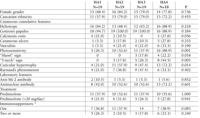
An additional analysis involving patients with DM showed that the serum HA level did not correlate with age, duration between disease diagnosis and initial symptoms, disease duration, disease status, serum muscle enzyme levels or the cumulative prednisolone dose (Table II). The serum AH serum level also did not correlate with gender, ethnicity, or auto-antibodies. However, the level correlated with cutaneous features, such as photosensitivity, “shawl” sign, “V-neck” sign and periungual telangiectasia (Table III).
Regarding drugs, the serum HA level did not corre-late with prednisolone or immunosuppressive
treat-ment (Table III).
dIscussIon
In the present study, we observed a high serum HA level in a large sample of patients with DM. Moreover, there was a tendency for HA to positively correlate with cutaneous manifestations.
Yoshinoya et al.8showed high levels of serum
gly-cosaminoglycans, including HA, in rheumatoid arthri-tis, and the serum HA level correlated specifically with disease severity.
Chang et al.15observed an accumulation of HA and
sulfate chondroitin specifically in cutaneous lesions of patients with lupus erythematosus or DM. These au-thors suggested that these glycosaminoglycans could be involved in the pathogenesis of cutaneous inflam-mation in these diseases.
Faaber et al.16studied the function of HA in patients
with systemic lupus erythematosus. These authors showed cross-reactivity of anti-DNA antibodies with HA and sulfate chondroitin, leading to anti-proteogly-can activity and consequently promoting inflammato-ry changes in these tissues, leading to arthralgia,
arthri-tAble I. deMogrAphIc, clInIcAl And lAborAtory feAtures of pAtIents wIth derMAtoMyosItIs And heAlthy IndIvIduAls Controls DM N=75 N=75 P Age (years) 44.2 ± 14.6 44.5 ± 12.0 0.905 Female gender 58 (77.3) 58 (77.3) 1.000 Caucasian ethnicity 54 (72.0) 52 (69.3) 0.720
Time between symptoms and diagnosis (mo) – 6.0 [3.0-12.0] –
Disease duration (years) – 4.0 [1.0-7.0] –
Clinical cumulative features
Constitutional symptoms – 25 (75.8) –
Articular involvement – 12 (36.4) –
Pulmonary involvement – 13 (39.4) –
Cutaneous involvement – 51 (60.0) –
Laboratory features
Creatine phosphokinase (U/L) 112.0 [86.0-164.0] 125.0 [72.0-275.0] 0.190
Aldolase (U/L) 3.6 [2.8-4.4] 4.0 [4.8-6.5] <0.001
Anti-Mi-2 antibody – 5 (6.7)
Antinuclear antibody – 41 (54.7) –
Hyaluronic acid (ng/mL) 133.0 [30.0-262.0] 329.0 [80.0-958.0] <0.001
tAble II. speArMAn correlAtIon between deMogrAphIc, clInIcAl And lAborAtory feAtures of pAtIents wIth derMAtoMyosItIs And seruM hyAluronIc AcId
r P
Age (years) 44.5 ± 12.0 0.2196 0.059
Time between symptoms and diagnosis (mo) 6.0 [3.0-12.0] -0.0571 0.629
Disease duration (years) 4.0 [1.0-7.0] -0.0643 0.584
Current disease status
Patient VAS 2 [0-5] 0.0021 1.000
Physician VAS 1 [0-4] 0.5190 1.000
MMT-8 80 (78-80) -0.0724 0.537
HAQ 0.14 [0-12.9] 0.1702 0.145
Laboratory features
Creatine phosphokinase (U/L) 125.0 [72.0-275.0] -0.0615 0.600
Aldolase (U/L) 4.0 [4.8-6.5] -0.0662 1.000
Treatment
Prednisolone: cumulative dose (g) 13.3 [8.0-23.2] 0.0254 0.930
VAS: visual analog scale; MMT-8: manual muscle testing; HAQ: healthy assessment quality. The results are expressed as the means ± standard
deviation, mean [interquartile 25-75th].
tAble III. correlAtIon between deMogrAphIc, clInIcAl And lAborAtory feAtures of pAtIents wIth derMAtoMyosItIs And seruM hyAluronIc AcId expressed In InterquArtIles
HA1 HA2 HA3 HA4
N=19 N=19 N=19 N=18 P
Female gender 13 (68.4) 16 (84.2) 15 (79.0) 14 (77.8) 0.736
Caucasian ethnicity 11 (57.9) 15 (79.0) 15 (79.0) 13 (72.2) 0.455 Cutaneous cumulative features
Heliotrope 16 (84.2) 13 (68.4) 12 (63.2) 16 (88.9) 0.218 Gottron’s papules 18 (94.7) 19 (100.0) 19 (100.0) 16 (88.9) 0.184 Calcinosis cutis 4 (21.0) 2 (10.5) 0 5 (27.8) 0.056 Cutaneous ulcers 1 (5.3) 3 (17.8) 2 (10.5) 5 (27.8) 0.253 Vasculitis 1 (5.3) 4 (21.0) 4 (21.0) 6 (33.3) 0.190 Photosensitivity 5 (26.3) 10 (52.6) 11 (57.9) 16 (88.9) 0.001 “Shawl” sign 0 0 3 (17.8) 4 (22.2) 0.018 “V-neck” sign 0 3 (17.8) 5 (26.3) 8 (44.5) 0.005 Cuticular hypertrophy 4 (21.0) 11 (57.9) 9 (47.4) 13 (72.2) 0.014 Raynaud’s phenomenon 4 (21.0) 7 (36.8) 9 (47.4) 6 (33.3) 0.402 Laboratory features Anti-Mi-2 antibody 2 (10.5) 1 (5.3) 1 (5.3) 1 (5.6) 0.852 Antinuclear antibody 8 (42.0) 10 (52.6) 10 (52.6) 13 (72.2) 0.601 Treatment Prednisolone 11 (57.9) 10 (52.6) 11 (57.9) 10 (55.6) 1.000 Prednisolone (>20 mg/day) 4 (21.0) 6 (31.6) 5 (26.3) 5 (27.8) 0.941 Immunosuppressors * One 7 (36.8) 11 (57.9) 14 7 (38.9) 0.083 Two or more 5 (26.3) 2 (10.5) 3 (17.8) 6 (33.3) 0.240
The results are expressed as percentages (%). AH: hyaluronic acid, expressed in interquartiles (HA1: 0-50 ng/mL; HA2: 51-329 ng/mL; HA3: 330-958; HA4: 959-15110 ng/mL).
*Immunosuppressors: azathioprine (2~3 mg/kg/day), methotrexate (20~25 mg/week, cyclosporine (2~3 mg/kg/day), mycophenolate mofetil (2~3 g/day), leflunomide (20 mg/day).
tis and skin rashes.
Elkayam et al.12observed that a high serum HA le
-vel was related to psoriatic arthritis, particularly to skin activity and not joint disease. HA turnover is increased in psoriasis associated with an elevated serum HA le -vel, and the turnover most likely occurs in the skin, as in suction blisters, in patients with active untreated psoriasis, which contain high concentrations of HA in the fluid. In another study, Lundin et al.14showed that
the HA concentration in the blister fluid from active skin lesions in psoriatic arthritis patients was greatly in-creased compared to the control group.
The role of serum HA in DM has been rarely des -cribed in the literature17,18. Kubo et al.17reported two
cases in which there was a positive correlation between the serum HA level and DM disease activity. In one of the DM patients analysed, the serum HA level decreased after using corticosteroid therapy. In the se -cond patient, who also suffered from cancer, the HA level decreased after the surgical resection of a mam-mary carcinoma and subsequent chemotherapy. These same authors observed in a subsequent study18,
in-volving 40 patients with DM, that the serum HA level was higher in DM patients compared to patients with systemic lupus erythematous, rheumatoid arthritis or systemic sclerosis. As a limitation, the authors did not include a control group and did not exclude DM pa-tients with neoplasia.
In our study, we observed a high serum AH level in a large sample of patients with defined DM. Different than previous, we excluded not only patients with can-cer associated myositis but also clini cally amyopathic DM, as well as chronic infections and/or current treat-ments (viral, bacterial or fungal) that could interfere with serum HA expression. Our data showed that HA did not correlate with laboratory features, drug treat-ment with prednisolone or current disease status. However, HA positively correlated with DM cutaneous features, such as photosensitivity, “shawl” sign, “V-neck” sign and peringual telangiectasia.
In healthy skin, HA is distributed mainly in the in-tercellular space of the papillary dermis, with intense staining and uneven distribution observed in the reticu lar dermis6. A prominent staining layer is found
below the epidermis in the basement membrane zone6.
HA has been shown to control the production and acti -vation of matrix metalloproteinase in fibroblast and keratinocyte cell cultures26. HA plays an important role
in regulatory processes, such as inflammation, wound healing and tumor progression4.
Increased HA production has been reported in diffe -rent skin diseases13. The proliferation and activation of
fibroblasts has been reported in fibrous and inflam-matory tissue in skeletal muscle27. This finding could
explain the DM physiopathological mechanism and ele vated serum HA level in active disease because HA plays an important role in the inflammatory process, associated with physiologic abundance in the skin.
As a limitation of the present study, our patients were receiving a consolidated treatment at the time of analysis. This fact may have prevented the identifica-tion of correlaidentifica-tion between an elevated serum HA lev-el and DM clinical manifestations and laboratory fea-tures. Other limitation is the anti-HA antibody assay, which was performed using ELISA/EIA kits (assay availability and also sensitivity and specificity of the method). More studies are necessary including also pa-tients without any treatments (corticosteroid and/or immunossuppressives).
conclusIons
In conclusion, we found an elevated serum HA level in DM. Furthermore, HA correlated specifically with some cutaneous features (photosensitivity, “shawl” sign, “V-neck” sign, and peringual telangiectasia), sugges ting that this glycosaminoglycan could be in-volved in modulating cutaneous inflammation in this population. More studies are necessary to understand the correlation between HA and patients with DM fea-tures.
correspondence to
Samuel Shinjo
Av. Dr. Arnaldo, 455, 3 andar, sala 3150 E-mail: samuel.shinjo@gmail.com
references
1. Drake LA, Dinehart SM, Farmer ER et al. Guidelines of care for dermatomyositis. Am Acad Dermatol. 1996; 34 (5 pt 1): 824--829.
2. Bohan A, Peter JB. Polymyositis and dermatomyositis. N Engl J Med 1975; 13: 344-347.
3. Koler RA, Montemarano A. Dermatomyositis. Am Family Phy-sician 2001; 64: 1565-1572.
4. Fraser JR, Laurent TC, Laurent UB. Hyaluronan in nature, dis-tribution, function and turnover. J Intern Med 1997;242: 27--33.
5. Jiang D, Liang J, Noble PW. Hyaluronan as an immune regula-tor in human diseases. Physiol Rev 2001; 91: 221-264. 6. Chang X, Yamada R, Yamamoto K. Inhibition of antitrombin by
hyaluronic acid may be involved in the pathogenesis of rheu-matoid arthritis. Arthritis Res Ther 2005; 7: R268-273.
7. Majeed M, McQueen F, Yeoman S, McLean L. Relationship bet-ween serum hyaluronic acid level and disease activity in early rheumatoid arthritis. Ann Rheum Dis 2004; 63: 1166-1168. 8. Yoshinoya S, Mizoguchi Y, Hashimoto Y et al. Serum
concen-tration of hyaluronic acid in healthy populations and patients with rheumatoid arthritis-relationship to clinical disease acti-vity of RA. Ryumachi 1991; 31: 381-390.
9. Yoshioka Y, Kozawa E, Urakawa H et al. Suppression of hyalu-ronan synthesis alleviates inflammatory responses in murine arthritis and in human rheumatoid synovial fibroblasts. Arth-ritis Rheum 2013;65: 1160-1170.
10. Goldberg RL, Huff JP, Lenz ME, Glickman P, Katz R, Thonar EJ. Elevated plasma levels of hyaluronate in patients with os-teoarthritis and rheumatoid arthritis. Arthritis Rheum 1991; 34: 799-807.
11. Yoshizaki A, Iwata Y, Komura K et al. Clinical significance of se-rum hyaluronan levels in systemic sclerosis: association with di-sease severity. J Rheumatol 2008; 35: 1825-1829.
12. Elkayam O, Yaron I, Shirazi I, Yaron M, Caspi D. Serum levels of hyaluronic acid in patients with psoriatic arthritis. Clin Rheu-matol 2000; 19: 455-457.
13. Lindqvist U, Phil-Lundin I, Engström-Laurent A. Dermal dis-tribution of hyaluronan in psoriatic arthritis; coexistence of CD44, MMP-3 and MMP-9. Acta Derm Venereol 2012; 92: 372-377.
14. Lundin A, Engström-Laurent A, Hallgren R, Michaelsson G. Circulation hyaluronate in psoriasis. Br J Dermatol 1985; 112: 663-671.
15. Chang LM, Maheshwari P, Werth S et al. Identification and molecular analysis of glycosaminoglycans in cutaneous lupus ery -thematosus and dermatomyositis. J Histochem Cytochem 2011; 59: 336-345.
16. Faaber P, Capel PJA, Rijke GPM, Vierwinden G, Van De Putted LBA, Koene RAP. Cross-reactivity of anti-DNA antibodies with proteoglycans. Clin Exp Immunol 1984; 55: 502-508. 17. Kubo M, Ihn H, Matsukawa A, Kikuchi K, Tamaki K.
Derma-tomyositis with elevated serum hyaluronate. Clin Dermatol 1999; 24: 275-278.
18. Kubo M, Kikuchi K, Yazawa N, Fujimoto M, Tamaki T, Tama-ki K. Increased serum concentration of hyaluronate in derma-tomyositis patients. Arch Dermatol Res 1998; 290: 579-581.
19. Rider LG, Giannini EH, Harris-Love M et al, International Myo-sitis Assessment and Clinical Studies Group. Defining Clinical Improvement in Adult and Juvenile Myositis. J Rheumatol 2003; 30: 603-617.
20. Harris-Love MO, Shrader JA, Koziol D et al. Distribution and severity of weakness among patients with polymyositis, der-matomyositis, and juvenile dermatomyositis. Rheumatology (Oxford) 2009; 48: 134-139.
21. Miller FW, Rider GL, Chung YL et al, International Myositis Outcome Assessment Collaborative Study Group. Proposed preliminary core set measures for disease outcome assessment in adult and juvenile idiopathic inflammatory myopathies. Rheumatology (Oxford) 2001; 40: 1262-1273.
22. Rider LG, Feldman BM, Perez MD et al. Development of vali-dated disease activity and damage indices for the juvenile idio-pathic inflammatory myopathies: I. Physician, parent, and pa-tient global assessments. Juvenile Dermatomyositis Disease Ac-tivity Collaborative Study Group. Arthritis Rheum 1997; 40: 1976-1983.
23. Ekdahl C, Eberhardt K, Andersson SI, Svensson B. Assessing di-sability in patients with rheumatoid arthritis: use of a Swedish version of the Stanford Health Assessment Questionnaire. Scand J Rheumatol 1988; 17: 263-271.
24. Alexanderson H, Lundberg IE, Stenstrom CH. Development of the myositis activities profile -validity and reliability of a self-administered questionnaire to assess activity limitations in pa-tients with polymyositis/dermatomyositis. J Rheumatol 2002; 29: 2386-2392
25. Cruellas MG, Viana V dos S, Levy-Neto M, Souza FH, Shinjo SK. Myositis-specific and myositis-associated autoantibody profiles and their clinical association in a large series of patients wtih polymyositis and dermatomyositis. Clinics 2013; 68: 909-914.
26. Isnard M, Legeais J-M, Renard G, Robert L. Effect of hyaluro-nan on MMp expression and activation. Cell Biol Int 2001; 25: 735-739.
27. Yamazaki M, Minota S, Sakurai H et al. Expression of transfor-ming growth factor-beta 1 and its relation to endomysial fibro-sis in progressive muscular dystrophy. Am J Pathol 1994; 144: 221-226.