Monte Real, Leiria, Portugal, 6-7 de fevereiro, 2015
S
HOULDER AND THORACIC SPINE MOBILITY ARE IMPAIRED IN PATIENTSWITH
C
HRONICO
BSTRUCTIVEP
ULMONARYD
ISEASENuno Morais 1, Joana Cruz 2,3 and Alda Marques 3
1 ESSLei, Instituto Politécnico de Leiria, Portugal; [email protected] 2 SACS, Universidade de Aveiro, Portugal; [email protected] 3 ESSUA, Universidade de Aveiro, Portugal; [email protected]
PALAVRAS CHAVE: Arm elevation, Impairment, Kinematic chain, Respiratory dysfunction
RESUMO: Patients with Chronic Obstructive Pulmonary Disease (COPD) often complain about
difficulties in performing activities above shoulders height. These difficulties have been associated with altered lung mechanics; however, musculoskeletal mechanisms may also contribute to restrict the biomechanics of the upper body quadrant, increasing the effort. The purpose of this research was to explore the capacity of this population to fully elevate the arms in the standing upright position and the contribution of the thoracic spine posture and mobility to such task. Fifteen patients with COPD and nineteen age-matched healthy controls volunteered to participate in this study. Sagittal alignment and range of motion (ROM) of the thoracic spine and shoulder joint were measured, using a computer software, in digital lateral photographs obtained in 3 different testing positions: arms at rest, arms parallel to the ground (90º of shoulder flexion) and full arm elevation. Patients with COPD showed significantly less shoulder flexion (~11º) and thoracic spine extension (~5º) ROM than their healthy counterparts in full arm elevation position. These findings suggest that this population may show mobility impairments of the upper body quadrant that possibly contribute for further deteriorating functionality in their daily living.
1 INTRODUCTION
Patients with Chronic Obstructive Pulmonary Disease (COPD) often complain about difficulties in performing activities above shoulders height, including dressing, bathing, shopping and many household tasks [1]. Part of these difficulties could be explained by altered lung mechanics in this arm position [2]. However, the presence of postural and mobility impairments (e.g., decreased mobility of the shoulder, scapular protraction, pectoralis muscles tightness) may also restrict the biomechanics of the upper body quadrant (i.e., head, thorax, cervical and thoracic spine and upper limb), thus increasing the effort of placing and moving the upper limb in space [3, 4]. Moreover, postural and mobility impairments are considered risk factors for
developing musculoskeletal pain [5, 6], which may also contribute to altered breathing mechanics and increased work of breathing [7], possibly aggravating the pulmonary symptoms of these patients. Nevertheless, little is known about the musculoskeletal mechanisms that may restrict arm elevation above shoulders height in this population. Given the reasons presented, shoulder and thoracic spine posture and mobility should be analyzed in patients with COPD to investigate for potential limitations in the kinematic chain of arm elevation.
The purpose of this study was to explore the capacity of this population to fully elevate the arms in the standing upright position and the contribution of the thoracic
spine posture and mobility to such task. This knowledge may help clinicians designing interventions to minimize the risk of developing upper quadrant pain, tailoring exercises accordingly, and improve patients’ functioning in daily living.
2 METHODS
2.1 P
ARTICIPANTSThis cross-sectional comparative study had the approval of the ethics committee of the Agrupamento de Centros de Saúde (ACES) de Aveiro, Aveiro, Portugal. Patients with COPD and healthy individuals were recruited in the community and informed about the purposes and measurement procedures of the study. Written informed consent was obtained from all participants before data collection. Patients were included if they were: (1) ≥ 18 years old; (2) clinically stable, i.e., without a respiratory exacerbation over the past month; (3) living in the community; (4) able to walk independently without an assistive device; and (5) able to follow simple instructions. Patients were excluded if they had: (1) a thoracic or abdominal surgery in the previous year; (2) a recent fracture of the ribs, clavicle or upper limb; (3) recurrent sub-luxation of the shoulder; (4) scoliosis; or (5) a previous mastectomy. They were also excluded if they presented other severe musculoskeletal (e.g., osteoporosis), systemic (e.g., rheumatoid arthritis), neurological or cardiovascular disorders that could interfere with the measurements, or cognitive impairment that could prevent them from understanding and following simple instructions. The eligibility criteria for the control group were the same as for the COPD group, with the addition of normal lung function.
2.2 M
EASURESSagittal alignment and mobility of the thoracic spine and shoulder joint were assessed using digital lateral photographs
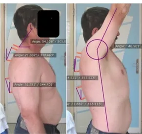
obtained in 3 different testing positions: arms at rest, arms parallel to the ground (90º of flexion) and full arm elevation (Fig. 1).
Fig. 1 Angular measurements performed with the arms at rest (left) and at full shoulder flexion (right), showing the upper and lower thoracic spine angles (º)
and shoulder range of motion (º).
A 12-megapixel digital camera (Canon PowerShot SX200, Canon Inc., Tokyo) with a 5.0-60.0 mm lens was set on a tripod with a multi-angle bubble level, to allow leveling the camera in horizontal and vertical planes. The camera was positioned at 1.50 m from a cross marked on the ground. Participants were instructed to remove their upper-body clothing and stand in the middle of the cross mark in the upright position and to remain with their arms alongside the trunk, feet slightly apart, body weight even on both feet and a horizontal gaze. One researcher attached 4 markers to the skin of the spinous processes of the 1st, 4th, 8th and 12th thoracic vertebrae. Sagittal alignment of the thoracic spine at rest and during arm elevation and range of motion (ROM) of full shoulder flexion were measured using a computer software (Osirix Imaging v5.0.2, Pixmeo SARL, Switzerland). Landmarks and angle definitions used to assess upper, lower and full thoracic spine (thoracic kyphosis) alignment and full shoulder flexion ROM were set according to previous studies [8, 9]. The angles considered for analysis were
those formed between the line joining the tip of 2 markers and the y-axis (vertical) as shown in Figure 1. Positive angles represented rotations toward the positive y-axis and negative angles represented rotations toward the negative y-axis. Full thoracic spine angle was the sum of upper and lower thoracic spine angles, in absolute values.
Repeated analysis of the same photographs showed coefficients of variation (CV) ranging from 0.60% to 1.80% and 0.60% to 1.90%, with standard errors of measurement (SEM) of 0.4° to 1.3° and 0.4° to 1.5°, for thoracic kyphosis measured in the neutral position and in full arm elevation, respectively [10], indicating very good repeatability.
Before data collection, all participants practiced the testing positions with one researcher and acquired a natural balanced upright posture according to a standardized protocol [11].
2.3 D
ATAA
NALYSISAn independent samples t-test was used to compare the ROM of full shoulder flexion between groups. Three two-way repeated measures analysis of variance (ANOVA) with interaction (group x arm position) were used to compare the effects of arm position on the (upper, lower and full) thoracic spine angles between groups. Significance level was set at 0.05. Effect sizes, Cohen’s d and Eta2 partial (η2
p), were
calculated to complement inferential statistics. Cohen’s d measures the size or the strength of the differences between two means. For d > 1 the effect is considered as very large; 1.0 ≥ d > 0.5 as large; 0.2 > d ≥ 0.5 as moderate; and d ≤ 0.2 as small. Eta2
partial measures the proportion of variation and error attributable to the factor excluding other factors from the total non-error variation. In other words, it describes the amount of variation of the dependent variables (thoracic spine angles) explained by the factors arm position and group. For
values η2
p > 0.5 the effect size is considered
as very large; 0.5 ≥ η2
p > 0.25 as large; 0.25
≥ η2p < 0.05 as moderate; and η2p ≤ 0.05 as
small [12]. Analyzes were conducted using SPSS v22.0 (IBM Corp., Armonk, NY) and G*Power v3.1.3 (G*Power Software Inc., Kiel, Germany).
3 RESULTS
Twenty-five patients with COPD were invited to participate in the study; however, 2 did not meet the eligibility criteria (i.e., one was unable to walk without an assistive device and the other presented thoracolumbar scoliosis), 3 refused to participate and 5 failed to attend the appointment. Therefore, 15 patients with COPD (67.9±9.7 years old, 13 ♂, FEV1 =
1.7±0.5 L [66.0±17.8pp], FVC = 2.6 ± 0.8 L [75.3±17.8pp)], BMI = 28.0±5.9 kg/m2)
and 19 age-matched healthy controls (68.63±9.86 years old, 7 ♂, FEV1 = 2.1±0.6
L [106.8±22.6pp], FVC = 2.4±0.6 L [97.0±21.1pp], BMI = 27.28±3.58 kg/m2)
completed the study.
The ROM of full shoulder flexion was significantly different between groups (COPD = 136.71º ± 11.91º vs. control = 147.87º ± 13.03º; mean difference [95% CI] = 11.1º [19.7º – 2.4º], p = 0.016, d = 0.89). Arm position had a significant effect on the upper, lower and full thoracic spine angles in both groups (p < 0.001, 0.443 < η2p <
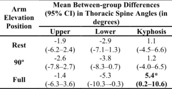
0.778). On average (95% CI), the upper and full thoracic spine angles of both groups gradually decreased from rest to full shoulder flexion, 12.8º (11.1º – 14.6º) and 7.2º (5.5º – 8.8º) respectively, whereas lower thoracic spine angle increased 5.8º (4.0º – 7.6º). The COPD group tended to present a greater magnitude in thoracic kyphosis (or less thoracic spine extension) during arm elevation than the control group (Table 1). However, differences were only significant for the full thoracic spine angle at full shoulder flexion (5.4º, p = 0.04; η2
p =
0.156; Tab. 1). Most of these differences were related to a more kyphotic (less
vertical) lower thoracic spine in the COPD group (Tab. 1).
Tab. 1 Mean differences (± 95% confidence intervals) in thoracic spine angles between patients with COPD
and controls in the 3 arm elevation positions tested.
Mean Between-group Differences (95% CI) in Thoracic Spine Angles (in
degrees) Arm
Elevation Position
Upper Lower Kyphosis Rest (-6.2–2.4) -1.9 (-7.1–1.3) -2.9 (-4.5–6.6) 1.1 90º (-7.8–2.7) -2.6 (-8.3–0.7) -3.8 (-4.0–6.5) 1.2 Full (-6.3–3.6) -1.4 (-10.3–-0.3) -5.3 (0.2–10.6) 5.4*
*p=0.04, interaction effect (group x arm elevation).
4 DISCUSSION
This study showed that patients with COPD present less ROM in arm elevation than their healthy counterparts. Moreover, arm position was found to influence the thoracic spine angles (upper, lower and full) in both groups. These results suggest that differences in full arm elevation between groups may be related to less mobility of the shoulder complex in patients with COPD. However, as these patients also presented less extension of the full thoracic spine at full arm elevation, this thoracic movement may also restrict arm elevation. Activities that require placing and moving the upper limb above shoulders height often involve complex interactions of the proximal body segments to assist the movements of the arm. These interactions occur either to provide a stable postural basis for arm movement or to participate in the kinematic chain in a synergic way during the action of the arm [3, 13-15]. The results of this study support this rationale by showing a significant relationship between arm elevation and thoracic spine extension. Irrespective of group differences, our findings are in accordance with previous research that examined shoulder and thoracic spine kinematics during arm elevation, using other instruments (e.g., motion tracking devices) [3, 14, 15], or different populations (e.g., healthy young adults) [3, 10, 14, 15].
The between-group differences in shoulder and thoracic spine mobility found in this study were related to the increased thoracic kyphosis angle (or less thoracic spine extension) in the COPD group. Increased thoracic kyphosis has been shown to alter shoulder girdle kinematics and reduce the capacity to actively and fully elevate the arms [13]. Changes in shoulder girdle kinematics include less upward rotation, external rotation and posterior tilting of the scapula, which can modify the operating length of the shoulder muscles and create a bony block between the acromion and the humerus, hence, impairing arm elevation and predisposing the upper quadrant to pain syndromes [13]. In this study, the relative contribution of each joint of the shoulder
complex (glenohumeral, and
sternoclavicular, acromioclavicular and scapulothoracic) to full arm elevation was not assessed due to instrumental constraints. Nevertheless, it seems likely that altered scapular kinematics may play a role in reducing arm elevation in this population. Future research in this field is therefore warranted. Less mobility of the thoracic spine (especially its lower segments) found among patients with COPD might also be explained by structural and functional changes in the rib cage and the diaphragm muscle (part of this muscle inserts in the lower thoracic-superior lumbar vertebral bodies) that commonly occur in this population [16]. Such changes stiffen the spine during arm elevation [17-20] and contribute to adaptive shortening of the accessory respiratory muscles (e.g., pectoralis major and minor), furthermore restricting and increasing the effort to elevate the arms [4].
Exercises to increase shoulder flexion ROM are often included in pulmonary rehabilitation programs to improve upper limb functioning [21]. Our findings support this practice, however, thoracic spine extension exercises should also be considered because of their potential to
improve arm elevation, which may reduce the effort to perform activities above shoulders height.
4.1 L
IMITATIONSSome limitations should be acknowledged. Physical performance often differs between men and women, thus results could have been influenced by the uneven distribution of gender in the two samples. However, several studies have suggested that gender is not a factor of variation in posture and kinematics of the upper quadrant [22, 23]. The analysis performed in this study support these observations as no significant differences were found in shoulder and thoracic spine mobility between men and women (data not shown in the text). Another limitation refers to the sample size. The number of participants examined in this study was relatively small. Therefore, caution should be taken when interpreting and generalizing data from this study. Measurement of the ROM of full arm elevation cannot be interpreted as or used in replacement of an exercise capacity test of the upper limb [24, 25]. Nevertheless, it may point to mobility impairments of the upper quadrant, alerting the clinician to perform a more detailed examination and look for other potential mechanisms that explain difficulties in performing activities above shoulders height in this population. Finally, other biomechanical mechanisms related to mobility of the upper quadrant, such as shoulder girdle kinematics and muscle length [26] and strength [27, 28] were not assessed. Given their potential to determine mobility of the upper quadrant, these should be included in future studies.
5
CONCLUSIONSPatients with COPD presented a reduced arm elevation capacity related to less mobility of the shoulder joint and thoracic spine that may contribute to further deterioration of their functionality in daily living.
REFERENCES
[1] A. Marques, et al., "Comprehensive ICF Core Set for Obstructive Pulmonary Diseases: validation of the Activities and Participation component through the patient’s perspective", Disability and Rehabilitation,Vol. 35, n. 20, pp. 1686-1691, 2013.
[2] Z.J. McKeough, J.A. Alison, and P.T.P. Bye, "Arm positioning alters lung volumes in subjects with COPD and healthy subjects", Australian Journal of Physiotherapy,Vol. 49, n. 2, pp. 133-137, 2003.
[3] F. Fayad, et al., "The trunk as a part of the kinematic chain for arm elevation in healthy subjects and in patients with frozen shoulder", Brain Research,Vol. 1191, n. 0, pp. 107-115, 2008.
[4] M.T. Putt, et al., "Muscle Stretching Technique Increases Vital Capacity and Range of Motion in Patients With Chronic Obstructive Pulmonary Disease", Archives of physical medicine and rehabilitation,Vol. 89, n. 6, pp. 1103-1107, 2008.
[5] P.M. Ludewig and J.P. Braman, "Shoulder impingement: Biomechanical considerations in rehabilitation", Manual therapy,Vol. 16, n. 1, pp. 33-39, 2011.
[6] J. Rose, et al., "Back pain and spinal deformity in cystic fibrosis", Am J Dis Child,Vol. 141, n. 12, pp. 1313-1316, 1987.
[7] P. Kolar, et al., "Postural Function of the Diaphragm in Persons With and Without Chronic Low Back Pain", J Orthop Sports Phys Ther,Vol. 42, n. 4, pp. 352-362, 2012. [8] S.J. Edmondston, et al., "Functional Radiographic Analysis of Thoracic Spine Extension Motion in Asymptomatic Men", Journal of manipulative and physiological therapeutics,Vol. 35, n. 3, pp. 203-208, 2012. [9] C.C. Norkin and D.J. White, Measurement of Joint Motion: A Guide to Goniometry. F. A. Davis Company, 2009.
[10] S.J. Edmondston, et al., "Clinical and radiological investigation of thoracic spine extension motion during bilateral arm elevation", J Orthop Sports Phys Ther,Vol. 42, n. 10, pp. 861-861, 2012.
[11] B. Greenfield, et al., "Posture in patients with shoulder overuse injuries and healthy individuals", J Orthop Sports Phys Ther,Vol. 21, n. 5, pp. 287-95, 1995.
[12] J. Maroco, Análise Estatística com Utilização do SPSS. Sílabo, Lda, 2007.
[13] M. Kebaetse, P. McClure, and N.A. Pratt, "Thoracic position effect on shoulder range of motion, strength, and three-dimensional scapular kinematics", Arch Phys Med Rehabil,Vol. 80, n. 8, pp. 945-50, 1999.
[14] D. Theodoridis and S. Ruston, "The effect of shoulder movements on thoracic spine 3D motion", Clinical Biomechanics,Vol. 17, n. 5, pp. 418-421, 2002.
[15] J. Crosbie, et al., "Scapulohumeral rhythm and associated spinal motion", Clinical Biomechanics,Vol. 23, n. 2, pp. 184-192, 2008.
[16] F.J. Jacono, "Control of ventilation in COPD and lung injury", Respiratory Physiology & Neurobiology,Vol. 189, n. 2, pp. 371-376, 2013.
[17] M. Decramer, "Hyperinflation and respiratory muscle interaction", European Respiratory Journal,Vol. 10, n. 4, pp. 934-941, 1997.
[18] P.W. Hodges and S.C. Gandevia, "Activation of the human diaphragm during a repetitive postural task", J Physiol,Vol. 522 Pt 1, n., pp. 165-75, 2000.
[19] D. Shirley, et al., "Spinal stiffness changes throughout the respiratory cycle", Journal of Applied Physiology,Vol. 95, n. 4, pp. 1467-1475, 2003.
[20] P. Kolar, et al., "Stabilizing function of the diaphragm: dynamic MRI and synchronized spirometric assessment", Journal of Applied Physiology,Vol. 109, n. 4, pp. 1064-1071, 2010.
[21] M.A. Spruit, et al., "An Official American Thoracic Society/European Respiratory Society Statement: Key Concepts and Advances in Pulmonary Rehabilitation", American Journal of Respiratory and Critical Care Medicine,Vol. 188, n. 8, pp. e13-e64, 2013.
[22] S. Raine and L.T. Twomey, "Head and shoulder posture variations in 160 asymptomatic women and men", Archives of physical medicine and rehabilitation,Vol. 78, n. 11, pp. 1215-1223, 1997.
[23] J.H. Groot and R. Brand, "A three-dimensional regression model of the shoulder rhythm", Clin Biomech (Bristol, Avon),Vol. 16, n. 9, pp. 735-43, 2001.
[24] E.M. Clini and N. Ambrosino, "Impaired arm activity in COPD: a questionable goal for rehabilitation", European Respiratory Journal,Vol. 43, n. 6, pp. 1551-1553, 2014. [25] T. Janaudis-Ferreira, et al., "How should we measure arm exercise capacity in patients with copd?: A systematic review", Chest,Vol. 141, n. 1, pp. 111-120, 2012.
[26] J.D. Borstad and P.M. Ludewig, "The effect of long versus short pectoralis minor resting length on scapular kinematics in healthy individuals", J Orthop Sports Phys Ther,Vol. 35, n. 4, pp. 227-38, 2005.
[27] R. Gosselink, T. Troosters, and M. Decramer, "Distribution of Muscle Weakness in Patients With Stable Chronic Obstructive Pulmonary Disease", Journal of Cardiopulmonary Rehabilitation and Prevention,Vol. 20, n. 6, pp. 353-360, 2000.
[28] E. Calik-Kutukcu, et al., "A comparison of muscle strength and endurance, exercise capacity, fatigue perception and quality of life in patients with chronic obstructive pulmonary disease and healthy subjects: a cross-sectional study", BMC Pulmonary Medicine,Vol. 14, n. 1, pp. 1-10, 2014.