Camila Tirapelli(a) Heitor Panzeri(a)
Rodrigo Gonçalves Soares(a) Oscar Peitl(b)
Edgar Dutra Zanotto(b)
(a) Department of Dental Materials and
Prosthesis, School of Dentistry of Ribeirão Preto, University of São Paulo, Ribeirão Preto, SP, Brazil.
(b) Vitreous Materials Laboratory, Department
of Materials Engineering, Federal University of São Carlos, SP, Brazil
Corresponding author: Camila Tirapelli
University of São Paulo, School of Dentistry of Ribeirão Preto, Department of Dental Materials and Prosthesis
Avenida do Café, s/n Ribeirão Preto - SP - Brazil CEP: 14040-090.
E-mail: [email protected]
Received for publication on Jun 16, 2010 Accepted for publication on Aug 07, 2010
A novel bioactive glass-ceramic for
treating dentin hypersensitivity
Abstract: Dentin hypersensitivity (DH) is a painful response to stimu-lus applied to the open dentinal tubules of a vital tooth. It’s a common oral condition, however, without an ideal treatment available yet. This work evaluated in vitro the effect of micron-sized particles from a novel bioactive glass-ceramic (Biosilicate) in occluding open dentinal tubules. A dentin disc model was employed to observe comparatively, using scan-ning electron microscopy (SEM), dentinal tubule occlusion by different products and deposition of hydroxyl carbonate apatite (HCA) on dentin surface by Biosilicate, after a single application: G1 - Dentifrice with po-tassium nitrate and luoride; G2 - Two-step calcium phosphate precipita-tion treatment; G3 - Water-free gel containing Biosilicate particles (1%); G4 - Biosilicate particles mixed with distilled water in a 1:10 ratio; all of them after 1, 12 and 24 hours of immersion in artiicial saliva. Fourier transform infrared spectroscopy (FTIR) was performed to detect HCA formation on dentin discs illed with Biosilicate after 2 minutes, 30 min-utes and 12 hours of immersion in artiicial saliva. SEM showed a layer of HCA formed on dentin surface after 24 hours by G4. G1, G2 and G3 promoted not total occlusion of open dentinal tubules after 24 hours. FTIR showed HCA precipitation on the dentin surface induced by Bio-silicate after 30 minutes. The micron-sized particles from the bioactive glass-ceramic thus were able to induce HCA deposition in open dentinal tubules in vitro.This inding suggests that Biosilicate may provide a new option for treating DH.
Descriptors: Biocompatible Materials; Dentin Sensitivity; Dentin.
Introduction
Dentin hypersensitivity (DH) is a common oral condition, but an ideal product or protocol for its treatment does not exist, and active manage-ment is a challenge. DH is caused when the luids within the dentinal tu-bules are subjected to changes (thermal, mechanical, osmotic). The move-ment in the luids stimulates a nerve receptor sensitive to pressure, which leads to the transmission of the stimuli. Consequently, dental products proposed to treat DH seek to interrupt the pulp neural response of pain and/or to block the sensitive mechanisms through occlusion of the open dentinal tubule.1-2
apatite (HCA) deposition in open dentinal tubules.3
Bioactive glasses and glass-ceramics are widely rec-ognized as one of the best clinical choices to improve bone regeneration,4 and the similarity of
composi-tion between bone, dentin and enamel led to the as-sumption that bioactive glasses and glass-ceramics could also be eficient for the regeneration of enamel and dentin. Indeed, the hypothesis was that glass-ceramics could treat DH by providing permanent occlusion of the open dentinal tubules through in situ deposition of a HCA-bonded layer.5-6
The irst experiments with Biosilicate corrobo-rated its bioactivity.7-8 In addition, crystallization
has been shown to signiicantly change the fracture characteristics of glass, which provided less-sharp, less-abrasive particles.9 Therefore, crystallization
hypothetically results in two advantages:
i. the dull particles can be safely added to any kind of formulation to be used in the oral environ-ment, and
ii. the particles can be easily inserted into dentin-al tubules because there are no edges to delect them away from the oriices.
The aim of this study was to test the hypothesis that Biosilicate could be an effective desensitizing agent for the treatment of DH. To test this hypoth-esis, we analyzed in vitro Biosilicate comparatively against commercial materials and evaluated three parameters:
i. dentinal tubule occlusion provided by the prod-ucts tested and deposition of HCA triggered by Biosilicate in open dentinal tubules;
ii. the effect of two different vehicles for the incor-poration of Biosilicate particles; and
iii. the reaction of Biosilicate formulations on dentin
discs after different time periods.
Materials and Methods
The research protocol was approved by the Eth-ics Committee of the School of Dentistry of Ribeirão Preto, USP, Brazil (process # 2003.1.654.58.7).
Preparation of the desensitizing agents based on Biosilicate
The desensitizing methods tested in this study, including product concept, desensitizing agent and
mechanisms of action are shown in Table 1. To sim-ulate home-use and professional-use products, we sought to establish the simplest and safest mode of application because safety and ease of application are two of the desirable characteristics of desensitiz-ing products. For simulation of home-use products, a water-free gel based on Carbopol and glycerin (Sigma Chemical Co., Saint Louis, USA) was used as a vehicle for the Biosilicate particles. The water-free gel containing 1% Biosilicate was formulated at the School of Pharmaceutical Sciences of Ribeirão Preto, USP, Brazil, and inserted into a sterilized (7 g) dentifrice tube (Embagel, Pharmaceutical tube, code 211, São Paulo, SP, Brazil). For simula-tion of the professional-use products, the Biosilicate particles were mixed with distilled water immedi-ately before application. A 1.5-mL tube (Microtubes safe-lock, Eppendorf Brazil, São Paulo, SP, Brazil) containing 0.15 mg of Biosilicate powder was illed with 1.35 mL of water, which provided a 1:10 mix-ture of Biosilicate powder in distilled water.
Preparation of dentin discs
This study employed the dentin disc model with a strict control methodology, which has been de-scribed in the literature.5 A total of 40 dentin discs
were obtained and stored in a vial with 100 ml of distilled water. Each disc was fractured at the center using wire cutters to provide test and control halves. Both parts of each disc were stored together in a 1.5-mL tube containing artiicial saliva.
Five control and experimental halves were ran-domly assigned to four groups (Table 1). Immedi-ately before the application of the proposed mate-rials, the control and test halves of each disc were etched in 6% citric acid for 2 minutes, washed with distilled water for two minutes and stored in label-paired tubes containing artiicial saliva. At no time during the procedures were discs allowed to dry.
Application of the product to the dentin discs
preserved in the test discs, and control discs were maintained in the 1.5-mL tubes with artiicial saliva for either 1, 12 or 24 hours. The materials were ap-plied to test discs positioned on a sterile glass sur-face as follows:
• G1: Approximately 0.15 mL of toothpaste was poured onto the test disc, and the product was dispersed over the dentin surface for 30 seconds using a micro-applicator (Microbrush Tube Se-ries, Fine size, MFA 400, Grafton, USA).
• G2: Solution 1 was rubbed on the test disc for ive seconds and kept moistened with the solu-tion for 30 seconds. With another micro-appli-cator, solution 2 was rubbed on the same test disc for two seconds and kept moistened for ten seconds in accordance with the manufacturer’s instructions.
• G3: One drop (about 0.2 mL) of gel from a tube was dropped on the test disc. The gel was gently dispersed on the dentin surface using a micro-applicator for 30 seconds.
• G4: Approximately 0.15 mL of the solution was gently applied to the test disc with a micro-appli-cator for 30 seconds.
After the application of each product for its re-spective duration, each test disc was gently washed with distilled water for 30 seconds and stored in a tube containing 1.5 mL of artiicial saliva for either 1, 12 or 24 hours. The discs remained in artiicial saliva for the mentioned periods, and after that, the
control and test halves were pulled out of the 1.5-mL tube and air dried in a desiccator (Eikonal do Brasil, São Paulo, SP, Brazil) at 36°C for 1 week. The discs were then mounted on aluminum stubs and photographed with an SEM (FEG2000, Philips, Oregon, USA).
FTIR analysis
To investigate the conversion of the Biosilicate into HCA on the dentin surface, dentin discs were analyzed by a FTIR spectrometer immediately after product application as well as 2 minutes, 30 minutes and 12 hours after application and immersion in artiicial saliva. Pure Biosilicate and dentin control discs were also analyzed with the FTIR spectrom-eter.
Results
Scanning electron micrographs
The experimental and control discs had compa-rable diameters and showed a similar pattern of tu-bule distribution and orientation. Figure 1 shows an overall view of the micrographs taken from the den-tin test discs after different periods of immersion in artiicial saliva. After 1 hour of immersion in saliva, the front surface of the open dentinal tubules did not show the same pattern. Indeed, some particles could be observed on the dentin surface of the G1 discs (image a) but not inside the dentinal tubules, which was probably due to the size and
morphol-Table 1 - The experimental groups of the study, including product concepts regarding their mode of use, desensitizing agents and proposed mechanisms of action.
Group Product
concept Desensitizing agent
Proposed mechanism of action
G1- Desensitizing dentifrice (Sensodyne, GSK Brasil, Rio de
Janeiro, RJ, Brasil).
Home-use 5% potassium nitrate; 1187 ppm of MFP (active fluoride)
Depolarizing effect on neural conduction of pain
G2 - Desensitizing solutions (Sensi Kill, DFL, Rio de Janeiro, Brasil).
Professional use
Solution 1: phosphate dipotassium, sodium fluoride, methylparaben and distilled water. Solution 2: calcium
chloride, sodium benzoate and distilled water.
Calcium phosphate deposition on the dentin surface - tubule occlusion
G3 - Experimental desensitizing gel
Vitrovita, São Carlos, SP, Brasil. Home-use Fully crystallized bioactive glass-ceramic (Biosilicate)
HCA layer formation on dentin surface - tubule
occlusion
G4 - Experimental desensitizing solution
Vitrovita, São Carlos, SP, Brasil.
Professional
use Fully crystallized bioactive glass-ceramic (Biosilicate)
HCA layer formation on dentin surface - tubule
ogy of the particles contained in the dentifrice for-mulation. Few or no particles can be observed on the surfaces of the G2 (image b), G3 (image c) or G4 discs (image d), probably due to the fact that G2 has a liquid constitution and Biosilicate has
micron-sized particles dispersed in the distilled water or gel, which were possibly inserted inside the dentinal tu-bules by gentle application. After 1 hour of immer-sion, the G2 discs (image b) showed dentinal tubules with diminished diameters, and the G3 (image c)
Figure 1 - SEM micrographs of the test discs at different durations of immersion in artificial saliva. The images on the left, middle and right columns show dentin discs where the products were applied after 1, 12 and 24 hours of immersion in artificial saliva, respectively. The images from the top to the bottom rows show dentin discs allocated in accordance with the experimental group: G1, G2, G3 and G4, respectively.
G2 G1
1 hour 12 hours 24 hours
G3
G4
A E I
B F J
C G K
and G4 (image d) experimental discs showed little (image c) to no tubule occlusion (image d). After 12 hours, the G1 discs (image e), displayed a large num-ber of particles on the dentin surface. The G2 discs at 12 hours (image f), however, were not changed compared with the G2 discs after 1 hour of immer-sion (image b). In the G3 discs, more particles were observed inside the dentinal tubules at 12 hours (image g) than at 1 hour (image c). Interestingly, a noticeable difference was observed in the dentinal tubule diameter of G4 discs at 12 hours compared with 1 hour (image h).
After 24 hours of immersion, the G1 disc surface showed some open dentinal tubules and a weak pat-tern of tubule occlusion (image i). Compared with the G2 discs at 12 hours (image f), the G2 discs at 24 hours (image j) showed a small decrease in the dentinal tubule diameter. Compared with the ind-ings in G3 discs at 12 hours (image g), tubule occlu-sion increased in the G3 discs after 24 hours (image k). Interestingly, the G4 discs showed total oblitera-tion of the dentinal tubules after 24 hours of immer-sion in artiicial saliva (image l).
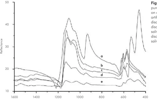
Fourier transformed infrared analysis The results of FTIR analysis plotted in Figure 2 show the Biosilicate reactions incorporated on
a dentin surface until complete conversion into a HCA layer. The mechanism of HCA formation on a monolithic glass-ceramic was described by Peitl
et al.,10 and Biosilicate powder undergoes the same
steps. The line a shows pure Biosilicate without reaction and the line c shows pure dentin. After 2 minutes in artiicial saliva we observe, at the line e, a mixture between the peaks from dentin and Bio-silicate. The main spectral peak assignments for the molecular vibrations of Biosilicate are observed at 460, 536, 930 nm, and at 1124, 602, 574 nmfor dentin. The double peaks at 602 and 574 nm are the most important P-O crystal vibrational bend mode associated to HCA. We clearly observe these peaks on pure dentin. Increasing time reactions to 30 minutes in the solution (line d), we observe the phosphate peaks, 602 and 574 nm, develop and the Biosilicate decrease as 460 and 930 nm. This is thus experimental evidence for the formation of HCA on Biosilicate surface. We conclude from these spectra that the Biosilicate formed a thin layer of HCA on dentin. Finally, after 12 hours (line b) we cannot observe the Biosilicate peaks, but only the identical spectra showed by pure dentin. Therefore, at this time it is not possible to distinguish the substrate and dentine from reacted Biosilicate.
10 20 30 40 50
R
e
fl
e
c
ta
n
ce
Wavenumber e d c b a
1600 1400 1200 1000 800 600 400
Figure 2 - FTIR spectrum. Line a:
pure Biosilicate; line b: Biosilicate
on dentin disc over 12 hours in
artificial saliva; line c: control dentin
disc; line d: Biosilicate on dentin
disc over 30 minutes in artificial
saliva; line e: Biosilicate on dentin
Discussion
Studies regarding desensitizing agents have indi-cated that, nowadays, the treatments currently used to block the sensitive mechanisms of DH pain could be improved to obtain easy, fast, non-invasive, du-rable relief of patient discomfort.
The irst aim of this study addressed the dentinal tubule occlusion of the tested products as well as the deposition of HCA triggered by Biosilicate in open dentinal tubules. Although only the treatment used in G4 triggered total occlusion, the treatments used in G1, G2 and G3 also promoted a decrease in the diameter of the dentinal tubules. In G1, a widely used desensitizing dentifrice was used. Although one study11 showed considerable dentinal tubule
occlusion by desensitizing dentifrices, in this study dentin surfaces were brushed for over two minutes twice a day for seven days, which probably created smear layer deposits in the tubules. In G2, the ilm of mineralized deposit on the dentin surface was not observed at 24 hours (dissolution or dislodgment were supposed) as it was for G4.
We also examined the ability of Biosilicate to oc-clude open dentinal tubules by inducing HCA for-mation. The FTIR analysis showed that HCA was formed after a reaction time of 30 minutes. It is im-portant to note that the test products were only ap-plied once in the present study, and they were gently applied with a micro-applicator for a few seconds to ensure that the materials covering and inside the dentinal tubules were exclusively the products tested and not a smear layer. The ine particle size was im-portant to allow a faster reaction and deposition of HCA inside the dentinal tubules and on the dentin surface.12 Considering the properties of bioglasses
and the images from this in vitro investigation, it is possible that the micron-sized particles of Biosili-cate, which ranged from 0.1-10 µm, became smaller as the contact time with the liquid increased. The present study also addressed the interplay between Biosilicate particles and two different vehicles. Re-lated to this, Biosilicate mixed with distilled water (1:10) performed much better than Biosilicate in the gel (1:100). The difference shown by the same par-ticulate bioactive material in occluding open den-tinal tubules and covering the dentin surface was
probably due to the percentage of incorporation of Biosilicate into the gel, which was much lower than the amount of powder mixed into distilled water. In addition, the cross-linked structure of the gel may have prevented Biosilicate particles from escaping. Only 1% Biosilicate was incorporated into the wa-ter-free gel because we were trying to simulate a dai-ly home-use product. The formulation of the Biosili-cate mixed with distilled water, however, was meant for professional use, and the ratio was established at 1:10. This difference in the amount of particles had an important effect on tubule occlusion, which has also been suggested by Lee et al.6
The present study also investigated how Biosili-cate formulations react on dentin discs after differ-ent durations. The best performance of the bioac-tive material occurred after 24 hours of immersion in artiicial saliva. This suggests that a bonded layer of HCA formed on the dentin surfaces, which resist-ed the dislodgment that immersion promotes. This hypothetically permanent and dificult-to-remove obliteration promoted by the biomaterial in the sub-strate surfaces (dentin surface and tubules) was cor-roborated by a recent study13 that evaluated the
bio-mechanical behavior of the tissue formed in tibial consolidation when Biosilicate was employed to ill bone defects. Because we investigated dentin discs immersed in artiicial saliva for up to 24 hours, we were able to observe how a single application of DH products affected the dentinal tubules and dentin disc surfaces over time.
The present study produced several interesting indings:
i. the products tested showed different patterns of dentinal tubule occlusion, and deposition of HCA triggered by Biosilicate occurred in open dentinal tubules;
ii. the type of vehicle and the amount of Biosilicate particles played an important role in desensi-tizing formulations containing Biosilicate (i.e., different patterns of interaction between dentin discs and Biosilicate particles were observed for G3 and G4); and
iii. the time needed for a homogeneous layer of HCA
con-irmation of the research hypothesis that micron-sized Biosilicate particles are capable of occlud-ing open dentinal tubules and could be used in DH treatments.
Further in vitro investigations with different ra-tios of particles incorporated into water-free gels are needed to determine the best proportion of Biosili-cate in home-use products. In addition, other stud-ies should test the bonded character of the HCA lay-er and investigate the hydraulic conductance in the dentinal tubules occluded with the Biosilicate par-ticles. A six-month clinical study was carried out be-cause of these in vitro results. That study conirmed the results of the present in vitro experiments (i.e.,
the eficacy of Biosilicate) and is being described in a forthcoming paper.
Conclusions
Micron-sized bioactive glass-ceramic (Biosilicate) particles were able to induce HCA deposition in open dentinal tubules, which suggests that the mate-rial could provide a new option for treating DH.
Acknowledgements
We would like to thank the FAPESP (contract number 04/05133-0 and 07/08179-9), and the CNPq (contract number 0400604/2004-3) for inancial support of this work.
References
1. Orchardson R, Gillam DG. Managing dentin hypersensitivity. J Am Dent Assoc. 2006 Jul;137(7):990-8.
2. Rösing KR, Fiorini T, Liberman DN, Cavgani J. Dentine hy-persensitivity: analysis of self care products. Braz Oral Res. 2009 June, 23 Suppl 1:56-63.
3. Zanotto ED, Ravagnani C, Peitl O, Panzeri H, Lara EH, in-ventors. Process and compositions for preparing particulate, bioactive or resorbable Biosilicate for use in the treatment of oral ailments. Patent: WO2004/074199. 2004 Feb. 20. 4. Hench LL. The story of Bioglass. J Mater Sci Mater Med.
2006 Nov;17(11):967-78.
5. Gillam GD, Tang JY, Mordan NJ, Newman HN. The effects of a novel Bioglass dentifrice on dentine sensitivity: a
scan-ning electron microscopy investigation. J Oral Rehabil. 2002 Apr;29(4):305-13.
6. Lee SY, Kwon HK, Kim BI. Effect of dentinal tubule occlu-sion by dentifrice containing nano-carbonate apatite. J Oral Rehabil. 2008 Nov;35(11):847-53.
7. Moura J, Teixeira LN, Ravagnani C, Peitl O, Zanotto ED, Beloti MM, et al. In vitro osteogenesis on a highly bioactive glass-ceramic (Biosilicate). J Biomed Mater Res A. 2007 Sep
1;82(3):545-57.
8. Roriz VM, Rosa AL, Peitl O, Zanotto ED, Panzeri H, Oliveira PT. Efficacy of a bioactive glass-ceramic (Biosilicate) in the
maintenance of alveolar ridges and in osseointegration of tita-nium implants. Clin Oral Implants Res. 2010 Feb;21(2):148-55.
9. Varner JR, Quinn GC, Wightman M. Fractography of Glasses and Ceramics V. New Jersey: Wiley & Sons; 2007. 484 p. 10. Peitl O, Zanotto ED, Hench LL. Highly bioactive P2°5
-Na2°-CaO-SiO2 glass-ceramics. J Non-Cryst Solids. 2001 Oct;292(1):115-26.
11. Arrais CA, Micheloni CD, Giannini M, Chan DC. Occlud-ing effect of dentifrices on dentinal tubules. J Dent. 2003 Nov;31(8):577-84.
12. Vollenweider M, Brunner TJ, Knecht S, Grass RN, Zehnder M, Imfeld T, et al. Remineralization of human dentin us-ing ultrafine bioactiveglass particles. Acta Biomater. 2007 Nov;3(6):936-43.