Assessment of tooth inclination in the
compensatory treatment of pattern II using
computed tomography
Objective: To evaluate changes in the inclination of anterior teeth caused by orthodon-tic treatment using a Straight-Wire appliance (Capelozza’s prescription II), before and after the leveling phase with rectangular stainless steel archwires. Methods: Seventeen adult subjects were selected who presented with facial pattern II, Class II malocclusion, referred for compensatory orthodontic treatment. Inclinations of anterior teeth were clinically assessed using CT scans at three different times, i.e., after the use of 0.020-in (T1), 0.019 X 0.025-in (T2) and 0.021 X 0.025-in (T3) archwires. Friedman’s analysis of variance was applied with 5% significance level to compare the three assessments (T1, T2 and T3). Results: It was noted that the rectangular wires were unable to produce any significant changes in inclination medians, except for a slight change in mandibular lateral incisors (p<0.05). On the other hand, variations in inclination were smaller when 0.021 X 0.025-in archwires were employed, particularly in maxillary incisors (P<0.001). Con-clusion: The use of rectangular 0.021 X 0.025-in archwires produces more homogeneous variations in the inclination of maxillary incisors, but no significant median changes. Abstract
Keywords: Computed Tomography. Orthodontic treatment. Tooth inclination.
IntROduCtIOn
The aim of the Straight-Wire technique is to ensure that teeth are optimally positioned by the end of treatment while reducing the need for bending orthodontic archwires. Since its incep-tion, several authors have suggested changes to the original prescription values.5 These changes
yielded new, unique prescriptions in the search for one that would fit all or most cases.
In the following years—before this technique became the most widely used worldwide—sev-eral authors claimed that most orthodontists had embraced this technique because they did not use larger-caliber archwires to finish their cases.12,13 Nonetheless, discussions were already
under way about the need for adjustments to compensate for the slack between archwire and bracket slot, even when thicker archwires were Liana Fattori*, Liliana Ávila Maltagliati Brangeli**, Leopoldino Capelozza Filho***
* MSc in Orthodontics, Umesp.
** MSc and PhD in Orthodontics, FOB-USP. Coordinator of the Specialization Program in Orthodontics, ABCD-SP. Invited Professor of the Masters Program in Orthodontics, USC-Bauru.
used, in order to move teeth to their planned position. Thus, when evaluating an orthodontic appliance, one should not just consider its pre-scription but also the archwire progression pro-tocol being employed. Moreover, professionals need to tailor the orthodontic treatment for each patient individually if satisfactory aesthetic and functional outcome are to be achieved.11
After assessing the inclinations of teeth of treated and untreated groups who had normal oc-clusion, Vardimon and Lambertz29 noted a
stan-dard deviation of ± 5°, indicating a considerable dispersion of inclination means in all teeth. There was no statistically significant difference between the two groups, except in the second mandibular molar. In contrast with the original Straight-Wire prescription, this study showed different values for maxillary incisors, +1° for central incisors and -1° for lateral incisors.
Any ideal preadjusted appliance featuring identical torques and angulations for all patients seems to be unacceptable. This conclusion was confirmed after examining the buccal surface of the teeth, determining the extent and frequency of changes in their contour and assessing incli-nation when brackets were bonded more incis-ally or gingivincis-ally on their buccal axis.13 As the
more posterior teeth were examined, wider variations were noted on their buccal surface both in the maxilla and mandible, however, all the teeth of the same individual presented ho-mogeneous variation.
In a comparison between the inclination of an-terior teeth in cases treated with fixed edgewise, Straight-Wire, Roth prescription appliances and normal occlusion cases, the upper anterior teeth of the latter individuals exhibited negative values, whereas the former displayed positive, or buccal inclinations.28 Inclinations found in subjects
treat-ed with MBT™ prescription were statistically different when compared with the “Six Keys to Normal Occlusion”.5 Significant individual
varia-tions were also observed.6 When Brazilians with
normal occlusion were compared with the origi-nal Straight-Wire5 values, the inclinations of the
vast majority were negative, with the sole excep-tion of the maxillary incisors.30
For compensatory treatment of patients with facial patterns whose basal bones present with acceptable discrepancies, attention is paid to the position that the teeth should occupy by the end of treatment. The focus point is the direction of the dental compensation based on malocclusion features, treatment goal and treat-ment prognosis.5,8 Three sets of prescriptions
have been described,8 one geared to the
treat-ment of cases with normal maxillomandibular relationship (pattern I), and two other prescrip-tions aimed at cases of maxillomandibular dis-crepancies (pattern II or III), where the anterior teeth require compensatory torque and angula-tion to achieve an optimal occlusion, despite the skeletal condition.
Dental compensation of maxillary and man-dibular incisors related to the anteroposterior relationship of the basal bones was evaluated in young Brazilians treated with standard Straight-Wire appliances, with orthodontic treatment without extractions and cases finished accord-ing to the Six Keys of Occlusion Normal.5 The
values found for the upper incisors were close to Andrews’ sample (+7.96° to +7°, respectively), but highly discrepant in mandibular incisors (+5.03° to -1°). Moreover, it was observed that as the basal bones extend positively (maxilla ahead of the mandible) maxillary incisors vary their inclinations lingually while mandibular in-cisors vary their inclinations buccally, suggesting that orthodontic treatment could be performed with fewer extractions since it allows a signifi-cant buccoversion of mandibular incisors.7
especially limiting in terms of inclination, when archwire progression stops before maximum cal-iber archwires are inserted, thereby preventing the features of a particular prescription from be-ing fully expressed.
For this reason, it seems important to assess whether inclinations produced in the anterior teeth during the final stages of orthodontic level-ing reflect the prescription values described by the bracket manufacturer.
Thanks to advances in dental imagining tech-nology, more accurate diagnoses are now possible that boast a high degree of reliability while pro-viding detailed images of structures in three-di-mensional tests with less radiation exposure.21,26,27
Computed tomography (CT) allows the recon-struction and visualization of anatomical areas in three dimensions, revealing information about size, shape and texture and has become an impor-tant tool for all areas of dentistry, providing reli-able linear15,18,20,23 and angular22,23 measurements.
A method for evaluating torques and angu-lations by means of computed tomography has been described,10 which faithfully depicts dental
structures and allows professionals to measure each individual tooth, in addition to facilitating the study of dental positioning15 and inclinations,
instrumental in the diagnosis, prognosis and analy-sis of finished orthodontic cases.16
MAtERIAL And MEtHOdS Sample selection
The sample for this prospective study com-prised individuals selected for orthodontic treat-ment in the departtreat-ment of graduate studies and met the following requirements: Permanent denti-tion, presenting with Angle Class II malocclusion without significant crowding (>2 mm); facial pat-tern II,9 but with enough facial pleasantness24 as to
contraindicate orthodontic-surgical treatment. A group of 17 individuals was selected, 10 males and 7 females, aged between 16 years and 5 months, and 52 years and 11 months; 16 Caucasians and 1
Afro-descendant. Nine patients had Class II, divi-sion 1 maloccludivi-sion and 8 had Class II, dividivi-sion 2 malocclusion. Volumetric Computed Tomography (VCT) examinations were performed to obtain the proposed measurements. VCT was preferred as it allows measurements of each individual tooth10
without superimposing images while providing im-ages without magnification.20,22
Methods
Orthodontic treatment protocol
Patients were subjected to compensatory orth-odontic treatment using Capelozza’s8 prescription
II brackets with 0.022 X 0.028-in slots (Abzil, São José do Rio Preto, Brazil). Treatment was provided by a single specialist from start (bonding) to fin-ish. Bonding was performed by implementing An-drews’ bracket placement technique2, i.e., using
the center of the clinical crown as reference. Sub-sequently, a strict archwire progression protocol (Table 1) was performed ensuring that alignment and leveling occurred gradually without the inter-vention or use of any additional mechanical re-sources. Therefore, any changes in tooth position would be directly related to the gradual increase in size of the leveling archwires.
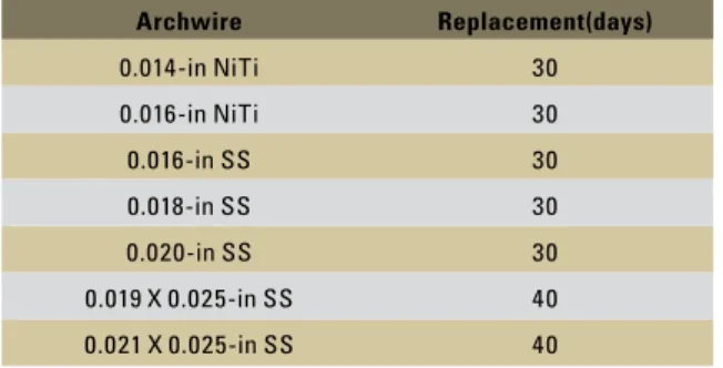
Archwire Replacement(days)
0.014-in NiTi 30
0.016-in NiTi 30
0.016-in SS 30
0.018-in SS 30
0.020-in SS 30
0.019 X 0.025-in SS 40
0.021 X 0.025-in SS 40
FIGURE 1 - Positive inclination. FIGURE 2 - Negative inclination.
CT image scanning
In order to perform the dental measurements, all sample patients were subjected to VCT scan-ning at three different times during the protocol described above:
» T1 - At the end of the leveling phase, using 0.020-in stainless steel (SS) archwire.
» T2 - At the end of the rectangular 0.019 X 0.025-in SS archwire period.
» T3 - At the end of the rectangular 0.021 X 0.025-in SS archwire period.
NewTom DVT-9000 Computed tomography equipment (NIM - Verona - Italy) was used to acquire the images. QR-DVT 9000 software was used for reformatting the images and measuring tooth inclinations.
Tooth inclination measurement
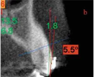
The method described by Capelozza, Fattori and Maltagliati10 was implemented.
To be considered optimal for this sample tooth inclination values (Figs 1 and 2) had to be close to those of the prescription described by the manu-facturer, taking into account a maximum allowed
slack of ± 3.9°.11 Therefore, the value of each
in-clination angle was analyzed in each subject at the three study times by adding or subtracting the value of the slack. Thus, each tooth was classified into one of three categories, within, above or be-low prescription values.
Statistical analysis
Analysis of systematic error was performed by paired t-test and random error was examined us-ing Dahlberg’s formula for all measurements, in 23.5% of the sample (n=4), 90 days after the first measurement. For random error, values above 1.5° were regarded as significant in terms of angular measurements, as suggested by Houston.19
Data normality was examined using the Shapiro-Wilk test (Table 2). Friedman’s analysis of variance was used to compare data between the different times (T1, T2 and T3) due to the fact that some data exhibited abnormal distribution or unequal varianc-es (Figs 3 - 8). Coefficient of variation was used to examine the variation between T1, T2 and T3.
10 -5 5 -10 0 -15 20 20 5 5 15 15 -5 -5 0 0 10 10 -10 -10 -15 5 20 20 -5 10 10 0 15 15 10 25 25 30 -10 5 5
-15 0 0
-20 -5 -5
-25 -10 -10
-30 -15 -15
T1
(0.020-in) Normal
T2
(0.019 X 0.025-in) Normal
T3
(0.021 X 0.025-in) Normal Capelozza Prescription
Friedman (P)
Med IQD p (SW) Med IQD p (SW) Med IQD p (SW) T1XT2XT3
Maxill. Canine -1.80 3.40 0.08 -2.75 4.63 0.44 -2.45 4.05 0.61 -5 0.99 (ns)
Maxill. Lat. Inc. 7.00 3.40 <0.01** 7.20 4.75 <0.01** 7.05 4.63 <0.01** 3 0.13 (ns)
Maxill. Cent. Inc. 5.75 5.73 0.03* 6.20 6.15 0.02* 6.65 4.93 0.04* 7 0.07 (ns) Mand. Canine -4.95 8.03 0.04* -6.10 5.48 0.09 -5.15 6.35 0.53 -11 0.44 (ns)
Mand. Lat. Inc. 4.70 4.08 0.05 5.60 3.00 0.02* 4.85 3.00 0.01* 4 0.013* (T1=T2) #T3
Mand. Cent. Inc. 6.00 5.15 0.19 7.50 4.68 0.02* 6.60 3.05 0.04* 4 0.15 (ns) TABLE 2 - Median (Med), Interquartile Deviation (IQD) and p value for the analysis of normality (Shapiro-Wilk) and for Friedman’s analysis at T1, T2 and T3.
*p<0.05/ **; p<0.01; SW= Shapiro-Wilk.
FIGURE 3 - Boxplot for maxillary canines (teeth 13 and 23). The solid line corresponds to Capelozza’s Prescription value (-5º). Median values and coefficient of variation between the groups were similar between the three times (T1=T2=T3).
FIGURE 6 - Boxplot for mandibular canines (teeth 33 and 43). The solid line corresponds to Capelozza’s Prescription value (-11º). Me-dian values were similar between groups (T1=T2=T3). Although the range of values ob-tained at T1 seems wider, no significant differ-ence was found.
FIGURE 4 - Boxplot for maxillary lateral incisors (teeth 12 and 22). The solid line corresponds to Capelozza’s Prescription value (+3º). Median values were similar between groups (T1=T2=T3). However the range of values obtained at T1 was significantly wider compared to the T3 group (p<0.01).
FIGURE 7 - Boxplot for mandibular lateral in-cisors (teeth 32 and 42). The solid line corre-sponds to Capelozza’s Prescription value (+4º). Median differences between groups T1≠T2 and T2≠T3. Variation between the groups was similar.
FIGURE 5 - Boxplot for maxillary central inci-sors (teeth 11 and 21). The solid line corre-sponds to Capelozza’s Prescription value (+7º). Median values were similar between groups (T1=T2=T3). However the range of values ob-tained at T1 was significantly wider compared to the T3 group (p<0.01).
FIGURE 8 - Boxplot for mandibular central in-cisors (teeth 31 and 41). The solid line corre-sponds to Capelozza’s Prescription value (+4º). Median values and coefficient of variation between the groups were similar between the three times (T1=T2=T3).
0.020-in 0.020-in 0.019x 0.025-in 0.019x 0.025-in 0.021x 0.025-in 0.021x 0.025-in 0.020-in 0.020-in 0.020-in 0.020-in 0.019x 0.025-in 0.019x 0.025-in 0.019x 0.025-in 0.019x 0.025-in 0.021x 0.025-in 0.021x 0.025-in 0.021x 0.025-in 0.021x 0.025-in maxillary canines maxillary lateral incisors maxillary central incisors
mandibular canines mandibular lateral incisors mandibular central incisors
Prescription Prescription
Prescription
Prescription Prescription
T1 T1 T2
RESuLtS
The systematic error test showed no statistically significant differences in none of the teeth at the three different times, with the sole exception of tooth 32, which showed a value of p=0.043 when the 0.021 0.025-in (T3) archwire was examined. No represen-tative value (> 1.5 °) was found for random error.
normality values (Shapiro-Wilk)
T1 (0.020-in archwire) and Capelozza’s Class II Prescription
Comparing tooth inclination values at T1 with the prescription, a prevalence of individual patient values was noted due to different measurements among individuals. This result was expected, since it referred to a phase of round wire use, and little changes in inclination were expected, as round archwires cannot express torque. Therefore, any change in inclination at this stage can be attrib-uted to adjustments in alignment and as a result of angular values built into lower anterior brackets. It should be noted, however, that both maxillary and mandibular central incisors exhibited median torque values that were close to the prescription used in the study. This finding suggests that in the presence of skeletal discrepancy, like that of the individuals in this sample, a natural compensation takes place, especially in mandibular teeth, which showed positive values close to the prescription, although such values were different from standard prescriptions, applicable to individuals with pro-portionate basal bones (-1°). Furthermore, maxil-lary teeth displayed values close to normal since prescription II features values that are identical with those of standard prescriptions, confirming that in pattern II malocclusions, increased com-pensation also occurs in the lower arch.8
T2 (0.019 X 0.025-in archwire) and Capelozza’s Class II Prescription
This detachment of prescription values from the median, observed during the first use of a rectangular wire, means that 0.019 X 0.025-in
archwires did not express the inclinations incor-porated into the preadjusted brackets but, on the contrary, yielded even higher values. This behav-ior may result from a greater vertical filling of the bracket slot by the archwire responsible for finishing alignment. The dental crowns are there-fore moved to a more buccal position (Fig 9) by a lack of available spaces but without expressing the torque values built into the prescription due to the amount of slack, which is enough to com-promise torque efficiency. Thus, one can assume that the main function of rectangular 0.019 X 0.025-in archwires is to finish leveling, and not to express numerically the angular inclination values present in the prescription, as previously believed. Therefore, if the expression of these torques in anterior teeth is desired, this archwire does not seem to be the most appropriate choice.
T3 (0.021 X 0.025-in archwire) and Capelozza’s Class II Prescription
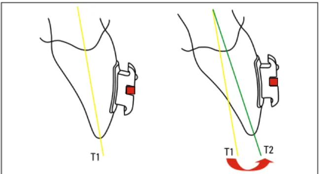
When this archwire was in use, many teeth still showed values that were different from the prescription. However, median inclination values were harmonized for all teeth, causing them to exhibit more similar values between the teeth of the same group, but in opposing quadrants. This fact is clinically significant because it represents movement toward symmetry.
Statistical analysis between the values found at each of the three times showed no statistically significant difference during the test between T1 and T2 (0.020 and 0.019 X 0.025-in) and be-tween T2 and T3 (0.019 X 0.025-in and 0.021 X 0.025-in). Statistically significant differences were found only between T1 and T3 (0.020-in and 0.021 X 0.025-in) for the following groups of teeth: maxillary central incisors (p=0.0023) and maxillary lateral incisors (p=0.0055).
Slack between bracket slot and archwire Taking into account the maximum slack for the 0.021 X 0.025-in archwire (± 3.9°),11 it
was found that after this archwire had done its job, the inclination values of all teeth exam-ined began to approach the torque values built into Class II brackets. It can be asserted that the prescription values tended to be expressed at this time. It was also found that, in terms of the slack between archwire and bracket slot, the percentage of teeth whose torque values approached the prescription values increased between times (Table 3).
Of the 204 teeth examined at T1, 52.9% (108 teeth) were within the prescription range, 13.2% (27 teeth) had values below the prescrip-tion and 33.8%, i.e., 69 teeth were above pre-scription. At T2, the values remained unchanged when compared with those that were above or below the prescription. As at T1, the same 52.9% (108 teeth) were found to be within the prescription range, with 38.7% above prescrip-tion values (79 teeth), while 17 teeth, i.e., 8.3% displayed lower values. At T3, however, a ten-dency was noted whereby the number of teeth within the prescription range rose to 59.8% (122 teeth). Those above prescription declined to 35.8% (73 teeth), and those below prescrip-tion decreased to 4.4% or 9 teeth.
The results displayed in Table 3 allow the following explanation. At T2 the number of teeth within the prescription was found to be
the same as at T1, which confirms the finding that smaller-caliber rectangular archwires are unable to fully express the torque values built into the bracket prescription. Nevertheless, there was an increase in the number of teeth whose values were above prescription, which can be explained by the action of leveling, as it causes greater proclination of anterior teeth by increasing the perimeter of the dental arches. These data confirm that 0.019 X 0.025-in arch-wires work primarily for leveling.
The values found at T3 indicate that 0.021 X 0.025-in archwires successfully express the bracket prescription. The number of teeth that reached the torque values built into the brackets increased from 52.9% at T1 and T2, to 59.8%, or 122 teeth, at T3. These data clearly confirm that 0.021 X 0.025-in archwires are the only ones capable of adequately expressing inclination val-ues, leading to a decrease in the number of teeth whose values were above and below the prescrip-tion (Fig 10).
Nonetheless, some teeth failed to exhibit in-clination values within the prescription’s range of tolerance which—for 0.021 X 0.025-in arch-wires—would be +4° of torque in the mandib-ular incisors, ±3.9º slack between bracket slot and archwire (Fig 11).
dISCuSSIOn
T1 T2 T3 T2
+4º
< 0º
T1 T2 T3
within prescription above below within prescription above below within prescription above below
13 14 3 0 12 5 0 11 5 1
12 5 10 2 6 11 0 7 10 0
11 9 4 4 11 4 2 12 4 1
21 13 2 2 12 4 1 13 4 0
22 10 6 1 11 6 0 9 8 0
23 9 8 0 12 5 0 11 5 1
43 3 10 4 3 10 4 7 10 0
42 12 2 3 11 3 3 13 3 1
41 9 5 3 7 8 2 11 4 2
31 7 7 3 7 8 2 10 5 2
32 10 3 4 9 5 3 13 3 1
33 7 9 1 7 10 0 5 12 0
Total 108 69 27 108 79 17 122 73 9
Percentage 52.9% 33.8% 13.2% 52.9% 38.7% 8.3% 59.8% 35.8% 4.4%
TABLE 3 - Number of teeth whose inclination values were within the prescription, considering a ± 3.9° slack, according to Creekmore11.
FIGURE 10 - Effect on tooth inclination from T1 to T2 and from T2 to T3. The inclination prescription influenced the effect of the 0.021 X 0.025-in archwire on the position of the teeth.
FIGURE 11 - Effect at T3 on the teeth whose values were below the pre-scription.
the misconception that ‘one prescription fits all cases’ and lay bare the need for bracket individ-ualization and a selective use of archwires and even so, the difficulties in controlling the results expressed in the final position of the teeth would not be easily surmounted.
Most orthodontists use a single prescription because they do not use larger-caliber archwires to finish their cases, which results in loss of control over the full expression of prescription,
especially in terms of inclination. This allows similar brackets to be used in different patients with distinct therapeutic goals.12
Andrews’5 standard prescription, however,
since the introduction of Straight-Wire that in-dividual prescriptions be employed using three torque values for the incisors in order to accom-modate compensable inter-maxillary Class I, II or III relationships. Interestingly, this concept has aroused very little attention in the vast universe of those who routinely use this technique.
In this study, assessment of inclinations in anterior teeth was performed as of the stage when round the 0.020-in stainless steel arch-wire stopped being used. The results were used as inclination reference for comparison with the effects produced by rectangular 0.019 X 0.025-in and 0.021 X 0.025-0.025-in archwires. The use of rectangular wires aimed to induce the highest possible expression of the inclinations built into the brackets and, therefore, they were kept in-serted for longer than the round wires, 40 and 30 days, respectively. It was only after this pe-riod that CT images were acquired.
It is important to stress that the slack be-tween a 0.019 X 0.025-in archwire and the bracket slot is 10.5º.11 Theoretically, this is a
very high value and a significant expression of the prescription can be therefore expected in the anterior teeth in terms of inclination. From this perspective, the 0.021 X 0.025-in archwire was the last to be used, with a 3.9º slack11 since
it is potentially better able to express the pre-scription. It was thus possible to assess and com-pare the behavior of all archwires and brackets, always taking into consideration the slack be-tween archwire and bracket slot.
The absence of statistically significant dif-ferences in the values of tooth inclination be-tween the three times (T1, T2 and T3) for most teeth analyzed in this study can be attributed to the similarity between the torque values in the prescription and those found in the first phase, when the round 0.020-in archwire was used. This fact has a direct bearing on the means and statistical results. Some individuals showed little difference between the three moments
(T1, T2 and T3) and others showed great differ-ences, which caused an increase in result variability. For both lateral and central maxillary inci-sors, the statistical differences found between the round archwire and the rectangular arch-wires that filled the bracket slot maximally can be ascribed to the fact that the prescription reading was based on these teeth, for most in-dividuals examined in this sample. In the Class II sample, the selection was made for both those subjects whose anterior teeth had buccal (Class II, division 1) and lingual (Class II, division 2) inclinations. By using rectangular 0.021 X 0.025-in archwires, these teeth reached values that differed from their initial values, as well as, from the values found when round archwires were used. This effect did not occur with any other tooth examined in this study.
CLInICAL COnSIdERAtIOnS
At this point in this article it seems impor-tant to highlight the clinical insights that can be inferred from the results. Much has been said about the individualization of orthodontic treatment by means of an accurate, differential and individualized diagnosis with a view to de-termining the best treatment plan for each indi-vidual. This concept encompasses the choice of orthodontic brackets, a key issue often neglect-ed by users of the Straight-Wire technique. This technique requires that brackets be chosen ac-cording to the final position of the teeth, which varies from patient to patient.
prescription values, inclinations varied widely between individuals, even at the three different assessment times.
Some teeth displayed a unique behavior, such as the maxillary central incisors. Inclina-tion values varied little at each time, regard-less of archwire size and its effect on anterior teeth. Despite the proclination tendency shown by 0.019 X 0.025-in archwires, torque val-ues for these teeth remained at around +7°, a value suggested by Andrews5 as ideal and used
in Capelozza’s prescription II. This finding re-garding the central incisors is also corroborated by another study in which, although the value (0.96º) was higher than the one found by An-drews, it is not clinically significant.7
This information reinforces the recommen-dation that a +7º torque be built into the Class II prescription for central incisors which, unlike the +2º prescription suggested by Andrews,5 do
not have their inclination values decreased. The argument in favor of maintaining +7º in max-illary incisor inclination, even in brackets de-signed for compensatory treatment of Pattern II malocclusions, stems from the need to give resistance to these teeth in the face of other mechanical resources used to treat this maloc-clusion, such as headgear and Class II elastics, thereby minimizing the tendency towards a more vertical position. Thus, any compensation for tooth inclinations occurs in the lower arch in order to prevent the negative aesthetic im-pact that takes place when maxillary teeth are inclined in an attempt to compensate for the facial pattern.8
A unique behavior was also noted in maxil-lary lateral incisors, which exhibited values well above those found in the sample of normal oc-clusions suggested by Andrews5 and above Class
II prescription values.8 This seems due to the
fact that the means were influenced by indi-viduals who presented with Class II, division 2 malocclusion.
In the maxillary canines a behavior was not-ed which differs from that found in other teeth during the transition of T1, T2 and T3. Clini-cally, it was observed that in each individual, the initial position of the canines tended to remain unchanged. Thus, if one of the teeth exhibited an inclination that was altogether different from its analogue in the opposite quadrant, such dif-ference in position was maintained despite the use of rectangular wires. This finding attests that the importance of canine position, and the impact it exerts on other teeth, especially in terms of inclination, cannot be overemphasized. The size of the root may have been the main obstacle to the full expression of the prescrip-tion inclinaprescrip-tion, despite the use of larger-caliber rectangular archwires.
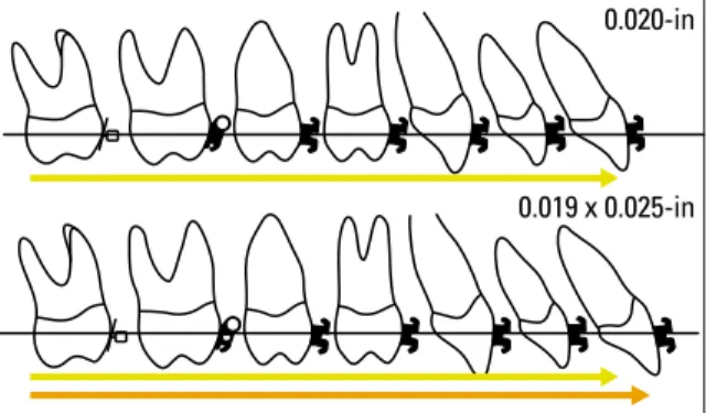
Also based on the results of this study, but now seen from a clinical perspective, it seems reasonable to emphasize that the 0.019 X 0.025-in archwire should be primarily regarded as a leveling archwire, since its major effect is to procline incisors (Fig 12), irrespective of the prescription built into the bracket.
FIGURE 12 - Proclination effect and increase in arch perimeter in the tran-sition from T1 to T2.
0.020-in
Therefore, to ensure that the inclination values of a given prescription are fully expressed, it is ad-visable to use larger-caliber rectangular archwires, e.g., 0.021 X 0.025-in in a 0.022-in slot. It would also be reasonable to assume that this wire should be maintained for a longer period of time to pro-duce a more effective prescription expression.11
As for the orthodontic treatment of the sam-ple, it must be emphasized that there was a re-duction, if not a complete correction,of overjet in these individuals, even without the use of any additional mechanical resources. This probably occurred in a compensatory manner, through changes in their inclinations.
The use of individualized prescriptions can be helpful in creating or maintaining the incli-nations and angulations necessary to achieve the planned movements, which ultimately compen-sate for pattern II malocclusions by facilitating, and not hindering, these movements.
When planning an orthodontic case, ortho-dontists envision the positioning of teeth, in terms of the desired inclinations and move-ments, with the purpose of attaining an in-terincisor relationship that is acceptable both esthetically and functionally. As regards the individuals selected for this scientific research, it is expected that the mandibular incisors will remain or become proclined, i.e., with a positive
inclination value. Upper incisors should also un-dergo proclination, in line with dental and fa-cial esthetics. Values, however, should not be too high but nominally equivalent to the values built into the prescription.
The use of the prescription can still be ad-vocated given the increased value used in the mandibular canine angulation. This angulation makes for lower incisor proclination. It should be emphasized once again that in the absence of mandibular canine proclination,which should be expected as compensation for Pattern II, the prescription would help achieve the best possi-ble positioning. On the other hand, in the pres-ence of an increased angular value, the prescrip-tion would ensure its maintenance.
COnCLuSIOnS
Based on the methodology used in this in-vestigation and the results it achieved, it seems reasonable to state that:
» The median inclinations found at T1, T2 and T3 were similar. Statistical significance was found only for mandibular lateral incisors.
1. Andrews LF. The Straight-Wire appliance: origin, controversy, commentary. J Clin Orthod. 1976 Feb;10(2):99-114. 2. Andrews LF. The Straight-Wire appliance: explained and
compared. J Clin Orthod. 1976 Mar;10(3):174-95.
3. Andrews LF. The Straight-Wire appliance: case histories – non-extraction. J Clin Orthod. 1976 Apr;10(4):282-303.
4. Andrews LF. The Straight-Wire appliance: extraction brackets and “classiication of treatment”. J Clin Orthod. 1976 May;10(5):360-79. 5. Andrews LF. Straight-Wire: o conceito e o aparelho. San Diego:
LA Well; 1989.
6. Bastia FMM. Estudo das angulações e inclinações dentárias obtidas no tratamento ortodôntico com a utilização da prescrição MBT™. [dissertação]. São Bernardo do Campo (SP): Universidade Metodista de São Paulo; 2005.
7. Cabrera CAG. Estudo da correlação do posicionamento dos incisivos superiores e inferiores com a relação antero-posterior das bases ósseas. Rev Dental Press Ortod Ortop Facial. 2005;10(6):59-74.
8. Capelozza L Filho, Silva OG Filho, Ozawa TO, Cavassan AO. Individualização de braquetes na técnica de straight wire: revisão de conceitos e sugestão de indicações para uso. Rev Dental Press Ortod Ortop Facial. 1999 jul-ago;4(4):87-106.
9. Capelozza L Filho. Diagnóstico em Ortodontia. Maringá: Dental Press; 2004.
10. Capelozza L Filho, Fattori L, Maltagliati LA. Um novo método para avaliar as inclinações dentárias utilizando a tomograia computadorizada. Rev Dental Press Ortod Ortop Facial. 2005 set-out;10(5):23-9.
11. Creekmore TD. JCO Interviews Dr. Thomas D. Creekmore on Torque. J Clin Orthod. 1979;13(5):305-10.
12. Dellinger EL. A scientiic assessment of the straight-wire appliance. Am J Orthod. 1978 Mar;73(2):290-9.
13. Germane N, Bentley BE Jr, Isaacson RJ. Three biologic variables modifying faciolingual tooth angulation by straight-wire appliances. Am J Orthod Dentofacial Orthop. 1989 Oct;96(4):312-9.
14. Gündüz E, Rodríguez-Torres C, Gahleitner A, Heissenberger G, Bantleon HP. Bone regeneration by bodily tooth movement: dental computed tomography examination of a patient. Am J Orthod Dentofacial Orthop. 2004 Jan;125(1):100-6.
15. Hamada Y, Kondoh T, Noguchi K, Iino M, Isono H, Ishii H, et al. Application of limited Cone Beam Computed Tomography to clinical assessment of alveolar bone grafting: a preliminary report. Cleft Palate Craniofac J. 2005 Mar;42(2):128-37.
16. Hatcher DC, Aboudara CL. Diagnosis goes digital. Am J Orthod Dentofacial Orthop. 2004 Apr;125(4):512-5.
17. Heiland M, Schulze D, Rother U, Schmelzle R. Midfacial imaging using digital volume tomography. Int Congr Ser. 2003 Jun;1256:1230-4.
REfEREnCES
18. Honda K, Arai Y, Kashima M, Takano Y, Sawada K, Ejima K, et al. Evaluation of the usefulness of the limited cone-beam CT (3DX) in the assessment of the thickness of the roof of the glenoid fossa of the temporomandibular joint. Dentomaxillofac Radiol. 2004 Nov;33(6):391-5.
19. Houston WJB. The analysis of errors in orthodontics measurements. Am J Orthod Dentofacial Orthop. 1983 May;83(5):382-90.
20. Lascala CA, Panella J, Marques MM. Analysis of the accuracy of linear measurements obtained by cone beam computed tomography (CBCT – NewTom). Dentomaxillofac Radiol. 2004 Sep;33(5):291-4.
21. Mah JK, Danforth RA, Bumann A, Hatcher D. Radiation absorbed in maxillofacial imaging with a new dental computed tomography device. Oral Surg Oral Med Oral Pathol Oral Radiol Endod. 2003 Oct;96(4):508-13.
22. Marmulla R, Wörtche R, Mühling J, Hassfeld S. Geometric accuracy of the NewTom 9000 Cone Beam CT. Dentomaxillofac Radiol. 2005 Jan;34(1):28-31.
23. Podesser B, Williams S, Bantleon HP, Imhof H. Quantitation of transverse maxillary dimensions using computed tomography: a methodological and reproducibility study. Eur J Orthod. 2004 Apr;26(2):209-15.
24. Reis SAB, Abrão J, Capelozza L Filho, Claro CAA. Análise Facial Subjetiva. Rev Dental Press Ortod Ortop Facial. 2006 set-out;11(5):159-72.
25. Rustmeyer P, Streubühr U, Suttmoeller J. Low-dose dental computed tomography: signiicant dose reduction without loss of image quality. Acta Radiol. 2004;45:847-53.
26. Schulze D, Heiland M, Schmelzle R, Rother UJ. Diagnostic possibilities of cone-beam computed tomography in the facial skeleton. Int Congr Ser. 2004;1268:1179-83.
27. Schulze D, Heiland M, Thurmann H, Adam G. Radiation exposure during midfacial imaging using 4- and 16-slice computed tomography, cone beam computed tomography systems and conventional radiography. Dentomaxillofac Radiol. 2004 Mar;33(2):83-6.
28. Ugur T, Yukay F. Normal faciolingual inclinations of tooth crowns compared with treatment groups of standard and pretorqued brackets. Am J Orthod Dentofacial Orthop. 1997;112(1):150-7.
29. Vardimon A, Lambertz W. Statistical evaluation of torque angles in reference to straight-wire appliance (SWA) theories. Am J Orthod. 1986;89:56-66.
30. Zanelato ACT. Estudo das angulações e inclinações dentárias em brasileiros, leucodermas com oclusão normal natural. [dissertação]. São Bernardo do Campo (SP): Universidade Metodista de São Paulo; 2003.
Contact address
Liana Fattori
Rua Primeiro de Maio, 188 / cj.111 – Centro CEP: 09.015-030 – Santo André/SP, Brazil
E-mail: [email protected] - [email protected] Submitted: September 2007