Camila Gornic*, Paula Paiva do Nascimento**, Camilo Aquino Melgaço***, Antonio Carlos de O. Ruellas****, Paulo José D’Albuquerque Medeiros*****, Eduardo Franzotti Sant’Anna******
Cephalometric analysis of the upper airways
of Class III patients subjected to orthosurgical
treatment
Objective: The aim of this study was to evaluate the effects of orthognathic surgery for man-dibular setback – with and without combined maxillary surgery – on the upper airways (UA). Methods: Immediate lateral preoperative and postoperative cephalometric radiographs of 17 Class III patients were evaluated. Measurements of airway space (AS) diameter were taken in the sagittal plane in the hypopharyngeal and oropharyngeal regions, and changes in hyoid bone position were also recorded. Paired t-test and Pearson’s coefficient were applied seeking for potential associations between skeletal and AS changes. Results: Significant AS reduction was noted in the hypopharyngeal region (mean= 3.10 mm, p= 0.024). The hyoid bone was displaced inferiorly and posteriorly, thereby reducing its distance to the anterior mandibular region. No quantitative correlation could be established between anteroposterior AS reduction and man-dibular setback. However, there was a strong correlation between initial AS diameter and the amount of reduction observed in the hypopharynx, but only moderate correlation with the oropharynx. Conclusions: Mandibular setback can cause significant UA narrowing, especially in the inferior-most portion (hypopharynx). Therefore, special attention should be given to UA evaluation when formulating an orthosurgical treatment plan since the potential deleterious ef-fects of these changes on functions of the patients should not be overlooked.
Abstract
Keywords: Orthognathic surgery. Mandibular setback. Airways. Oropharynx. Hypopharynx.
* Graduate, School of Dentistry – Federal University of Rio de Janeiro (UFRJ).
** MSc student in Orthodontics, Department of Pediatric Dentistry and Orthodontics, School of Dentistry - UFRJ. *** MSc in Orthodontics and PhD student, Department of Pediatric Dentistry and Orthodontics, School of Dentistry - UFRJ. **** PhD in Orthodontics and Adjunct Professor, Department of Pediatric Dentistry and Orthodontics, School of Dentistry - UFRJ. ***** PhD in Dentistry, UFRJ. Head Professor of Surgery, School of Dentistry - UERJ.
****** PhD in Orthodontics, School of Dentistry - UFRJ. Adjunct Professor, Department of Pediatric Dentistry and Orthodontics, School of Dentistry - UFRJ. intROduCtiOn
The upper airway (UA) – whose major com-ponent is the pharynx – consists of a tube that extends from the nostrils to the larynx (glottis).
This structure can be didactically divided into: Nasopharynx (superior-most region, related to the choanae), velopharynx (posterior region of the uvula), oropharynx (base of tongue) and
How to cite this article: Gornic C, Nascimento PP, Melgaço CA, Ruellas
ACO, Medeiros PJD, Sant’Anna EF. Cephalometric analysis of the upper air-ways of Class III patients subjected to orthosurgical treatment. Dental Press J Orthod. 2011 Sept-Oct;16(5):82-8.
» The authors report no commercial, proprietary, or inancial interest in the
hypopharynx (inferior-most region, posterior to the hyoid bone). The UA walls consist only of soft tissue and, therefore, is not supported by any rigid structure such as bone or cartilage. Thus, the mechanical support that ensures the con-tinued opening of this structure in opposition to negative pressure during inhalation move-ments results from tension and contraction of the muscles that surround it.11 This mechanism
is due in large part to muscular insertions in the genial tubercles, which enable the mandible to be closely involved in the function and support of the tongue and related soft tissues. The ge-nioglossus, geniohyoid and infrahyoid muscles are linked to the oropharynx and hypopharynx. The tongue muscles play an important role in maintaining airway opening, since they form the anterior wall of the pharynx in that segment.
Several procedures have been developed in the hope of increasing the pharyngeal airway space in patients with obstructive sleep apnea syndrome (OSAS). The goal would be to pull the tongue muscles anteriorly through mandibu-lar advancement, thereby decreasing airway re-sistance to enhance air flow efficiency.14
On the other hand, one might question wheth-er the opposite procedure, i.e., mandibular setback, could cause any reduction in pharyngeal airway space. This is a pertinent question since mandibu-lar setback surgical technique is widely used in the treatment of Class III dentofacial deformities.
The effects of orthognathic surgery on the airways of healthy patients have not been fully explained in the literature, especially with re-gard to the risk of these patients developing OSAS in the postoperative period.
The purpose of this study was to assess and quantify, by means of cephalometric analysis, changes in the airways caused by orthognathic surgery involving mandibular setback, more specifically in regions directly related to the mandible, corresponding to the oropharynx and hypopharynx.
MAtERiAL And MEtHOdS Sample
Seventeen patients who had been subjected to orthodontic treatment in preparation for surgi-cal correction of Class III dentoskeletal deformities were selected for this study. The surgeries were per-formed in the Department of Oromaxillofacial Sur-gery of the Pedro Ernesto State Hospital in 2006.
Six adult men and 11 adult women whose max-illofacial growth had already ceased were evaluated.
Surgical technique
Among the patients, 14 underwent com-bined orthognathic surgery for maxillary ad-vancement and mandibular setback, whilst the remaining patients (3) were subjected to man-dibular setback only. The surgical technique consisted of Le Fort I type maxillary osteotomy with rigid internal fixation. In performing man-dibular setback, the technique of choice was bilateral vertical osteotomy with maxilloman-dibular fixation for a period of 2 to 3 weeks de-pending on the degree of occlusal stability.
Cephalometric analysis
Preoperative lateral cephalometric X-rays ob-tained up to 7 months prior to surgery as well as immediate postoperative radiographs taken up to one week after surgery were evaluated. Pre- and postoperative radiographs were obtained using the same device and in accordance with the stan-dards of the lateral cephalometric technique.
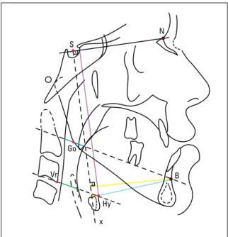
All cephalograms were traced on acetate pa-per and all measurements were pa-performed by the same examiner. Upper airway references were ob-tained according to the methodology advanced by Frohberg et al,3 which can be seen in Figure 1.
Soft tissue measurements
S
Go
N
Hy
x
Vr B
of the mandibular alveolar process) to the gonion (Go - point determined by the intersection of the bisector of the angle formed by the lines tangent to the posterior and inferior borders of the mandible and the gonial angle), and the anterior and poste-rior walls of the pharynx. This measure intends to quantify the changes undergone by the oropharynx after surgery. Point B was selected as a reference as it constitutes the principal region of muscle insertion in the mandible, i.e., the genial tubercles.
2) Airway diameter in the hypopharynx region (green): Distance between the points defined by the intersection between the line extending from the most superoanterior point of the hyoid bone to the most anteroinferior point of the third cervical verte-bra, and the anterior and posterior walls of the phar-ynx. This measure aims at quantifying the changes undergone by the hypopharynx after surgery.
Additional measures
3) Mandibular position (yellow): Measures the shortest distance between point B and a line per-pendicular to SN (sella-nasion) passing through S (here called x). This measure discloses the amount of mandibular setback achieved through surgery.
4) SNA - Angle formed by the SN and NA lines (nasion-point A), where A is the deepest point on the contour of the premaxilla, which determines the de-gree of maxillary retrusion relative to the cranial base. 5) SNB - Angle formed between the SN and NB lines (nasion-point B), which expresses the degree of mandibular protrusion relative to the cranial base.
6) GoGn-SN - Angle determined by the inter-section between the line joining the gonial (Go) and gnathion (Gn) points, and the SN line.
7) FMA - Angle determined by the intersection of the mandibular plane (line passing through the menton (Me) and tangent to the lower border of the mandible in the region of the gonial angle), with the Frankfort horizontal plane (junction of the Porium, Po, and orbitale, Or, points).
These last two angular measures (GoGn-SN and FMA) help to diagnose potential mandibular rotations in the vertical direction that can result from surgery, consequently inducing changes in the oropharyngeal reference plane.
8) Hy-SN (purple), the smallest distance between the most anterosuperior point on the body of the hy-oid bone (Hy) and the SN line, revealing the vertical position of the hyoid bone relative to the cranial base.
9) Hy-x (orange), the smallest distance between Hy and line x, enabling the evaluation of the antero-posterior position of the hyoid bone.
10) Hy-B (light blue), distance between the Hy and point B, reflecting the relationship between the hyoid bone and the anterior region of the man-dibular body.
RESuLtS
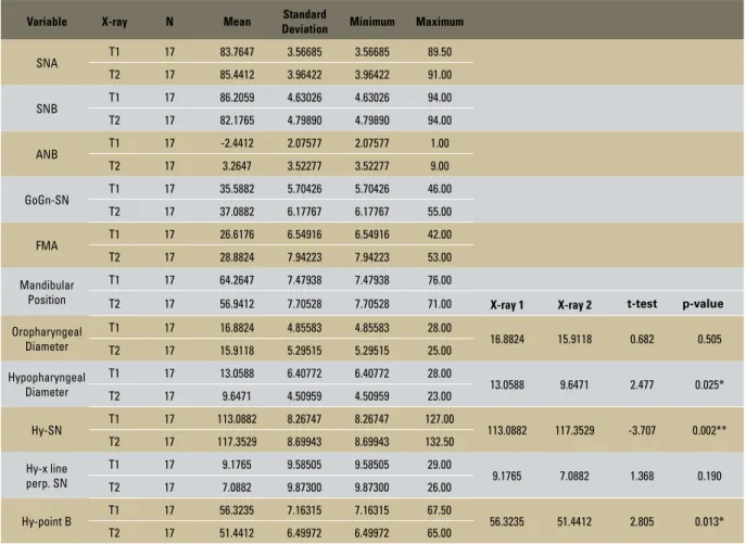
TABLE 1 - Descriptive statistics of cephalometric variables obtained from preoperative (T1) and postoperative (T2) radiographs.
* Correlation at 0.05 significance level. ** Correlation at 0.01 significance level.
Variable X-ray N Mean Standard
Deviation Minimum Maximum
SNA T1 17 83.7647 3.56685 3.56685 89.50
T2 17 85.4412 3.96422 3.96422 91.00
SNB T1 17 86.2059 4.63026 4.63026 94.00
T2 17 82.1765 4.79890 4.79890 94.00
ANB T1 17 -2.4412 2.07577 2.07577 1.00
T2 17 3.2647 3.52277 3.52277 9.00
GoGn-SN T1 17 35.5882 5.70426 5.70426 46.00
T2 17 37.0882 6.17767 6.17767 55.00
FMA T1 17 26.6176 6.54916 6.54916 42.00
T2 17 28.8824 7.94223 7.94223 53.00
Mandibular Position
T1 17 64.2647 7.47938 7.47938 76.00
T2 17 56.9412 7.70528 7.70528 71.00 X-ray 1 X-ray 2 t-test p-value
Oropharyngeal Diameter
T1 17 16.8824 4.85583 4.85583 28.00
16.8824 15.9118 0.682 0.505
T2 17 15.9118 5.29515 5.29515 25.00
Hypopharyngeal Diameter
T1 17 13.0588 6.40772 6.40772 28.00
13.0588 9.6471 2.477 0.025*
T2 17 9.6471 4.50959 4.50959 23.00
Hy-SN T1 17 113.0882 8.26747 8.26747 127.00 113.0882 117.3529 -3.707 0.002**
T2 17 117.3529 8.69943 8.69943 132.50
Hy-x line perp. SN
T1 17 9.1765 9.58505 9.58505 29.00
9.1765 7.0882 1.368 0.190
T2 17 7.0882 9.87300 9.87300 26.00
Hy-point B T1 17 56.3235 7.16315 7.16315 67.50 56.3235 51.4412 2.805 0.013*
T2 17 51.4412 6.49972 6.49972 65.00
TABLE 2 - Pearson’s correlation (r) between different variables.
Change in oropharyngeal diameter
Change in
hypopharyngeal diameter Hy-SN Hy-x Hy-point B
Mandibular Setback
r 0.162 0.067 0.263 0.338 -0.261
p 0.535 0.800 0.307 0.184 0.312
Initial airway space diameter
r -0.526 -0.728
p 0.030* 0.001**
Hy-SN
r -0.389 -0.266
p 0.123 0.302
Hy-x
r 0.224 0.102
p 0.388 0.697
Hy-point B
r -0.127 0.001
p 0.626 0.997
diSCuSSiOn
In the cases analyzed in this study, the man-dible experienced a mean setback of 7.32 mm af-ter surgery. The impact on the airway space was evidenced by a mean reduction of 0.97 mm in the oropharynx and 3.41 mm in the hypopharynx. The initial mean diameters of these spaces were 16.88 mm and 13.05 mm, respectively.
This reduction was statistically significant only in the region of the hypopharynx (P=0.025), as shown by the paired t-test (Table 1). In a study8 where the smallest diameter in the
pos-terior region of the tongue (which in most cases corresponds to the hypopharynx) was measured, the same result was found: Airway space reduc-tion, in agreement with other authors.12
Despite this reduction in the hypopharynx, this study was unable to establish a significant quantitative correlation between this reduction and the mandibular setback, i.e., mandibular set-back causes airway narrowing, but not propor-tionally. However, there was a strong correlation between the initial diameter and the amount of AS reduction in the hypopharynx (r= -0.728, p<0.01). In the oropharyngeal region there was moderate correlation (r=-0.526, p<0.05) be-tween the initial diameter and the reduction observed in the airway after surgery. Thus, pa-tients presenting extensive airway space in the preoperative period tend to experience greater AS reduction after mandibular setback surgery.
Because most patients underwent combined orthognathic surgery, one should take into ac-count the influence of maxillary surgery on the final position of the mandible. This influence can be observed by a change in mandibular plane angulation, as assessed by measuring GoGn-SN. The correlation found between this measure and changes in the oropharynx (r=0.511, p<0.05) in-dicates a direct link between this space and man-dibular rotation, i.e., when GoGn-SN increases (clockwise mandibular rotation) the diameter of the oropharynx is likely to increase as well.
This finding may explain the variability found in the behavior of oropharyngeal soft tissues, where it was found that 57% of patients who underwent mandib-ular setback associated with maxillary intervention showed an increase in oropharyngeal space. Oth-erwise, three patients who underwent mandibular setback alone experienced only a reduction in this space. Another explanation for this variation may be ascribed to a difficulty in standardizing tongue position during radiograph acquisition, since such position is very unstable and constantly changes in response to physiological movements.²
A study evaluating patients who underwent mandibular setback surgery alone found a reduc-tion in airway space (in the mandibular plane) in all cases, establishing a correlation between these two variables (r=0.52).5
Another significant finding was that the hyoid bone was displaced inferiorly (mean=4.26 mm) in response to mandibular setback. Horizontally, bone movement varied with a slight prevalence of posterior displacement (2.08 mm), following the mandibular movement. Other authors4,7,12
also observed inferior and posterior displace-ment of the hyoid bone. In contrast, anterior dis-placement of this bone has also been reported.15
Reduction in the distance between the hy-oid bone and the anterior mandible region (mean=4.88 mm) was also a significant finding, which raised the question of how the suprahy-oid musculature might behave. Changes in this muscle’s tone are likely to take place over time to compensate for the decrease in this distance. The largest reduction found for this distance was 18 mm, corresponding to 27% of the original dis-tance exhibited by this particular individual.
Magnetic resonance evaluation of airway be-havior after orthognathic surgery has shown no signs of edema in patients’ airways in the imme-diate postoperative period.10 It is therefore safe
to assert that the airway changes observed in this study were not masked by tissue edema but result in fact from movements induced by bone surgery. This study focused on assessing changes in the immediate postoperative period. Other authors, however, followed their patients for longer peri-ods: 3 months, 6 months, 1 year and up to three years. After one year, no significant tendency was found showing that airway soft tissues tend to re-turn to their initial dimensions.5 Another study,7
which used computed tomography, also dem-onstrated this same result. A two-year or longer follow-up actually suggests that changes occur in the airways over time after mandibular setback surgery.13 Moreover, some authors argue that the
hyoid bone tends to return to its original position, probably because mandible position tends to re-lapse forward and upward even though the hyoid bone does not quite extend as far as the mandible.4
Although there are many studies on this sub-ject in the literature, any comparison between them is complicated by the use of a wide range
of different methodologies, which are still rather limited due to the nature of airway tissues and their dynamic structure. Further studies are war-ranted to clinically evaluate the impact of changes observed in cephalometric radiographs. Are such changes sufficient to cause dysfunction or are they within the scope of physiological limits? The answer to this question requires that patients be interviewed for possible symptoms of discomfort, breathing or swallowing difficulties, snoring, ap-nea or other symptoms that might exert a genuine impact on the health of these individuals.
COnCLuSiOnS
Contact address
Eduardo Franzotti Sant’Anna Av. Brigadeiro Trompowsky, s/n
CEP: 21.949-900 - Ilha do Fundão - Rio de Janeiro/RJ, Brazil E-mail: [email protected]
1. Chen F, Terada K, Hua Y, Saito I. Effects of bimaxillary surgery and mandibular setback surgery on pharyngeal airway measurements in patients with Class III skeletal deformities. Am J Orthod Dentofacial Orthop. 2007;131(3):372-7.
2. Eggensperger N, Smolka K, Johner A, Rahal A, Thüer U, Iizuka T. Long-term changes of hyoid bone and pharyngeal airway size following advancement of the mandible. Oral Surg Oral Med Oral Pathol Oral Radiol Endod. 2005;99(4):404-10.
3. Frohberg U, Naples RJ, Jones DL. Cephalometric comparison of characteristics in chronically snoring patients with and without sleep apnea syndrome. Oral Surg Oral Med Oral Pathol Oral Radiol Endod. 1995;80(1):28-33.
4. Gu GM, Gu G, Nagata J, Suto M, Anraku Y, Nakamura K, et al. Hyoid position, pharyngeal airway and head posture in relation to relapse after the mandibular setback in skeletal Class III. Clin Orthod Res. 2000;3(3);67-77.
5. Hochban W, Schürmann R, Brandenburg U. Mandibular setback for surgical correction of mandibular hyperplasia - does it provoke sleep-related breathing disorders? Int J Oral Maxillofac Surg. 1996;25(5):333-8.
6. Jacobson A. Airway changes after orthognathic surgery as assessed by cone-beam computed tomography [abstract]. Am J Orthod Dentofacial Orthop. 2007;132(5):712.
7. Kawamata A, Fujishita M, Ariji Y, Ariji E. Three-dimensional computed tomographic evaluation of morphologic airway changes after mandibular setback osteotomy for prognathism. Oral Surg Oral Med Oral Pathol Oral Radiol Endod. 2000;89(3):278-87.
REfEREnCES
8. Liukkonen M, Vähätalo K, Peltomäki T, Tiekso J, Happonen RP. Effect of mandibular setback surgery on the posterior airway size. Int J Adult Orthodon Orthognath Surg. 2002;17(1):41-6.
9. Lowe AA, Fleetham JA, Adachi S, Ryan CF. Cephalometric and computed tomographic predictors of obstructive sleep apnea severity. Am J Orthod Dentofacial Orthop. 1995;107(6):589-95.
10. Meisami T, Musa M, Keller MA, Cooper R, Clokie CM, Sàndor GK. Magnetic resonance imaging assessment of airway status after orthognathic surgery. Oral Surg Oral Med Oral Pathol Oral Radiol Endod. 2007;103(4):458-63. Epub 2006 Oct 27.
11. Rajagopal MR, Jerry P. Applied anatomy and physiology of the airway and breathing. Indian J Anaesth. 2005;49(4):251-6. 12. Samman N, Tang SS, Xia J. Cephalometric study of the
upper airway in surgically corrected Class III skeletal deformity. Int J Adult Orthodon Orthognath Surg. 2002;17(3):180-90.
13. Saitoh K. Long-term changes in pharyngeal airway morphology after mandibular setback surgery. Am J Orthod Dentofacial Orthop. 2004;125(5):556-61.
14. Silverstein K, Costello BJ, Giannakpoulos H, Hendler B. Genioglossus muscle attachments: an anatomic analysis and the implications for genioglossus advancement. Oral Surg Oral Med Oral Pathol Oral Radiol Endod. 2000;90(6):686-8. 15. Tselnik M, Pogrel A. Assessment of the pharyngeal airway
space after mandibular setback surgery. J Oral Maxillofac Surg. 2000;58(3):282-5.