Arno Locks1
Angle Class II, division 2 malocclusion with deep overbite
Angle Class II, division 2, malocclusion is characterized by a Class II molar relation associated with retroclined or vertical positioning of the upper incisors and in general an overbite. This clinical case was presented to the Brazilian Board of Orthodontics and Facial Orthopedics (BBO) as part of the requirements for becoming a BBO Diplomate .
Keywords: Angle Class II malocclusion. Corrective orthodontics. Overbite.
» The author reports no commercial, proprietary or financial interest in the products or companies described in this article.
» Patients displayed in this article previously approved the use of their facial and in-traoral photographs.
Contact address: Arno Locks Rua Presidente Coutinho, 311 – conj. 1101 l CEP: 88.015-230- Florianópolis/SC – Brazi Email: [email protected]
1 MSc in Orthodontics, UFRJ. PhD in Orthodontics, UNESP. Post-Doc at the
University of Aarhus, Denmark. Professor of Orthodontics, UFSC. Diplomate from the Brazilian Board of Orthodontics and Facial Orthopedics (BBO).
How to cite this article: Locks A. Angle Class II, division 2 malocclusion with deep overbite. Dental Press J Orthod. 2012 Nov-Dec;17(6):160-6.
Submitted: July 19, 2012 - Revised and accepted: August 08, 2012 HISTORY AND ETIOLOGY
Caucasian female patient, 12 years old, attended the office reporting that the pediatric dentist had asked for the extraction of four first premolars as treatment for malocclusion.
The chief complaint, reported by the mother and patient, was the search for better smile esthetics, mainly due to the lack of space for all the teeth, al-though they were not wishing for any extractions.
DIAGNOSIS
On extra-oral clinical examination, symmetrical facial pattern, lip competence, slightly convex pro-file, with well-developed pogonion and good naso-labial angle were observed (Fig 1). The patient was
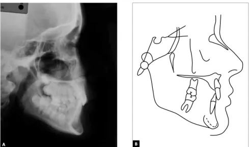
in the final stage of the mixed dentition, tooth #75 presenting clinical features of ankylosis (Fig 1). In the dental casts analysis, a Class II, Division 2 mal-occlusion was observed, slightly asymmetric, with overbite and a pronounced curve of Spee. A 0.5 mm overjet and the lower midline shifted 2 mm to the left were also observed, as well as upper and lower anterior crowding (Fig 2). In the cephalometric analysis, a Class II skeletal pattern was observed (ANB = 5°) with uprighting of the upper and lower incisors (1.NA = 1, 1.NB = 11), as it can be verified in Figure 3 and Table 1. In the initial panoramic radio-graph all permanent teeth were observed, including the third molar crypts and teeth #75 and #85 in the final process of root resorption (Fig 4).
A má oclusão de Classe II divisão 2 de Angle é caracterizada pela relação molar de Classe II associada a um posiciona-mento vertical ou retroinclinado dos incisivos superiores e, geralmente, a uma sobremordida exagerada. O presente caso clínico foi apresentado à Diretoria do Board Brasileiro de Ortodontia e Ortopedia Facial (BBO) como parte dos requisitos para a obtenção do título de Diplomado pelo BBO.
Figure 1 - Initial facial and intraoral photographs.
A B
Figure 3 - Initial profile cephalometric radiograph (A) and cephalometric tracing (B).
Figure 4 - Initial panoramic radiograph.
TREATMENT GOALS
The treatment goals were correction of Class II skeletal pattern, by reducing the SNA angle, anterior posterior mandibular growth monitoring, obtaining space for the canines and upper incisors, correction of lower crowding, curve of Spee and overbite, and ves-tibularization of the upper and lower incisors, in order to obtain better dental function and esthetics at the end of treatment.
TREATMENT PLAN
Figure 5 - Final facial and intraoral photographs.
space maintenance until the complete eruption of teeth #35 and #45, and favoring incisor projection. Then, the Edgewise conventional fixed appliance would be placed, including teeth #35 and #45 when possible.
EVOLUTION OF TREATMENT
First, as planned, the Kloehn headgear was in-stalled with recommended use of 12 hours a day. Af-ter opening spaces, the fixed appliance for corrective treatment was installed with conventional Edgewise technique, slot 0.022 x 0.028-in. A twist flex 0.015-in archwire was 0.015-installed 0.015-in the upper arch. Then, stainless steel archwires 0.014 to 0.020-in were used for alignment and leveling. After opening space for tooth #13, a superimposed coaxial wire was placed. In the lower arch, the “lip bumper” was immediately installed and the extraction of the second molars was required in order to allow for the eruption of teeth #35 and #45. The full fixed appliance was placed as soon as the occlusal condition allowed it, in order to correct the curve of Spee and overbite.
For finishing, an 0.019 x 0.025-in archwire was used, as planned, with individualized bends and torques ac-cording to need.
After obtaining the expected results, a wraparound removable appliance was used as retention in the up-per arch during one year, for 24 hours a day, followed by nighttime use for another two years. After this period, the use on alternate nights was indicated for four months, followed by weekly use for the next four months, after this use was suspended . In the lower arch a 0.032-in stainless steel intercanine fixed retain-er was used.
TREATMENT RESULTS
A B
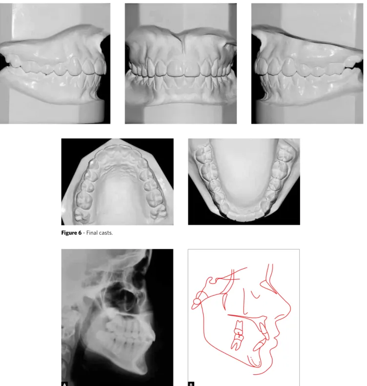
Figure 6 - Final casts.
Figure 7 - Final profile cephalometric radiograph (A) and cephalometric tracing (B).
The intraoral photographs show the correction of overbite and incisor inclination (Fig 5). In the lower arch, the leveling of the curve of Spee was obtained and also the maintenance of intercanine and intermolar distances. Despite the increased inclination of the low-er incisors, the clinical evaluation showed healthy plow-eri- peri-odontium because the patient presented, at the begin-ning of treatment, a good thickness of attached gingiva.
A B
Figure 8 - Final panoramic radiograph.
Measurements Normal A B A/B difference
Skeletal pattern
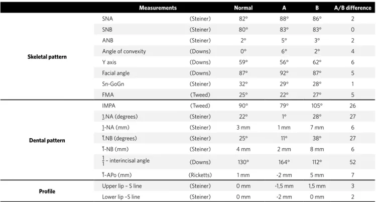
SNA (Steiner) 82° 88° 86° 2
SNB (Steiner) 80° 83° 83° 0
ANB (Steiner) 2° 5° 3° 2
Angle of convexity (Downs) 0° 6° 2° 4
Y axis (Downs) 59° 56° 62° 6
Facial angle (Downs) 87° 92° 87° 5
Sn-GoGn (Steiner) 32° 29° 28° 1
FMA (Tweed) 25° 22° 27° 5
Dental pattern
IMPA (Tweed) 90° 79° 105° 26
1.NA (degrees) (Steiner) 22° 1° 28° 27
1-NA (mm) (Steiner) 3 mm 1 mm 7 mm 6
1.NB (degrees) (Steiner) 25° 11° 38° 27
1-NB (mm) (Steiner) 4 mm 2 mm 8 mm 6
1
1 – interincisal angle (Downs) 130° 164° 112° 52
1–APo (mm) (Ricketts) 1 mm -2 mm 5 mm 7
Profile Upper lip – S line (Steiner) 0 mm -1,5 mm 1,5 mm 3
Lower lip –S line (Steiner) 0 mm -2 mm 0 mm 2
Table 1 -Initial (A) and final (B) cephalometric measurements.
1. Brodie AG. Late growth changes in the human face. Angle Orthod. 1953;23(3):146-57. 2. Coben SE. Growth and Class II treatment. Am J Orthod. 1966;5:5-26.
3. Creemore TD. Inhibition or stimulation of the vertical growth of the facial complex, its significance to treatment. Angle Orthod. 1967;37(4):285-97.
4. Mays RA. A cephalometric comparison of two types of extraoral appliance used with the edgewise mechanism. Am J Orthod. 1969;55(2):195-6.
5. Downs WB. Analysis of the dentofacial profile. Angle Orthod. 1956;26:191-212. 6. RIckets RM. Cephalometric synthesis: an exercise in starting objective and planning
treatment with tracings of the head roentgenogram. Am J Orthod. 1960;46:647-75.
REFERENCES
CONCLUSION
The Angle Class II malocclusion is very common among patients seeking orthodontic treatment.7
Dif-ferent authors1,2,3 observed spontaneous reduction of
ANB angle as a result of normal growth of the subject, although it is not sufficient for self-correction of sag-ittal facial dysplasia. Thus, the intervention of the or-thodontist is very important, still in the growing phase, making possible the achievement of a pleasant profile in adulthood. In cases of maxillary protrusion with high SNA angle, the use of Kloehn headgear is a good option, since this appliance has a strong influence on
the maxillary growth pattern.4 The results of
orthope-dic intervention in the maxilla are well known in the scientific field and properly described in studies using implants.8 In this clinical case, the incisor projection
was of paramount importance for overbite correction, reduction of tooth-arch discrepancy and leveling the curve of Spee. In cases of Class II division 2, it is very important to increase the incisor inclination to give more stability in the overbite correction. Thus, it can be confirmed that the objectives were fully achieved, as the case presents itself with appropriate function, health and esthetics.
7. Locks L et al. Prevalência das maloclusões de Angle em uma clínica de Ortodontia. Rev SBO. 1997;3(4):123-5.
8. Melsen B. Effects of cervical anchorage during and after treatment: an implant study. Am J Orthod. 1978;73(5):526-40.
9. Angle EH. Classification of malocclusion. Dent Cosmos. 1899;41(3):248-64. 10. Bittencourt MAV, Machado AW. Prevalência de má oclusão em crianças entre 6-10