Jonas Capelli Junior*, Rhita Cristina Cunha Almeida**
Orthosurgical treatment of patients in the
growth period: At what cost?
Awaiting growth to end prior to starting orthosurgical treatment is sometimes extremely difficult for patients suffering from severe facial deformities. In cases of substantial skel-etal disharmonies surgery may be indicated during active growth phase when the patient is psychosocially, aesthetically and/or functionally compromised. To indicate this therapy, orthodontic criteria, such as mild intramaxillary discrepancy and the possibility of preop-erative preparation without major dental repositioning, must be met. A second orthosurgi-cal treatment will probably prove necessary after growth has ended. This treatment should not be considered as routine, but rather as a therapeutic option in carefully selected cases.
Abstract
Keywords: Orthodontics. Orthognathic surgery. Psychosocial impact.
* PhD in Orthodontics, Associate Professor of Orthodontics, Rio de Janeiro State University (UERJ). Director of the Brazilian Board of Ortho-dontics and Facial Orthopedics (BBO).
** Specialist and MSc in Orthodontics, UERJ. PhD student in Orthodontics, UERJ. Clinical instructor in Orthodontics, UERJ.
IntroductIon
Awaiting the end of growth before subjecting a patient to an orthosurgical procedure always proves exhausting for both the professional and the patient. The professional feels pressured by the patient, who expresses his/her dissatisfac-tion with his/her face and occlusion. Moreover, issues emerge regarding the best moment for surgery and about the possible effects of early surgery on the residual facial growth.
Approximately 98% of all facial growth ceases by the age of 15 years in girls and 17 or 18 years in boys.1,2 However, in Class III skeletal disharmonies, mandibular growth sometimes even exceeds these ages.3 The data show that orthosurgical patients spend much of adolescence and even early adulthood living with a facial deformity. Appearance is a crucial factor in social relations. Adolescents with signifi-cant dentofacial deformities are considered less at-tractive and so end up as a target of discrimination.3
How to cite this article: Capelli Junior J, Almeida RCC. Orthosurgical treat-ment of patients in the growth period: At what cost? Dental Press J Orthod. 2012 Jan-Feb;17(1):159-77.
» The authors report no commercial, proprietary, or inancial interest in the
Thus, early orthognathic surgery can in some cases prevent future psychosocial problems.
In addition, postponing surgery until adult-hood in very severe cases may exacerbate prob-lems related to pain, speech, breathing, occlu-sion, articulation and masticatory function.3
According to Medeiros and Medeiros4 there are two situations in which orthognathic sur-gery in a growing patient becomes desirable: In the event of a progressive deformity which, if untreated, threatens to reach serious propor-tions; and when the dentofacial deformity is severe and is causing substantial psychosocial damage to the patient.
Some factors must be evaluated before sub-jecting patients to orthognathic surgery. One of these is the temporomandibular joint (TMJ). If the TMJ is not stable and healthy, surgical results may also be unstable, causing dysfunction and joint pain.5 Studies showed that the number of patients who reported joint pain or discomfort rose from 36% prior to surgery to 88% two years after surgery.6 Furthermore, condylar resorption was detected in 30% of patients, resulting in fa-cial deformity and malocclusion relapse.6 Other pathological joint conditions that can influence surgery are condylar hyperplasia, condylar hy-poplasia, idiopathic condylar resorption, osteo-chondroma, rheumatoid arthritis and even sys-temic lupus erythematosus.3
The tongue is also an important factor in the growth and development of the jaws. Microglos-sia can result in underdeveloped jaws and mac-roglossia can result in their overdevelopment. The tongue usually reaches its maximum size by eight years of age.7 One should assess whether it interferes with the patient’s speech, chewing, breathing and treatment stability. Surgical reduc-tion of the tongue can increase the stability and predictability of surgical outcomes.3
Although difficult to predict, determining vectors and amount of growth is important for planning.3 The differential growth of the jaws
can occur in 3 dimensions. A comprehensive un-derstanding of growth trends in each facial type provides clinicians with relevant information re-garding what to expect from sequential growth.3
MAndIBuLAr dEForMItIES Mandibular hypoplasia
Mandibular retrusion that results in a skel-etal Class II malocclusion may display either a normal or deficient mandibular growth.8 When there is normal mandibular growth the dishar-mony may be caused by a mandibular size small-er than the maxilla or the mandible may be in a more retruded position relative to the position of the maxilla, but with similar growth patterns. Thus, the same skeletal and occlusal Class II re-lationship is maintained throughout the growth period.8 This deformity could therefore be cor-rected during growth, yielding fairly stable re-sults. Surgical results are expected to last with a healthy TMJ and with no disruption of growth direction by means of a surgical procedure.3
When mandibular growth deficiency is pres-ent, malocclusion tends to worsen with growth, since the maxilla outgrows the mandible.9 Should one opt for surgical correction during growth it is likely that the residual growth will result in a new skeletal and dental Class II maloc-clusion as an uneven growth pattern will remain between the maxilla and the mandible.9 In these cases, early surgery is indicated in the presence of severe deformities that can affect patient func-tion or cause him/her to experience psychosocial problems.3 Early surgery in these circumstances can improve the quality of life of the patient, but it is important to warn him/her that a second surgery will probably be required.
Mandibular hyperplasia
Mandibular hyperplasia is defined as a pro-truded mandibular position resulting in a skeletal and dental Class III malocclusion.3
posi-tion of the tongue must be evaluated since man-dibular retrusion surgery decreases oral cavity space whereas macroglossia or tongue thrusting can result in surgical relapse.10
Mandibular hyperplasia cases can also ex-hibit a normal growth pattern between the max-illa and mandible, with the mandible positioned more anteriorly relative to the maxilla. Moreover, the mandible may be larger than the maxilla, or the two may present unequal growth patterns, with the mandible growing more rapidly than the maxilla.3
When the growth pattern of the jaws is simi-lar, virtually the same Class III relationship is maintained during growth. Thus, the early cor-rection of this disharmony enhances the chance of result stability.3
In patients with accelerated mandibular growth, facial deformity becomes increasingly severe. An increase in mandibular growth rates usually results from condylar hyperplasia, often begins at the peak of pubertal growth and can continue beyond the normal growth period until the beginning of the twentieth year of age. This growth can be uni- or bilateral, with vertical or horizontal prevalence.3
In this type of malocclusion one may decide to postpone surgery until growth is complete, which can lead to functional problems, facial de-formity, joint pains and psychosocial problems.11 In addition, allowing the deformity to be fully expressed can preclude an ideal treatment in the future. The possible consequences of an exces-sive mandibular growth are mandibular deforma-tion and compensatory changes in the maxilla, dentoalveolar structure and soft tissue, thereby hindering surgical outcome prognosis. Cases of unilateral growth can also present with severe asymmetries and joint dysfunctions.3
Another treatment option could be early surgery of the mandible, positioning it posteri-orly with an overcorrection.3 A second surgery will probably be required, but the patient will at
least enjoy some improvement in function and aesthetics. When one decides to perform early surgery, preoperative preparation should be car-ried out as fast as possible, just enough to allow the patient to be operated on. Furthermore, ex-tending the postoperative orthodontic treatment period is not recommended. Thus, when growth ceases and the need arises for further orthodon-tic preparation, the patient will not be exhausted from the first orthodontic treatment and the two treatment periods will not take place sequential-ly. Besides, it is important to avoid time-consum-ing dental movements, which are normally asso-ciated with root resorption and which can render the second orthodontic treatment unfeasible.
Some authors will indicate a third treatment option,i.e., eliminating any residual mandibular growth with a high condylectomy and correct-ing the skeletal deformity. A high condylectomy will remove the active growth center, preventing future mandibular growth.3,12
MAXILLArY dEForMItIES Maxillary hypoplasia
as optimally as possible, thus averting the re-placement of an unfavorable Class III condition by an unfavorable Class II condition. Growth prognosis is necessarily uncertain.
Vertical maxillary hyperplasia
Maxillary vertical hyperplasia or maxillary vertical excess is defined as excess in the verti-cal growth of the maxilla which may or may not result in an anterior open bite.13 This deformity can be corrected during the growth period with fairly predictable results. The vertical growth of the maxilla will continue after surgery in the same proportion as before the surgery,14 but the postoperative occlusal outcome will probably be preserved. The facial growth vector will continue downwards and backwards. Le Fort I osteotomy is not recommended as it may compromise the anteroposterior growth of the maxilla.3
cLInIcAL cASES
Surgeries performed at an early age are indi-cated in a number of cases presenting with facial deformities such as cleft lip/palate, facial microso-mia, condylar ankylosis, among other conditions.
Thus, clinicians tend to agree that patients with severe facial deformities, which render the patient’s social coexistence unbearable, should be subjected to early surgery. However, the indication of early surgery for Class III cases is controversial. Professionals should bear in mind that patients react differently to the same malocclusion and a facial pattern lacking in harmony can be well tol-erated by some, but can cause severe psychosocial disorders in others. Surgical patients should there-fore be assessed individually to decide whether or not it would be convenient to perform surgery before the end of the growth period.
One should assess the patient’s chief com-plaint, i.e., whether he or she has a clear notion of what is wrong with his/her face, how long he/ she has been aware of this problem, what they expect from the treatment, to what extent the
problem interferes with their daily routine and if someone is compelling them to seek treatment. Patients who have a precise idea of the problem and realistic expectations about the treatment are better indicated for early surgery.15 One should exercise caution with patients who are unable to identify where exactly the skeletal dis-harmony is located and hold very high expecta-tions regarding the treatment as they may not be psychologically prepared for the treatment and are therefore highly likely to be disappointed.
case 1
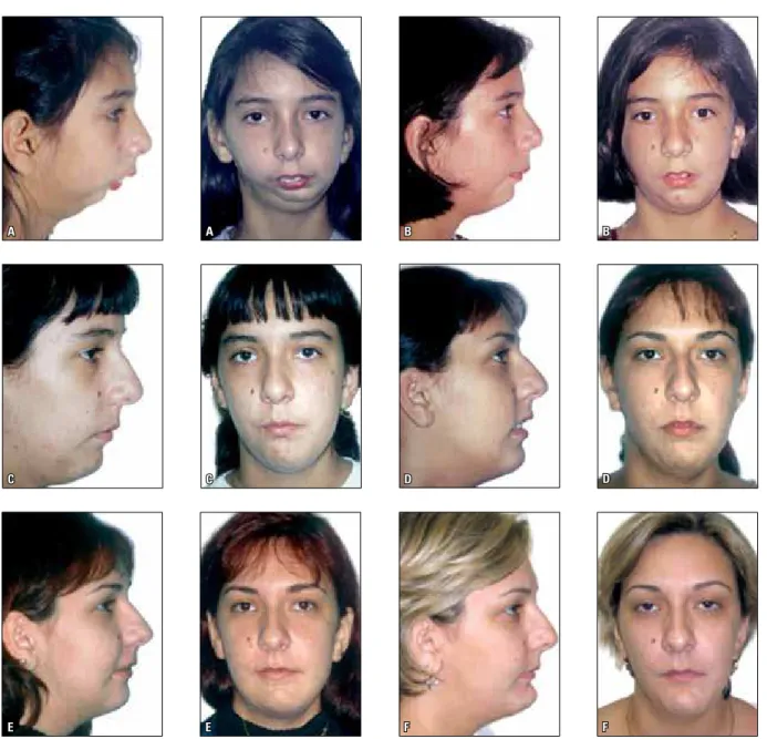
A female patient with severe facial deformity caused by condylar ankylosis.16 She presented with facial asymmetry and a deficient lower facial third, lip incompetence, limited mouth opening and man-dibular deviation during mouth closing (Fig 1A). There were functional problems both in speech and chewing. From a dental standpoint, there was impaction of the canine and second left premolar, agenesis of left upper first molar, widespread lack of space and unfavorable root inclinations.
When the patient was 11 years old a costo-chondral graft was placed between the mandibu-lar body and an ankylosed joint, in addition to an arthroplasty and a sagittal osteotomy to move the lower-right side anteriorly (Fig 1B). The man-dibular deviation was corrected, the retrognathic mandible was greatly reduced and the right side of the face showed a significant aesthetic gain. A 25 mm mouth opening and better lip positioning were achieved.
When she was 15 years old, four years after the first surgery, there was ankylosis relapse and graft resorption. A new arthroplasty was carried out and the lower left canine, which was impact-ed, was removed.
A B
D C
C
E E F F
D B A
E
Functional analysis showed mouth opening limi-tation, difficulty in swallowing and speaking, tongue protrusion, mandibular deviation to the left during mouth opening and left condylar an-kylosis with an abnormal mandibular ramus. One could also observe an increase in the lower third of the face, high smile line and lip incompetence.
Preoperative preparation required extraction of the first upper premolars and mandibular right first premolar. The mandibular first premolar sub-stituted the canine, which had previously been extracted. Combined maxillary and mandibular surgery was performed, substantially improving the patient’s facial aesthetics (Figs 1E, 1F and 3).
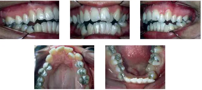
FIGURE 2 - Initial intraoral photographs.
case 2
A female patient aged 14 years, presenting with a skeletal Class III malocclusion, Class III dental relationship, mandibular excess interfer-ing with facial aesthetics, and maxillary atresia. The patient was still going through her growth period and reported extreme dissatisfaction
with her face, which negatively impacted on her normal social interaction (Fig 4).
The patient was forewarned of all surgical risks and about the need for a second surgery after the end of the growth period. It was de-cided then to perform a quick preoperative preparation (Fig 5) and vertical osteotomy to
FIGURE 4 - Initial facial and intraoral photographs.
FIGURE 6 - Facial and intraoral postoperative photographs.
achieve a 9 mm mandibular setback (Fig 6). The orthodontic appliance was removed after the surgery until it was time for the second treatment. The patient was monitored and at age 18 once again a skeletal Class III relation-ship, end-on bite and concave profile were ob-served (Fig 7).
FIGURE 7 - Facial and intraoral photographs at age 18 showing again a skeletal disharmony caused by postoperative growth.
FIGURE 9 - Initial facial and intraoral photographs. case 3
A female patient, aged 14 years with a dental and skeletal Class III relationship and concave profile. The patient had a maxillary deficiency in the trans-verse and anteroposterior direction, and mandibular excess in the anteroposterior direction (Fig 9).
The patient’s mother reported that the patient was very shy and was encountering serious psy-chosocial problems as a result of her face, which did not meet socially acceptable aesthetic stan-dard. The patient was informed about the future need of another surgical procedure due to the fact that she was still experiencing active growth.
The complexity index17 for the case, according
to the standards of the Brazilian Board of Ortho-dontics, was 61, significantly above 25, which is already considered a high degree of complexity.
FIGURE 10 - Facial and intraoral preoperative photographs.
case 4
A male patient, 13 years of age, dental and skeletal Class III relationship, presenting with maxillary atresia and mandibular anteroposterior excess, facial asymmetry and insufficient space for the eruption of impacted teeth (Fig 13).
A slightly more time-consuming preoperative preparation was carried out since it was necessary to create space and orthodontically erupt the
im-pacted teeth (Fig 14). Additionally, a combined surgery comprising a 7 mm maxillary advance-ment and 8 mm mandibular setback was per-formed (Figs 15 and 16).
The patient’s growth was monitored and by age 18 the dental relationship had been restored to a Class III condition, with incisors in an end-on relationship. Nevertheless, the patient reported that she was pleased with the outcome (Fig 17).
FIGURE 13 - Initial facial and intraoral photographs.
FIGURE 16 - Final facial photographs.
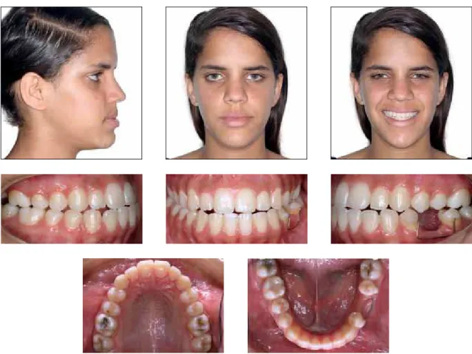
FIGURE 18 - Initial facial and intraoral photographs. case 5
A 14-year-old female patient presenting with a dental and skeletal Class III, anteropos-terior and vertical mandibular excess and max-illary atresia, as well as over-retained primary teeth (Fig 18). The complexity index17 for the
A B
B
A C
C FIGURE 19 - Final intraoral photographs.
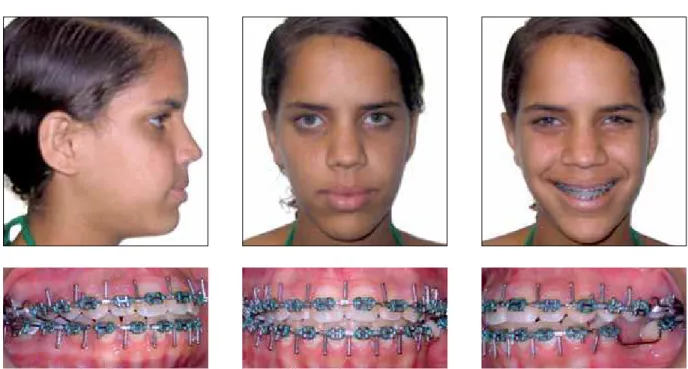
FIGURE 21 - Initial facial and intraoral photographs. case 6
A female patient, aged 12 and presenting with a Class III dental and skeletal pattern, concave pro-file with a sharply protruded mandible (Fig 21). The patient also presented with anterior and posterior crossbite, torsiversion of tooth #15, spaces in the lower arch and retroclined lower incisors. Maxillary expansion was performed with the aid of a Hyrax palatal separator and brief preoperative preparation, disregarding the closure of existing spaces (Fig 22).
The patient was subsequently subjected to maxillary advancement and mandibular setback orthognathic surgery. Two miniplates were then in-serted in the anterior region of the mandible as an-chorage for the closure of posterior spaces (Fig 23).
After space closure, the appliance and miniplates were removed, disclosing a rather significant aes-thetic and functional gain after these few months of treatment (Fig 24).
FIGURE 22 - Preoperative orthodontic preparation.
FIGURE 23 - Postoperative photographs showing anterior miniplates as an aid in closing spaces in the lower arch.
concLuSIonS
The patient’s type of facial deformity and growth vector should be carefully studied, as they will affect the surgical outcome. Patient and fam-ily must be aware of the expected results of early surgery as well as the risks and potential complica-tions of a non-traditional approach. Professionals should understand the uniqueness of each indi-vidual case and be aware that some patients with facial deformities need to be subjected to surgery during growth in order to ensure for such patients
function, aesthetics and a psychological attitude in line with their normal development. Difficulties in persuading these patients to undergo a second orthosurgical treatment have been observed. Most patients’ complaints are not sufficient to justify a second procedure. If, on the one hand, this may frustrate professionals when they realize that the case would indeed require a second procedure, on the other hand it becomes clear that early surgery was beneficial and added to the well being and sat-isfaction of the patient.
1. Broadbent BH Sr, Broadbent BH Jr, Golden WH. Bolton standards of dentofacial developmental growth. St. Louis: CV Mosby; 1975.
2. Van der Linden F. Facial growth and facial orthopedics. Surrey, UK: Quintessence; 1986.
3. Wolford LM, Karras SC, Mehra P. Considerations for orthognathic surgery during growth, Part I: Mandibular deformities. Am J Orthod Dentofacial Orthop. 2001;119(2):95-101.
4. Medeiros PJ, Medeiros PP. Cirurgia ortognática para o ortodontista. 2ª. São Paulo: Ed. Santos; 2004. 5. Reiche-Fischel O, Wolford LM. Changes in
temporomandibular joint dysfunction after orthognathic surgery. J Oral Maxillofac Surg. 1996;54:84.
6. Fuselier JC, Wolford LH, Pitta M, Talwar RM. Condylar changes after orthognathic surgery with untreated TMJ derangement. J Oral Maxillofac Surg. 1998; 56:61-62. 7. Profit WR, Mason RM. Myofunctional therapy for
tongue-thrusting: background and recommendations. J Am Dent Assoc. 1975;90:403-11.
8. Emrich RE, Brodie AG, Blayney JR. Prevalence of Class I, Class II and Class III malocclusions (Angle) in an urban population: an epidemiological study. J Dent Res. 1965; 44:947.
9. Huang CS, Ross RB. Surgical advancement of the retrognathic mandible in growing children. Am J Orthod. 1982;82:89-102.
10. Wolford LM, Cottrell DA. Diagnosis of macroglossia and indications for reduction glossectomy. Am J Orthod Dentofacial Orthop. 1996;110:70-7.
rEFErEncES
11. Bruce RA, Haywood JR. Condylar hyperplasia and mandibular asymmetry: a review. J Oral Surg. 1968;26:281-2.
12. Wolford LM, LeBanc J. Condylectomy to arrest disproportionate mandibular growth. Proceedings of the American Cleft Palate Association Annual Meeting; 1986 May 16-19; New York, NY. Chapel Hill (NC): American Cleft Palate Association;1986.
13. Wolford LM, Karras SC, Mehra P. Considerations for orthognathic surgery during growth, Part 2: Maxillary deformities. Am J Orthod Dentofacial Orthop. 2001;119(2):102-5.
14. Mogavero FJ, Buschang PH, Wolford LM. Orthognathic sugery effects on maxillary growth in patients with vertical maxillary excess. Am J Orthod Dentofacial Orthop. 1997; 111:288-96.
15. Cunningham SJ, Feinmann C. Psychological assessment of patients requesting orthognathic surgery and the relevance of body dysmorphic disorder. Br J Orthod. 1998;25(4):293-8. 16. Motta A, Louro RS, Medeiros PJ, Capelli J Jr. Orthodontic
and surgical treatment of a patient with an ankylosed temporomandibular joint. Am J Orthod Dentofacial Orthop. 2007;131(6):785-96.
17. Cangialosi TJ, Riolo ML, Owens SE Jr, Dykhouse VJ, Mofitt AH, Grubb JE, et al. The ABO discrepancy index: a measure of case complexity. Am J Orthod Dentofacial Orthop. 2004;125(3):270-8.
contact address
Jonas Capelli Junior
Av. 28 de Setembro, 157 - Vila Isabel
Zip code: 20.551-030 - Rio de Janeiro/RJ, Brazil E-mail: [email protected]