Article
Printed in Brazil - ©2014 Sociedade Brasileira de Química0103 - 5053 $6.00+0.00
A
*e-mail: [email protected]
ThermoFMN - A Thermofluor Assay Developed for Ligand-Screening as an
Alternative Strategy for Drug Discovery
Ricardo A. P. Pádua, Giovani P. Tomaleri, Renata A. G. Reis, Juliana S. David,
Valeria C. Silva, Matheus P. Pinheiro and Maria Cristina Nonato*
Laboratório de Cristalografia de Proteínas, Faculdade de Ciências Farmacêuticas de Ribeirão Preto, Universidade de São Paulo, Av. Café S/N, Monte Alegre, 14040-903 Ribeirão Preto-SP, Brazil
A técnica de Termofluor constitui uma importante ferramenta na identificação de moléculas protótipos a fármacos. No presente trabalho, foi desenvolvido um método alternativo para a técnica de Termofluor, chamado ThermoFMN, que explora o grupo prostético flavina mononucleotídeo (FMN) como sonda fluorescente. A validação do método foi feita através do monitoramento da fluorescência do FMN para diferentes alvos macromoleculares na presença de uma biblioteca aleatória de ligantes. Além disso, fármacos com eficácia comprovada tiveram seus perfis de inibição seletiva avaliado. Além de demonstrar que o rendimento quântico do FMN fornece intensidade adequada para detecção, nossos resultados revelam que o método de ThermoFMN utiliza-se de baixas concentrações de proteína e é compatível com uma vasta quantidade de tampões e aditivos químicos. A metodologia apresentada nesse trabalho propõe uma estratégia alternativa na busca por ligantes para proteínas dependentes de FMN, como uma importante ferramenta no desenvolvimento de novas terapias contra doenças negligenciadas.
Thermofluor has become a well-known and widely practiced methodologyfor screening of ligands that enhance stability and solubility of proteins, and also a powerful tool for hit identification in early drug discovery. In the present work, we developed an alternative Thermoflour method, named ThermoFMN, which explores the endogenous prosthetic group flavin mononucleotide (FMN) of flavoproteins as the fluorescent probe. Validation of ThermoFMN method was achieved by monitoring fluorescence signal of FMN of several drug targets in the presence of an unbiased library of ligands. In addition, drugs with known efficacy had their selective inhibition profile evaluated. Besides demonstrating that FMN signal provides sufficient fluorescence intensity for detection, our results revealed that ThermoFMN assay requires low concentration of protein samples and is compatible with a wide range of chemical reagents. The methodology presented here proposes an alternative strategy in the search for ligands of FMN-binding drug targets, therefore an important tool for the development of new therapies against neglected diseases.
Keywords: FMN, Thermofluor, drug targets, ThermoFMN, drug discovery, dihydroorotate
dehydrogenase
Introduction
The term neglected diseases refers to a group of tropical infections which used to be endemic to specific low-income populations in developing regions of Africa, Asia and the Americas. However, the latest statistical data available indicate that this reality has changed and many of those diseases have spread to new areas of the world, striking regions once thought safe, increasing the number of infected people and giving rise to greater genetic
diversity in terms of their related pathogens.1 This new
demographic distribution together with inadequate public health infrastructure and lack of investment from both public and private health sectors constitute large barriers to be overcame when searching for effective strategies to treat, control and eradicate this group of poverty-promoting
chronic infectious diseases.2
hampered a major role of the Academia in the process of drug development, and run contrary to the free-thinking basic research, thought and developed in the academic environment.
However, when it comes to neglected diseases, the research developed at the university, especially for those located in endemic countries like Brazil, is expected to contribute to change these devastating public health scenario.
For example, drug discovery for neglected diseases is carried out using both target-based and phenotypic
approaches.3 Target-based drug discovery has been largely
used in the pharmaceutical industry, but careful selection
of targets is still required.4 Unfortunately, there are still
very few fully validated drug targets in the vast field of neglected diseases, i.e., where there is a registered drug for which the elucidated mode of action depends on the inhibition/activation of the proposed target. Thus, academic concentrated efforts are still necessary for the identification, characterization and validation of new drug targets.
In addition, target-based drug discovery involves
screening libraries of compounds, in silico and in vitro,
and further optimization of lead compounds in order to achieve better potency, selectivity and pharmacological properties. As such, the development and/or improvement of techniques for target-based ligand screening are of extreme importance.
As an example, the fluorescence-based thermal stability assay (Thermofluor), originally developed by Pantoliano and coworkers in 2001, has nowadays become a
well-known and widely practiced methodologyfor screening
of ligands.5 Thermofluor monitors ligand effects on
temperature-dependent protein unfolding. Besides vastly used for ligand screening in modern drug discovery, by screening factors such as pH, buffer type, ionic strength and random solubilizing agents, Thermofluor also plays an important role in the identification of ideal chemical environment to enhance stability and solubility of proteins, allowing better conditions for sample manipulation,
characterization and storage.6,7
The probes commonly used in the Thermofluor assay give rise to a fluorescence signal when binding to exposed hydrophobic areas of unfolded proteins. Probes such as Nile red, 1,8-ANS, bis-ANS exploit hydrophobic and electrostatic interactions with proteins and display
characteristic excitation and emission spectra.8 In particular,
the most popular dye for thermal shift assays is SYPRO Orange with an excitation at 492 nm and fluorescence emission at 610 nm, after binding to protein hydrophobic patches.
A major limitation for the use of this methodology, due to the use of hydrophobic fluorophores, is their incompatibility with detergents, very needed to avoid both ligand and protein aggregation. Thus, alternative strategies have been developed to allow the use of Thermofluor techniques. For example, the screening of optimal buffers and detergents for purified membrane proteins can be
performed by using the thiol-specific fluorochrome N
-[4-(7-diethylamino-4-methyl-3-coumarinyl)phenyl]maleimide (CPM). This screen uses the chemical reactivity of the native cysteines embedded in the protein interior as a sensor
for the overall integrity of the folded state.9 In addition,
alternative dye-free methods have also been proposed to allow the use of thermoflour methodology. For example,
Warne et al. reported the development of microscale assay
to measure the thermal stability of a membrane protein
depending on 3H labeled ligand binding.10 Also, Forneris
et al. demonstrated that flavin adenine dinucleotide (FAD)
can be used as a probe to monitor protein unfolding.11
In the present work, we present an alternative dye-free method for Thermofluor (differential scanning fluorimetry), named ThermoFMN, which explores the endogenous prosthetic group FMN of flavoenzymes as a fluorescent probe. Flavoenzymes are involved in a wide range of biological processes including energy production, biodegradation, chromatin remodeling, DNA repair, apoptosis, protein folding, detoxification, neural development and biosynthesis and thus represent an
important group of potential drug targets.12
The efficacy of our method was validated by testing a wide range of ligands against potential macromolecular
targets for drug development including Leishmania major
dihydroorotate dehydrogenase (LmDHODH), Trypanosoma
cruzi dihydroorotate dehydrogenase (TcDHODH), all
targets for the development of trypanosomatid-related
diseases,13 and human dihydroorotate dehydrogenase
(HsDHODH), which besides used in cross-validation assays, also constitutes a validated target for the treatment
of proliferative, autoimmune and inflammatory diseases.14
Our results show that FMN quantum yield observed within the range of temperature tested and under a broad spectrum of chemical compounds provides sufficient fluorescence intensity to determine the melting temperatures of FMN-binding proteins. ThermoFMN assay proved to be a useful tool for screening of selective ligands that enhance stability and solubility of protein targets, as well as for hit identification in early drug discovery.
Experimental
Chemicals and materials
All buffers and salts used were HPLC grade and were acquired from J. T. Baker and Sigma. Solubility & Stability Screen was acquired from Hampton Research (HR2-072).
Protein Production
Leishmania major DHODH and Trypanosoma cruzi
DHODH have been expressed and purified as previously
described.15,16 For the cross-validation experiments, human
DHODH (HsDHODH) was subcloned into pET-28a expression vector (Novagen). A pet22-b construct encoding HsDHODH was kindly provided by Prof Jon Clardy from Harvard Medical School and used as a template.
HsDHODH was digested with NdeI/XhoI restriction
enzymes and the digestion product was purified using the QIAquick Gel Extraction Kit (Qiagen). The purified 1104 bp fragment was ligated into the pET-28a(+) vector, previously linearized with the same restriction enzymes to form a pET-28a(+)-HsDHODH construct. This
construction produces human DHODH as an N-terminal
and C-terminal six-histidine fusion protein. The ligated
mixture was first transformed into E. coli DH5α for plasmid
propagation. Plasmid DNA was extracted using Wizard Plus SV Minipreps (Promega). Sequence of the cloned gene was confirmed by DNA sequencing. The expression
plasmid was then transformed into E. coli BL21(DE3)
for protein production. Thus, a single colony of E. coli
BL21(DE3) cells harboring the pET-28a(+)-HsDHODH plasmid was inoculated into 20 mL Luria-Bertani broth
(LB) supplemented with 30 µg mL–1 kanamycin at 37 °C
overnight with shaking (250 rpm).
The overnight culture was then diluted to 1:100 with LB
containing 30 µg mL−1 kanamycin and grew at 37 °C till OD
600
reached 0.5-0.6. The cells were induced by the addition of
0.5 mmol L−1 isopropyl β-D-thiogalactopyranoside (IPTG)
for 6 h at 25 °C with shaking (250 rpm). Finally, the cells were harvested by centrifugation at 7,000 g for 10 min at 4 °C. A cell pellet from 2 L of culture was lysed in 15 mL
of lysis buffer (50 mmol L−1 sodium phosphate, pH 8,
300 mmol L−1 NaCl, 10 mmol L−1 imidazole, 1% Triton
X-100 and 1 mmol L−1 phenylmethylsulfonylfluoride
(PMSF)). The cells were further disrupted by sonication at 15 × 30 s bursts (with 30 s intervals between each burst) on ice. After 30 min of stirring at 4 °C, the insoluble debris were separated by centrifugation at 16,100 g for 30 min at 4 °C. The clear supernatant containing HsDHODH was loaded on a 1 mL column Ni-NTA agarose affinity resin
(Qiagen) equilibrated with 50 mmol L−1 sodium phosphate,
pH 8, buffer containing 300 mmol L−1 NaCl, 10 mmol L−1
imidazole and 0.1% Triton X-100. The column was then washed with a step gradient of imidazole from 10 to
50 mmol L−1 in equilibration buffer (50 mmol L−1 sodium
phosphate, pH 8, 300 mmol L−1 NaCl plus imidazole). The
washes consisted of 50 column volumes of equilibration
buffer in 10 mmol L−1 imidazole, followed by 50 column
volumes in 25 mmol L−1 imidazole, and 50 column
volumes in 50 mmol L−1 imidazole. The target protein was
eluted with 30 column volumes of the equilibration buffer
containing 250 mmol L−1 imidazole. The yield of pure
HsDHODH was typically 3.5 mg L−1 of culture.
Protein extinction coefficient
The concentration of the enzyme targets was estimated based on their molar extinction coefficient. Initially, 500 µL of pure protein samples were scanned within the visible region of the spectrum and the maximum absorbance
found around 450 nm recorded (Absmaxenz). The scanned
samples were then heated to 80 °C for 5 min followed by centrifugation at 16,100 g for 10 min. The yellow supernatant containing free FMN was separated from the insoluble fraction, scanned, and the maximum absorbance at 450 nm recorded. The enzyme extinction coefficient was determined considering equimolar concentrations of free and enzyme-bound FMN (equation 1). The FMN
extinction coefficient of εFMN = 12,500 mol
−1 Lcm–1 was
used as reference:
(1)
ThermoFMN assay
Protein melting temperature (Tm) determination was
performed by monitoring protein unfolding using FMN as the fluorescent probe. ThermoFMN monitors the fluorescent signal of FMN upon its releasing from the protein target as a function of temperature. ThermoFMN assay was
performed on AgilentMx3005P QPCR System equipped
with a FAM SYBr green I filter with excitation and emission wavelengths of 492 nm and 516 nm, respectively.
For the thermal stability assay each condition tested
required an aliquot of 10 µL from 10 µmol L−1 stock of
purified protein mixed with 10 µL of different screening solutions in 96-well plates sealed with optical quality
sealing tape (Microseal® ‘B’ seal from BIO-RAD). Plates
denaturation the plates were heated from 25 to 95 °C with stepwise increments of 1 °C and a 1 min hold step for every point, followed by the fluorescence reading.
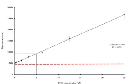
Considering that the experimental volume is fixed, two parameters may influence the signal strength: the FMN concentration and the mechanism of interaction between protein and FMN. Thus, in order to optimize the fluorescence signal-to-noise ratio, an initial assay was performed by monitoring the fluorescence signal
at different concentrations of free FMN (25 µmol L−1,
12.5 µmol L−1, 6.25 µmol L−1, 3.125 µmol L−1,
1.5625 µmol L−1, 0.78125 µmol L−1, 0.290625 µmol L−1
and 0.195325 µmol L−1). The reference experiment for
determining the size of the noise was performed by monitoring the fluorescence signal of water-containing wells. A signal-to-noise ratio of two was obtained using
5 µmol L−1 FMN final concentration (Figure S1). This
concentration was then used as reference for TcDHODH, LmDHODH and HsDHODH protein samples, for the remaining of this work.
Thus, previously to ThermoFMN experiment, TcDHODH, LmDHODH and HsDHODH were dialyzed
against 50 mmol L−1 HEPES pH 7.2, 150 mmol L−1
NaCl and concentrated to 10 µmol L−1 based on the
molar extinction coefficient. The commercially available Solubility & Stability Screen (Hampton Research) was used for evaluating the effects of different buffer and chemical additives on both quantum yield and protein melting temperatures during ThermoFMN assay. The control experiment was prepared by adding the protein buffer instead of the additive compound. In addition, the effects of known DHODH inhibitors (A77 1726, brequinar)
on protein unfolding were tested at 40 µmol L−1 final
concentration. The control experiment was prepared by adding DMSO instead of the ligand.
The fluorescence data were processed according to the
procedure described by Niesen et al..17
Results and Discussion
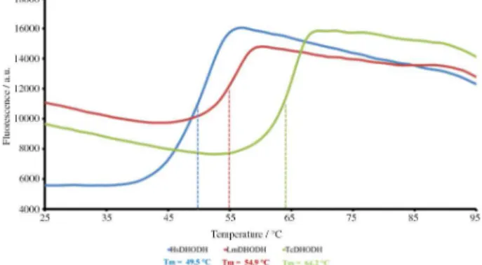
In the present work we developed the ThermoFMN assay, a methodology dedicated to evaluate ligand effects on thermostability of FMN-binding proteins. By monitoring the environment-sensitive fluorescence intensity of the cofactor flavin mononucleotide (FMN) as a function of temperature, ThermoFMN enables the identification of ligands that by interacting with the protein molecules lead to changes in protein thermostability.
Since the fluorescence of FMN cofactor is quenched by the protein environment when the protein is properly folded, the increase in FMN signal in response to temperature
elevation can be directly correlated to protein unfolding and consequent cofactor release. The melting curve,
i.e., fluorescence intensity versus temperature, usually
displays a sigmoid-like shape and changes in slope and
melting temperature (Tm), temperature where the protein
is 50% unfolded, are used to provide valuable information regarding the influence of chemical environment on structural order and conformational flexibility of FMN-binding proteins.
The proposed methodology was evaluated by using the enzyme dihydroorotate dehydrogenase (DHODH) as model. DHODH is a FMN-dependent enzyme that catalyzes the oxidation of S-dihydroorotate to orotate in the fourth
and only redox step in de novo biosynthesis of pyrimidine
nucleotides.18 Inhibition of dihydroorotate dehydrogenase
has been considered an attractive therapeutic approach to
cancer, autoimmune and parasitic diseases.13,19,20 DHODH
monomer shows a typical α/β barrel fold of with a central
barrel composed of eight parallel β strands surrounded
by eight α-helices. On the basis of sequence similarity,
subcellular location as well as their preferences for oxidizing substrates, the DHODHs are divided in two major
classes, named class 1 and class 2.21 Class 1 and class 2
are structurally and mechanistically distinct, features that allow the design of class-selective inhibitors.
In the present work, human DHODH and the homologous
enzymes from the protozoa parasites Leishmania major and
Trypanosoma cruzi were screened against a random set of
chemical factors including pH, buffer type and chemical additives. LmDHODH and TcDHODH are cytosolic enzymes, dimeric members of class 1 DHODHs and have been extensively exploited as potential drug targets against Leishmaniasis and Chagas diseases, infections found
among the most neglected diseases.13,16,18,19,22,23 Different
to the parasitic enzymes, the human DHODH homologue, used for cross-validation assays, is a membrane-anchored mitochondrial enzyme, monomeric and member of class
2 DHODHs. In addition to the α/β barrel, HsDHODH
presents a N-terminal extension of approximately 40
residues that folds into two α-helices connected by a short
loop and plays a role in membrane anchoring.24,25
Previously to the ThermoFMN assay, the value of the fluorescence signal-to-noise ratio for sensitivity of FMN detection was estimated using free FMN as reference (Figure S1). A signal-to-noise ratio of two could be found
for concentrations around 5 µmol L−1.
In fact, protein concentration of 5 µmol L−1 was
signal still depends on the quantum efficiency of the FMN itself as well as on its chemical environment and solvent accessibility. Therefore, protein concentration should be considered an important parameter to be optimized during ThermoFMN assay.
The characteristic molar extinction coefficients (ε)
for each protein sample were determined and protein concentration was then estimated monitoring the absorbance of protein-bound FMN as described by equation 1. As a result of different chemical environments, the height and the position of the maximum absorption peak was found to
differ among DHODHs: εLmDHODH = 11,722 mol−1 L cm−1 at
458 nm for LmDHODH, εTcDHODH = 12,333 mol−1 Lcm−1 at
460 nm for TcDHODH and εHsDHODH = 14,259 mol−1 L cm−1
at 453 nm for HsDHODH (Figure 1).
Although DHODHs were, in principle, used only as models for the development of our assay, the results obtained with ThermoFMN have provided valuable information on the characterization and screening of selective ligands for our protein targets. The single sigmoid-like curves obtained for DHODHs indicate a single melting transition state and the a sigmoidal fit allowed the determination of the characteristic melting temperatures
of 49.5 °C for HsDHODH, 54.9°C for LmDHODH and
64.2 °C for TcDHODH (Figure 2).
Structural comparison among DHODHs reveals that FMN is found non-covalently bound to the protein molecule and the short-range chemical environment of the flavin moiety is highly conserved. In fact, the prosthetic FMN group is located on the top of the barrel and stabilized by a set of conserved hydrogen bonds that involves the
residues Ala20, Lys44 (replaced by Gly in HsDHODH),
Ser45, Asn128, Lys165, Val194 (replaced by Ile in TcDHODH
and Thr in LmDHODH), Asn195, Gly223, Gly251, Gly272 and
Thr274 (Figure 3 and Figure S2). In addition, LmDHODH
and TcDHODH are profoundly similar, sharing 75% of sequence identity (Figure S2) with a very similar tendency
for amino acidcomposition and displaying on average a
rmsd of 0.61 Å when superposing the Ca atoms (PDB code
3GYE and PDB code 3C3N).16,22 Our analysis, together
with the fact that the characteristic melting curves for LmDHODH, TcDHODH and HsDHODH display large differences in terms of absolute fluorescence, melting temperature and slope (Figure 2) indicate that differences in long-range chemical environment significantly govern the fluorescence profile of a FMN binding protein.
Unfortunately, even after very careful inspection of chemical and stereo properties of LmDHODH and TcDHODH tertiary structures, we could not provide structural basis to explain the ten degrees difference in
Tm found when comparing LmDHODH and TcDHODH
melting curves. Site-direct mutagenesis experiments are currently under way and will be extensively used to map
Figure 1. UV/Visabsorption spectra for HsDHODH, LmDHODH and
TcDHODH (25 µmol L−1). Chemical environment of FMN affects the peak of maximum absorbance found to be 458 nm for LmDHODH, 460 nm for TcDHODH and 453 nm for HsDHODH.
Figure 2. Melting curves for HsDHODH, TcDHODH and LmDHODH .
The sigmoidal fitting against the data was used to determine the melting temperature found as 49.5 °C, 54.9°C and 64.2 °C for HsDHODH, LmDHODH and TcDHODH, respectively.
Figure 3. Stereoscopic three-dimensional representation of the FMN
the network of chemical interactions that contribute to enhance protein thermostability.
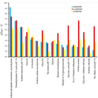
Stability and solubility screen were evaluated for HsDHODH, LmDHODH and TcDHODH. DHODH denaturation curves were analyzed and fitted to a sigmoid to determine the protein melting temperature for each assay. ThermoFMN proved to be compatible with the vast majority of the 96 conditions tested, including the presence of detergents, salts, chaotropic agents, known to help stabilizing and/or to reduce protein aggregation. Nevertheless, special attention should be given to acidic compounds, high concentration of reducing agents and bromide salts that showed to quench the fluorescence signal of FMN and should then be avoided.
Analysis of ThermoFMN data in terms of melting temperature and the protein-unfolding transient slope reveals that same set of seventeen compounds were found to be the best stabilizing agents for all three DHODHs tested (Figure 4): sodium phosphate monobasic monohydrate, sodium sulfate decahydrate, sodium chloride, DL-maleic acid pH 7.0, sucrose, D-sorbitol, xylitol, betaine
monohydrate, methyl-α-D-glucopyranoside,
D-(+)-trehalose dehydrate, sodium malonate, tacsimate, succinic
acid, triethylamine N-oxide dehydrate, choline acetate and
glycerol. Not surprisingly, the great majority of compounds identified fit into one of two general categories: salts and organic osmolytes, natural molecules well known for
protecting cells against adverse conditions.26
In general, organic osmolytes can uniformly affect all
proteins by interacting with the peptide backbone.27 The
ability of osmolytes to protect protein folding is due to their solvophobic effect on the peptide backbone; i.e., by disfavoring contact with the backbone, shielding it from the solvent.28
Salts have long been known to alter protein solubility.29
However, the beneficial effects of salts depend on protein-specific surface charge distribution, type and concentration of the salts. At low concentrations, salts can stabilize proteins through nonspecific electrostatic interactions, dependent only on the ionic strength of the medium. At high concentrations, however, salts can exert different effects on proteins by shielding the protein with multi-ions charges. Those charges can help protein molecules interact, aggregate, and even precipitate.
In addition to shielding effects, we should not rule out the possibility that shift in melting temperatures are due to direct ligand binding to protein pockets, as already observed in the crystal structures of DHODHs in complex
with glycerol,14,16,22 sulfate,16 and succinate molecules.30
In particular, the significant improvement of thermal stability observed for malonate, malate and succinate
molecules deserves special attention due to their relative similarity with fumarate, the natural substrate for class 1A DHODHs. Kinetic and structural studies of the DHODH protein targets in presence of these substrate analogues are currently under way. Our results will contribute not only to elucidate the mechanism of action of DHODHs, but also in the identification of new chemical entities that bind to DHODH and can be used to the development of drug prototypes.
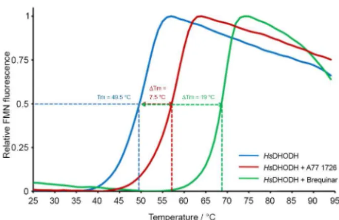
As a prove of concept, the antiproliferative agents
brequinar and teriflunomide (A771726),14,31,32 both known
selective inhibitors of class 2 DHODHs, had their inhibition profile evaluated by ThermoFMN assay (Figure 5). Brequinar was shown to be one of the strongest inhibitor
of human DHODH with Ki values in the 5-8 nmol L−1
range, however failed in clinical trials as anticancer and
immunosuppressant agent.33 A771726, the active metabolite
of leflunomide (Arava), also a potent inhibitor of DHODH
with an Ki value of 19 nmol L−1 is currently used in
medicine to treat autoimmune diseases such as rheumatoid arthritis or multiple sclerosis and have been investigated
in treatments of cancer, virus, and parasite infections.33
Our results showed that activity in the nano molar range of brequinar and A77 1726 against HsDHODH reflected on its increased thermostability. In fact, a large positive
shift in the melting temperature of ∆Tmof7.5 °C and 19
°C for A77 1726 and brequinar, respectively, could be observed for HsDHODH (Figure 5). On the contrary, as expected, no change in either slope or melting temperature Figure 4. Shift in melting temperatures (∆Tm) in the presence of the buffers
could be observed for LmDHODH and TcDHODH, data that correlate with the reported kinetic values. In addition, the correlation between binding constants and protein
thermostability has been previously reported.34-37 For
example, the correlation between ligand binding constant
(Kd) and the Tm has been stablished,34,35 where Kd has
been calculated using Thermofluor assays by varying the ligand concentration and using the constant of protein
unfolding and protein concentration.36,37 Additionally, the
Kd determination of tightly bound ligands (< 10 nM), which is poorly achieved by isothermal titration calorimetry (ITC), have already been precisely resolved by thermal
shift assays.36 The characterization of enzyme inhibition
mechanisms has also been accomplished by comparing the thermal shift of proteins in the presence of substrate
with varying concentrations of inhibitors of interest.38
Furthermore, insights on ligand potency and selectivity can
also be obtained.35 This arises since protein stabilization
can occur through ligand binding at a site not linked to enzyme activity.
Altogether, those results support the idea that this reference methodology is not only effective on identifying ligands but can also provide insights on their potency and selectivity, features that are crucial for cross-validation assays.
Conclusion
FMN-containing and FAD-containing enzymes correspond to the large group of flavoproteins found in archaea, bacteria, and eukaryotes and involved in the
regulation of many fundamental cellular processes.39
Thus, enzymes that utilize the redox-active isoalloxazine ring system (FMN and FAD) as coenzymes constitute an important group of proteins to be characterized and
explored as drug targets; knowledge that could result in new strategies to tackle chronic diseases.
In this study we described the development of ThermoFMN assay, an alternative dye-free methodology
for the conventional Thermofluor technique.5 ThermoFMN
exploits the presence of FMN flavin group as the fluorescence probe that can be used for monitoring effects of ligand binding on unfolding temperature of FMN-binding proteins. The thermal information gained by investigation of the protein stability under different buffer compositions can be effectively used to guide purification, biophysical characterization and identification of ligands to be used as prototypes for drug development.
ThermoFMN assay supports the use of a single filter pair optimized for excitation and emission of SYBR Green I (with excitation maxima at 492 and emission spectra at 516 nm, respectively), and display excellent sensitivity with minimum amounts of protein samples.
ThermoFMN assay was evaluated against the enzymes
dihydroorotate dehydrogenase from Leishmania major,
Trypanosoma cruzi and Homo sapiens. The characteristic
melting curves for LmDHODH, TcDHODH and HsDHODH showed to be highly reproducible and the monitoring of
melting temperatures (∆Tm) against a random set of buffers
and additives demonstrated a large compatibility with the majority of the chemical compounds tested.
In addition, our assays against the antiproliferative
agents, brequinar and teriflunomide,14 known DHODH
inhibitors, demonstrated that ThermoFMN assay can also be used to determine ligand selectivity, very useful for cross-validation assays.
In summary, the low cost, fast, simplicity, low sample-consuming and the general applicability of ThermoFMN, make this technique attractive for identification of positive hits of FMN-binding proteins and constitute a new tool to be extensively used in the process of development of new therapies against neglected diseases.
Supplementary Information
Supplementary data are available free of charge at http:// jbcs.sbq.org.br as PDF file.
Acknowledgments
We would like to thank Prof Jon Clardy from Harvard Medical School for providing us with the DNA construct used to subclone human DHODH.
Funding was provided by FAPESP (Fundação de Amparo à Pesquisa do Estado de São Paulo), grants 2012/25075-0 (MCN), 2008/11644-8 (RAPP), 2011/14269-6 (GPT), Figure 5. Effect of antiproliferative agents on thermal stability of human
2011/23504-9 (RGR), 2007/08703-0 (MPP) and CNPq (Conselho Nacional de Pesquisa), grant 159061/2012-1 (VCS) .
References
1. d’Alessandro, E. D. L.; Giraldi, G.; Clin. Ter. 2011, 162, E93. 2. Marchal, B.; Van Dormael, M.; Pirard, M.; Cavalli, A.;
Kegels, G.; Polman, K.; Acta Trop. 2011, 120, S177. 3. Gilbert, I. H.; J. Med. Chem. 2013, 56, 7719.
4. Renslo, A. R.; McKerrow, J. H.; Nat. Chem. Biol. 2006, 2, 701. 5. Pantoliano, M. W.; Petrella, E. C.; Kwasnoski, J. D.; Lobanov,
V. S.; Myslik, J.; Graf, E.; Carver, T.; Asel, E.; Springer, B. A.; Lane, P.; Salemme, F. R.; J. Biomol. Screening 2001, 6, 429.
6. Seabrook, S. A.; Newman, J.; ACS Comb. Sci. 2013, 15, 387. 7. Shi, S.; Semple, A.; Cheung, J.; Shameem, M.; J. Pharm. Sci.
2013, 102, 2471; Reinhard, L.; Mayerhofer, H.; Geerlof, A.; Mueller-Dieckmann, J.; Weiss, M. S.; Acta Crystallogr., Sect. F: Struct. Biol. Cryst. Commun. 2013, 69, 209.
8. Ruvinov, S. B.; Yang, X. J.; Parris, K. D.; Banik, U.; Ahmed, S. A.; Miles, E. W.; Sackett, D. L.; J. Biol. Chem. 1995, 270, 6357.
9. Alexandrov, A. I.; Mileni, M.; Chien, E. Y. T.; Hanson, M. A.; Stevens, R. C.; Structure 2008, 16, 351.
10. Warne, T.; Serrano-Vega, M. J.; Tate, C. G.; Schertler, G. F. X.;
Protein Expres. Purifi. 2009, 65, 204.
11. Forneris, F.; Orru, R.; Bonivento, D.; Chiarelli, L. R.; Mattevi, A.; FEBS J. 2009, 276, 2833.
12. Joosten, V.; van Berkel, W. J. H.; Curr. Opin. Chem. Biol. 2007,
11, 195.
13. Pinheiro, M. P.; Emery, F. S.; Nonato, M. C.; Curr. Pharm. Des.
2013, 19, 2615.
14. Liu, S. P.; Neidhardt, E. A.; Grossman, T. H.; Ocain, T.; Clardy, J.; Structure Fold Des.. 2000, 8, 25.
15. Feliciano, P. R.; Cordeiro, A. T.; Costa, A. J.; Nonato, M. C.;
Protein Expression Purif. 2006, 48, 98.
16. Pinheiro, M. P.; Iulek, J.; Nonato, M. C.; Biochem. Biophys. Res. Commun. 2008, 369, 812.
17. Niesen, F. H.; Berglund, H.; Vedadi, M.; Nat. Protoc. 2007, 2, 2212.
18. Nonato, M. C.; Costa-Filho, A. J. In The Dihydroorotate Dehydrogenases; Hille, R.; Miller, S.; Palfey, B., eds.; de
Gruyter: Berlin, Germany, 2013, ch. 14.
19. Vyas, V. K.; Ghate, M.; Mini-Rev. Med. Chem. 2011, 11, 1039.
20. Baumann, P.; Mandl-Weber, S.; Voelkl, A.; Adam, C.; Bumeder, I.; Oduncu, F.; Schmidmaier, R.; Mol. Cancer Ther.
2009, 8, 366.
21. Jones, M. E.; Annu. Rev. Biochem. 1980, 49, 253.
22. Cordeiro, A. T.; Feliciano, P. R.; Pinheiro, M. P.; Nonato, M. C.;
Biochimie 2012, 94, 1739.
23. Cheleski, J.; Rocha, J. R.; Pinheiro, M. P.; Wiggers, H. J.; da Silva, A. B. F.; Nonato, M. C.; Montanari, C. A.; Eur. J. Med. Chem. 2010, 45, 5899.
24. Couto, S. G.; Nonato, M. C.; Costa-Filho, A. J.; Biochem. Biophys. Res. Commun. 2011, 414, 487.
25. Couto, S. G.; Nonato, M. C.; Costa-Filho, A. J.; Biophys. J.
2008, 94, 1746.
26. Harries, D.; Rosgen, J. In Biophysical Tools for Biologists; Correia, J. J.; Detrich, H. W.,eds.; Elsevier: Oxford, UK,2008, ch. 22.
27. Bolen, D. W.; Baskakov, I. V.; J. Mol. Biol. 2001, 310, 955.
28. Ishibashi, M.; Sakashita, K.; Tokunaga, H.; Arakawa, T.; Tokunaga, M.; J. Protein Chem. 2003, 22, 345.
29. Hamada, H.; Arakawa, T.; Shiraki, K.; Curr. Pharm. Biotechnol.
2009, 10, 400.
30. Inaoka, D. K.; Sakamoto, K.; Shimizu, H.; Shiba, T.; Kurisu, G.; Nara, T.; Aoki, T.; Kita, K.; Harada, S.; Biochemistry 2008, 47,
10881.
31. Chen, S. F.; Perrella, F. W.; Behrens, D. L.; Papp, L. M.; Cancer Res. 1992, 52, 3521.
32. Davis, J. P.; Cain, G. A.; Pitts, W. J.; Magolda, R. L.; Copeland, R. A.; Biochemistry 1996, 35, 1270.
33. Munier-Lehmann, H.; Vidalain, P.-O.; Tangy, F.; Janin, Y. L.;
J. Med. Chem. 2013, 56, 3148.
34. Celej, M. S.; Montich, C. G.; Fidelio, G. D.; Protein Sci. 2003,
12, 1496.
35. Bullock, A. N.; Debreczeni, J. E.; Fedorov, O. Y.; Nelson, A.; Marsden, B. D.; Knapp, S.; J. Med. Chem. 2005, 48, 7604. 36. Zubriene, A.; Matuliene, J.; Baranauskiene, L.; Jachno, J.;
Torresan, J.; Michailoviene, V.; Cimmperman, P.; Matulis, D.;
Int. J. Mol. Sci. 2009, 10, 2662.
37. Matulis, D.; Kranz, J. K.; Salemme, F. R.; Todd, M. J.;
Biochemistry 2005, 44, 5258.
38. Lea, W. A.; Simeonov, A.; PLoS One 2012, 7, (4): e36219. 39. Macheroux, P.; Kappes, B.; Ealick, S. E.; Febs J. 2011, 278,
2625.
Submitted on: March 20, 2014 Published online: July 1, 2014
Supplementary Information
0103 - 5053 $6.00+0.00S
I
*e-mail: [email protected]
ThermoFMN - A Thermofluor Assay Developed for Ligand-Screening as an
Alternative Strategy for Drug Discovery
Ricardo A. P. Pádua, Giovani P. Tomaleri, Renata A. G. Reis, Juliana S. David,
Valeria C. Silva, Matheus P. Pinheiro and Maria Cristina Nonato*
Laboratório de Cristalografia de Proteínas, Faculdade de Ciências Farmacêuticas de Ribeirão Preto, Universidade de São Paulo, Av. Café S/N, Monte Alegre, 14040-903 Ribeirão Preto-SP, Brazil
Figure S1. Fluorescence intensity versus FMN concentration. Data was fit to the equation y = kϕI0εbx,where the intensity of emitted light, y, is described
by the relationship where ϕ is the quantum efficiency, I0 is the incident radiant power, ε is the molar absorptivity, b is the path length of the cell, and x
is the FMN molar concentration.1 The level of background noise of was estimated in 4960 a.u. close to the measured fluorescence signal of empty wells
Figure S2. Structure-based sequence alignment of selected DHODHs proteins: LmDHODH (PDB ID 3GYE), TcDHODH (PDB ID 3C3N), and HsDHODH
(PDB ID 1D3G). Residue numbering is based on LmDHODH and HsDHODH. Residues that are fully conserved are highlighted in dark gray box. Residues highly, little or non-conserved are colored in white, light gray and black, respectively. Secondary structures of LmDHODH (top) and HsDHODH (bottom) are also labeled. Residues that directly bind orotate, FMN, and both, are indicated by cyan filled circules, lime green triangles, and red stars, respectively. The structural alignment was performed using COOT and graphically displayed using ESPript.2,3
References
1. Guilbault, G. G.; Practical Fluorescence, vol. 3, 2nd ed.; CRC Press: New York, USA, 1990.