165
REV. HOSP. CLÍN. FAC. MED. S.PAULO 54(5):165-168, 1999 SEPTEMBER-OCTOBER
From the Department of Clinical Medicine, University of São Paulo School of Medicine, São Paulo - Brazil.
PRIMARY BILIARY CIRRHOSIS AND MYOPATHY: AN
UNCOMMON ASSOCIATION
Bruno Cupertino Migueletto, Abrahão Elias Hallack Neto, Elaine Zamora Domingues, Pedro Paulo Neves de Castro, Hartmut Stocker, Sueli K. Nagahashi Marie, Luiz Pedro Meireles and Milton Hideaki Arai
RHCFAP/ 2985
MIGUELETTO, B. C. et al. - Primary biliary cirrhosis and myopathy: an uncommum association. Rev. Hosp. Clín. Fac. Med. S. Paulo 54 (5):165-168, 1999.
SUMMARY: Primary biliary cirrhosis (PBC) is a cholestatic liver disease, which is characterized by a chronic inflammatory destruction of intrahepatic bile ducts. It is a rare disorder whose precise etiology is still to be elucidated. Even though the liver is the principal target of PBC, other organ systems also might be affected. Muscular involvement has rarely been described in this disease, and in the majority of cases, muscular weakness has been interpreted as polymyositis. We report the case of a 48-year-old woman suffering from classic PBC, in association with a myopathy whose histological features are distinct from the cases reported before. We also performed a MEDLINE research for PBC and concomitant muscular diseases.
DESCRIPTORS: Primary biliary cirrhosis. Myopathy.
Primary biliary cirrhosis (PBC) is a rare disease, characterized by chronic cholestasis. Its etiology is still not entirely understood, but clinical, laboratory and histopathologic findings strongly suggest an autoimmune pro-cess. Histologically the disorder is characterized by an inflammatory process that leads to the destruction of intrahepatic bile ducts and results in fibrosis of the portal tracts and finally in hepatic failure. Up to 84% of patients with PBC also suffer from at least one more disorder classified as auto immune, with Sjogren Syndrome, auto immune thyroiditis, and systemic sclerosis being the most frequent ones. Besides the liver, which is the principal target of PBC, other organs might also be affected. However, muscular damage has rarely been reported in patients with PBC, and in these cases the nature of the myopathy has been poorly characterized.
We report the case of a patient with histologically proven PBC with associated muscular disease. We
de-scribe the histologic features of this myopathy.
A MEDLINE research yielded twenty case reports on PBC in asso-ciation with muscular involvement, which was generally found to be polymyositis.
CASE REPORT
Our patient, a 48-year-old white woman, sought medical assistance be-cause of proximal muscular weakness of upper and lower extremities that had started four years earlier. She also com-plained of generalized prurigo as well as darkening of her skin, principally of her trunk and forearms, and hypertri-chosis of her arms, which had been troubling her for two years. Eight years
earlier, she had undergone total hyster-ectomy and bilateral oophorhyster-ectomy because of ovarian cysts. Estrogen re-placement was initiated and maintained ever since. In 1995 osteoporosis was diagnosed.
The patient reported that she did not drink alcohol on a regular basis and that she had not received any trans-fusions.
On physical examination she did not show any stigmas of chronic liver disease. She had no fever, she did not have jaundice, and her liver was normal in size. Muscular strength was grade 4 in proximal muscles of upper and lower extremities. There were no further alterations on physical exam.
166
REV. HOSP. CLÍN. FAC. MED. S.PAULO 54(5):165-168, 1999 SEPTEMBER-OCTOBER
dl, (952-1538); IgA 703 mg/dl (153-359) IgM 617 mg/dl (73-171).
Erythrocyte sedimentation rate was 78 mm.
Immunofluorescence was positive showing a granular cytoplasmic pattern (1:160).
Anti-DNA, SSA, SSB, SM, and RNP autoantibodies were absent. Anti-smooth muscle and anti-liver-kidney antibodies could not be detected.
Serum complement was normal. Serologic testing for viral hepatitis was negative.
Total serum bilirubin and fractions were within normal range. There were no alterations in coagulation tests.
Serum albumine was 4.4 mg/dl TSH was 0.9 um/L (0.25-4.0). Antimitochondrial antibodies (AMA) were present (titre>1:320).
ECG showed sinus rhythm with frequent atrial and ventricular pre-mature complexes.
Chest X-ray was normal.
Bone densitometry of the lumbar spine and of the neck of femur confir-med the diagnosis of osteoporosis. (T index lumbar spine:-3.83 DP/ T index neck of femur -4.06 DP)
On abdominal ultrasonography, the patient’s liver was found to be homo-geneously enlarged without evidence of congestion of the bile tree.
Liver biopsy (Microphotograph 1) showed moderate fibrous expansion of the portal tracts with preservation of liver architecture. Portal tracts were densely infiltrated with lymphocytes a few plasmocytes, and a significant number of neutrophils, with focal invasion of the portal tracts. Prolife-ration of bile ductules was evident in the periphery of portal tracts, and there was discreet lobular inflammatory activity.
The histopathologic findings were compatible with chronic hepatobiliary disease in moderate activity, suggesting primary biliary cirrhosis.
Focussing on the patient’s
muscu-Fig. 1 - Liver biopsy: proliferation of bile ductules infiltrated with lymphocytes.
167
REV. HOSP. CLÍN. FAC. MED. S.PAULO 54(5):165-168, 1999 SEPTEMBER-OCTOBER
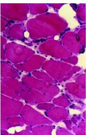
lar weakness, we had an electromyo-graphy performed, which revealed chronic myopathy affecting all extre-mities with a clear proximal distri-bution pattern. The findings were com-patible with polymyositis. A biopsy specimen (microphotograph 2) was ob-tained from the left brachial biceps. On examination we found accentuated variation of muscle fiber size with the presence of ragged, rounded, degener-ating fibers, as well basophilic necrotic fibers with subsarcolemmal vacuoles. Angular fibers were present with nuclei condensed into aggregates, a phenom-enon known as nuclear clumps. We also noted fibers with centrally located nuclei. There was proliferation of con-nective tissue both in endomysium and perimysium.
When stained for oxidative reac-tions, some fibers showed disarrange-ment. Occasional fibers showed in-creased granulation in the SDH stain that indicated mitochondrial prolifera-tion. Numerous fibers were identified by the acid phosphatase reaction, indi-cating lysosomal activity. A few regen-erating fibers reacted positively when stained with the alkaline phophatase reaction. We noted a slight predomi-nance of type one fibers.
The final diagnosis was PBC with muscular involvement.
We initiated treatment with cho-lestyramine 4.0 g, p.o. three times a day, and ursodeoxycholic acid 300 mg, p.o. b.i.d., as well as calcium car-bonate, vitamin D, and alendronate for osteoporosis. The patient was dis-charged from hospital and is conti-nuing treatment on an outpatient basis.
DISCUSSION
PBC is a rare, progressive, cho-lestatic liver disease of unknown etiology. Morphologically it is charac-terized by the destruction of intra-hepatic bile ductules. PBC is frequently
associated with other autoimmune disorders. The most frequent ones are Sjogren syndrome, rheumatoid ar-thritis, and systemic sclerosis (CREST syndrome).
In this article we present the case of a 48-year-old patient with PBC and a coexisting myopathy. The diagnosis of PBC was based on clinical data, on elevated levels of serum alkaline phosphatase, gGT, and IgM, as well as the presence of circulating antimi-tochondrial antibodies (AMA). The diagnosis was histologically confirmed by liver biopsy.
Concerning the patient’s muscular weakness, we found elevated creatine kinase, alterations in EMG, and finally a highly pathologic muscular histology. Muscular disease in patients with PBC has been poorly defined. There are few articles in medical literature that deal with this subject. In 1993 Varga and co-workers described two cases of asymptomatic PBC with a syndrome of severe progressive myo-pathy, cardiomyomyo-pathy, and gastro-intestinal dysmotility. They also re-viewed literature focussing on this rare association. They found most of the previous reports describing focal mononuclear inflammatory infiltrates generally localized in the perivascular spaces, in addition to other alterations compatible with inflammatory myopa-thy.
In our case, there was little perivas-cular and endomysial inflammation except for rare fibers with mito-chondrial proliferation. These findings are symptomatic of a mixture of neurogenic and primary myogenic muscular disorder.
In spite of the patient’s proximal muscular weakness, elevated levels of creatine kinase, and the results of EMG that all suggested polymyositis, mus-cular biopsy did not reveal any charac-teristics of active inflammatory muscu-lar disease.
The findings reveal a chronic
process that is compatible with the long duration of the muscular disease. Since our patient underwent biopsy four years after the onset of her muscular disease, we must consider the possibi-lity of a different histologic pattern at the beginning of the disease. On possibility includes a more distinct inflammatory pattern.
The coexistence of PBC and mus-cular disease raises the possibility of a common pathogenic mechanism for both processes. PBC is associated with various immunologic abnormalities. Patients typically have elevated levels of IgM, circulating immune com-plexes, and 90% of patients are positive for antimitochondrial antibodies.
Antimitochondrial antibodies may also be detected in patients with diffuse muscle weakness and mitochondrial abnormalities. In these cases, though, the specificity of these antibodies has not been defined yet, and the signifi-cance of this finding in diffuse muscle weakness and mitochondrial abnor-malities is still to be elucidated.
According to Varga2 and
co-workers, antimitochondrial antibodies directed against specific proteins present in the skeletal muscle and other organs could be responsible for the development of muscular disease in patients with PBC.
In this case, other primary and secondary myopathies have to be excluded. Autoimmune thyroiditis is very frequent in patients with PBC; therefore, hyperthyroidism and hypo-thyroidism have to be ruled out as causes of muscular weakness. Other causes such as drug-induced myo-pathies, hyperparathyroidism, hyper-adrenalism, and other inflammatory myopathies should be considered too.
post-168
REV. HOSP. CLÍN. FAC. MED. S.PAULO 54(5):165-168, 1999 SEPTEMBER-OCTOBER
menopausal women, its pathogenic mechanism is unclear, and there is no consensus on how to treat this con-dition. Besides her liver disease bilat-eral oophorectomy represents a further predisposing factor for osteoporosis in our patient. Treatment with estrogens is usually avoided because of its well-known cholestatic effect.
However, in a non-controlled, retrospective trial performed in 1994, Crippin and colleagues found that oestrogen replacement in patients with PBC and osteoporosis did not worsen cholestasis either clinically or bio-chemically.
Another observation from this study was that calcium and vitamin D were not sufficient for reverting bone loss in patients with PBC.
In 1999 Olsson et al.5, who
analyzed the effect of estrogens on osseous density in nine women with osteoporosis and PBC, found treatment with calcium and vitamin D to be effective and safe. The small number of patients in these trials, however, makes it difficult to recommend oestrogen treatment for all women with osteoporosis and PBC. In our case, we opted for treating osteoporosis with calcium, vitamin D, and alendronate
Treatment of myopathy generally requires the use of corticoids. In this case, however, benefits of an increased muscular strength have to be consi-dered against the risks of aggravating the patient’s osteoporosis in addition to the other complications this treatment may cause.
This article describes the case of a patient with PBC and myopathy, an infrequent association, complicated by osteoporosis. The physiopathology of this association is still not understood but may be related to the presence of antimitochondrial antibodies.
RESUMO RHCFAP/2985
MIGUELETTO, B. C. e col. - Cirrose biliar primária e miopatia: uma rara associação. Rev. Hosp. Clín. Fac. Med. S. Paulo 54 (5):165-168, 1999.
A cirrose biliar primária (CBP) é uma doença hepática colestática crô-nica de etiologia desconhecida e rara.
Apesar do principal órgão acometido na CBP ser o fígado, outros órgãos podem também ser afetados. O acome-timento muscular raramente tem sido relatado em pacientes com CBP e na maioria destes casos, a fraqueza mus-cular tem sido interpretada como Polimiosite. Nós relatamos o caso de uma mulher de 48 anos com CBP e
miopatia com achados histopatológicos distintos dos casos anteriormente descritos e fizemos uma revisão da literatura sobre o acometimento mus-cular na CBP.
DESCRITORES: Cirrose biliar primária. Miopatia.
REFERENCES
1. CULP KS, FLEMING CR, DUFFY J et al. - Autoimmune associations in primary biliary cirrhosis. Mayo Clin Proc 1982; 57:365-370. 2. VARGA J, PATTERSON TH, MUÑOZ S et al. - Myopathy with
mitochondrial alterations in patients with primary biliary cirrhosis and antimitochondrial antibodies. Arthr Rheum 1993; 36(10):1468-75.
3. SIMPSON WG & NICKL NJ - Polymyositis associated with primary biliary cirrhosis. J Ky Med Assoc 1995; 93 (3):93-94.
4. CRIPPIN JS, JORGENSEN RA, DICKSON ER et al. - Hepatic osteodystrophy in primary biliary cirrhosis: effects of medical treatment. Am J Gastr 1994; 89(1):47-49.
5. OLSSON R, MATTSSON LA, OBRANT K et al. - Estrogen-progestogen therapy for low bone density in primary biliary cirrhosis. Liver 1999; 19(3):188-92.