INTRODUCTION
Both stable and radioactive isotopes have many important applications in the aerospace area. Nuclear propulsion, based in very compact and highly enriched 235U demanding nuclear reactors, has been pointed as having great potential for deep space navigation (Bennet, 2006). Electricity generation in spacecrafts that travel far from the sun are in general based on radioisotope thermoelectric generator (Bennet, 2006; Flicker et al., 1964). Inertial sensors (accelerometers and gyrometers) QHHGVSHFLDOORZORVVHVZDYHJXLGHVDQG¿EHUVZKLFKFDQ be produced by combining different Si isotopes (Kato and Lamont, 1977). Lithium niobate is an optical material broadly used in electro-optics devices and circuits used in inertial optical platforms: lithium niobate with low content of 6Li is shown to be less sensitive to cosmic radiation and to have a longer operational life than the ordinary LiNb (Riley, 1999). Magnetic and magneto-optical sensors responsivity can be improved if isotope contents are considered (Itoh et al., 1999; Kamada et al., 2009).
Isotopes are separated in many different ways, depending on WKHVSHFL¿FLVRWRSHWKHGHVLUHGDPRXQWWKHSURGXFWLRQSURFHVV and application. The most used isotope separation methods are those based on centrifuges (Beams and Haynes, 1936; Kholpanov et al., 1997), gas diffusion (Naylor and Backer, 1955), electromagnetic methods (Martynenko, 2009), thermal diffusion (Furry et al., 1939; Rutherford, 1986), aerodynamic method (Becker et al., 1967), ion exchange and chemical separation (Calusaru and Murgulescu, 1976; Kim et al., 2001), plasma centrifuge (Prasad and Krishnan, 1987; Del Bosco et al., 1987), ion cyclotron resonance – ICR (Dolgolenko and Muromkin, 2009; Louvet, 1995), atomic vapor laser isotope separation – AVLIS (Schwab et al., 1998; Paisner, 1988), and molecular LIS – MLIS (Schwab et al., 1998; Jensen et al., 1982).
The main differences between two distinct isotopes are mass, nuclear volume and nuclear spin. Most of the isotope separation processes are based on mass difference, however, the methods based on lasers, generally called LIS methods rely on the subtle difference on electromagnetic radiation absorption spectra (Mack and Arroe, 1956). The Institute for Advanced Studies (IEAv) has studied isotope separation, both in MLIS and AVLIS, mainly in uranium enrichment for nuclear fuel production (Schwab et al., 1998).
Generation of an Atomic Beam by Using Laser Ablation for
Isotope Separation Purposes
Juliana Barranco de Matos1, Márcio de Lima Oliveira1, Emmanuela Melo de Andrade Sternberg1, Marcelo Geraldo Destro2, Rudimar Riva2, Nicolau André Silveira Rodrigues2*
1Instituto Tecnológico de Aeronáutica – São José dos Campos/SP – Brazil 2Instituto de Estudos Avançados – São José dos Campos/SP – Brazil
Abstract: Atomic vapor laser isotope separation has been studied at the Institute for Advanced Studies for nuclear purposes since 1982, and recently it has been questioned about its potentialities for the aerospace area. Many applica-tions from nuclear propulsion to electricity generation and space navigation have been found, which justify the study of isotope separation for aerospace applications. 2ne of the Ney process, and the ¿rst step for atomic vapor laser isotope separation, is the production of a neutral vapor jet. This paper discussed the potentiality of using laser ablation as a tool to generate neutral metal vapor jet for isotope separation purposes. The basis for the discussion is a set of experimental results obtained at the Institute for Advanced Studies. The experiments were described, the results were analyzed using basic theoretical treatment found in the literature, and it was concluded that laser ablation is a potential tool for the generation of a neutral vapor jet for atomic vapor laser isotope separation purposes.
Keywords: Laser ablation, Laser isotope separation, Neutral jet generation.
Received: 02/05/12 Accepted: 22/08/12 *author for correspondence: [email protected]
The isotope separation based on AVLIS process follows three steps: production of a neutral atomic beam; selective photoionization of the desired isotope, and collection of the photoionized atoms.
7KH¿UVWVWHSGHSHQGVRQWKHLVRWRSHWREHVHSDUDWHGD simple resistive heating can cause evaporation in low-melting temperature materials, whereas, for high melting temperature materials, electron beam heating is generally used (Schiller et al., 1983). However, there are refractory materials for which even electron beams cannot produce the desired vapor.
Short-laser pulses can remove a fraction of a target surface, generating a plume made of neutral atoms, ions, clusters, and GURSOHWVRIOLTXL¿HGPDWHULDO$PRUXVRet al., 1999; Capitelli et al., 2004; Noll, 2012). Such process is called laser ablation. Near the target surface, both atoms and ions are in excited states and tend to decay to the ground or metastable state as the plume expands (Kools et al., 1992). As far as the neutral fraction can be separated from the rest of the plume, laser ablation could be used as a neutral jet source for AVLIS.
All the references mentioned in the last paragraph deal with the behavior of the plasma in the target vicinity. However, the interest for isotope separation is far from the target surface, where the plume is not collisional anymore and most of the ions and neutral atoms have decayed to the ground or metastable states.
This paper presents an experimental study in order to investigate the assumption that laser ablation can be used to prepare a neutral atomic jet for AVLIS purposes. Firstly, laser ablation is discussed in the thermal regime, and the relevant parameters are presented. The experiments are described and the results are analyzed using basic theoretical models found in the literature. It is concluded that, under our experimental conditions, laser ablation is indeed a potential method for neutral jet production for further isotope separation, at least when tiny amounts of material are desired.
LASER ABLATION
Laser ablation is the general designation for the material removal, from a solid or liquid surface, by short laser pulses. In this paper, only the so-called thermal laser ablation will be considered since it corresponds to the present experimental conditions (Amoruso et al., 1999). If a laser pulse illuminates an area A of a solid target, a fraction aA of the pulse will be absorbed. If the target is metallic or a strong absorber, the laser energy will be absorbed in a very thin layer with the thickness given by the optical penetration length. For instance,
for metals illuminated by light in the visible, the optical penetration length is typically much smaller than the radiation wavelength (Born and Wolf, 2002). It is reasonable to suppose that all the laser pulse energy is absorbed in the target surface and transmitted to the target volume through heat conduction. During the pulse duration IJ, the heat penetrates the sample a depth given by the thermal diffusion length (Eq. 1):
,
LD= 4κT (1)
where,
țLVWKHWKHUPDOGLIIXVLRQFRHI¿FLHQW
Usually, in thermal ablation experimental conditions, the diameter of the illuminated area is much larger then LD, DQG WKH KHDW FRQGXFWLRQ FDQ EH WUHDWHG DV D µVHPLLQ¿QLWH solid uniformly illuminated’ problem and the target surface temperature, at the end of the laser pulse, will be as in Eq. 2 (Duley, 1976):
( ) ,
T 0 T 2a IKA /
0
0 1 2
r lx
= + ` j (2)
where,
T0: is the target temperature before the laser pulse, I0: is the laser intensity (power/illuminated area), and K: is the thermal conductivity.
The mass mE removed from one single laser pulse can be estimated by using calorimetry and neglecting the solid-liquid phase transformation and the temperature dependence on the VSHFL¿FKHDWcE as in Eq. 3:
, m
c T L a E
E V
Afp D =
+ (3)
where,
İP: is the pulse energy,
ǻT: is the temperature variation from the room until the boiling temperature, and
LV: is the vaporization enthalpy.
EXPERIMENTS
These experiments were performed in vacuum (~10-5 mbar), and three different sensors were used: a mass spectrometer, a SLH]RHOHFWULFSRO\YLQ\OLGHQHGLÀXRULGH3'9)¿OPDQGDQ electrostatic probe. Three different lasers were used with the characteristics given in the Table 1.
A set of experiments of emission spectroscopy was accom-plished in air. In such cases, a fraction of the light emitted by the plume was collected by a quartz lens, coupled to 500 um FRUHRSWLFDO¿EHUGHOLYHUHGWRDQ2FHDQ2SWLFVVSHFWURPHWHU model HR4000 and later analyzed.
Mass spectrometry
In this set of experiments, the measuring device was a Pfeiffer quadrupole mass spectrometer model QMS-300, placed either at 10 or 25 cm from the target, with the ion collector placed orthogonally to the plume pathway. The lasers used in these experiments were the CuHBr and the CVL. Stainless steel, nickel, copper, tungsten, and tantalum targets ZHUHDQDO\]HGLQWZRGLIIHUHQWPHDVXUHPHQWVWKH¿UVWZLWKWKH
mass spectrometer ionization sector turned off and the second with the ionization sector turned on. With the ionization off, the ions captured by the mass spectrometer were only those produced in the laser ablation. With the ionization sector on, ions produced by impact of neutral atoms with electrons were added to those produced by laser ablation. For the stainless steel sample and the ionization sector turned off, peaks for Fe and Cr were observed, the main components of steel, and only singly ionized single atoms were present in the spectrograms, or rather, there were no peaks corresponding to ions doubly or highly ionized, not even for particles with the double of the unitary mass. For the ionization sector turned on, the spectra remained relatively the same, only the amplitude increased by a factor of about two. Although it is not possible to obtain quantitative information from this factor, because it is not clear which fraction of the neutral atoms are ionized in the ionization sector, it indicates that the ion and neutral populations have roughly the same order of magnitude. The same behavior was observed with all the remaining samples: only peaks due to single atoms singly ionized were observed. It is known that the plume generated by laser ablation is always followed by droplets; however, they were not seen in the mass spectrograms because the droplet mass was much above the mass spectrometer upper limit (300 amu). Thus, in our experimental conditions, except for the droplets, the monitored plume was mainly made of single atoms (neutral or singly ionized).
PVDF sensor
The PVDF is a polymer that exhibits pyro and piezoelectric SURSHUWLHV DQG 39') ¿OPV DUH VXLWDEOH IRU WLPHRIÀLJKW 72) FKDUDFWHUL]DWLRQ LQ ODVHU DEODWLRQ H[SHULPHQWV ,W LV very convenient to measure the plume drift velocity (center of mass velocity) and translational temperature both for neutral or ionized atoms. It is well-accepted that the plume generated by laser ablation of single element targets, far from the target surface, is mainly made of a bunch of atoms, which expand according to a maxwellian velocity distribution with a drift YHORFLW\VLQFHWKH39')¿OPUHVSRQGVWRWKHSOXPHDVD SUHVVXUHWUDQVGXFHULWV72)VLJQDOLVJLYHQE\(T*LmR et al., 2004):
. exp
S t
t k T
m
t v 1
2 l
B Z
5 0
2
v -
-^ h ; ` jE (4)
&RQVLGHULQJWKH39')VLJQDODQG¿WWLQJWR(TLWLV possible to obtain the drift velocity v
0 and the translational temperature T
z. These experiments were performed with a Table 1. Laser parameters
Parameter CVL CuHBr 1G<$* Wavelength (nm) 511/578 355 Pulse width (ns) 40 35 25 Repetition rate (kHz) 5 16 2 Pulse energy (mJ)* 2.5 1 0.23 Peak power (kW)* 60 29 9.2 Average power (W)* 12.5 16 0.46 Illuminated area (cm2) 1.1 × 10⁻⁴ 1.5 × 10⁻⁵ 6.3 × 10⁻⁶
Beam quality – M2 16.7 6 1.7
Peak power density (W/cm2)** 5.3 × 10⁸ 2.0 × 10⁹ 1.5 × 10⁹
Fluency (J/cm2) 22 66 37
HyBrID-copper laser and Fig. 2 provides a typical PVDF
72)VLJQDOZLWKWKHVHQVRUSODFHGFPIDUIURPDWXQJVWHQ
sample; the solid lines indicate the PVDF signal and the
GDVKHGOLQHUHSUHVHQWVWKHEHVW¿WWLQJWR(T7DEOHJLYHV VRPHW\SLFDO¿JXUHVREWDLQHGGXULQJWKHVHH[SHULPHQWV
Electrostatic probe
The electrostatic probe is convenient to measure drift velocity, translational temperature of ions, ion density, electron
GHQVLW\DQGWHPSHUDWXUH&KXQJ2QHHOHFWURVWDWLFSUREH PDGHRIDPPORQJDQGȝPGLDPHWHUWXQJVWHQZLUH
placed transversely to the plume, at distances ranging from 4 to 20 cm from the target, was used to study the plasma generated
E\DEODWLRQRIWXQJVWHQWDUJHWV)LUVWO\WKH72)VLJQDORIWKH
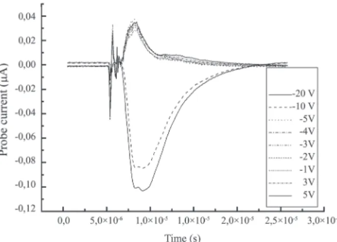
ions was studied with the probe polarized at a -10 V voltage. Later, the electrostatic probe was used to evaluate the charge densities and electron temperature. Figure 3 shows the probe current signal time behavior for different probe electric potentials in experiments with the CuHBr laser and W targets. The noise around 5 × 10⁻⁶s is from laser electric discharge pulses, and it was taken as reference for triggering the oscilloscope. The peak values of the probe current curves were plotted against the polarization voltage, giving rise to the Langmuir curve. From this curve, the charge densities
and electron temperature were evaluated, considering the hydrodynamic expansion approach (Koopman, 1971). This experiment was repeated with aluminum and tungsten targets, ablated by CVL laser, and for copper samples, ablated by the
1G<$*RQH7\SLFDOUHVXOWVDUHVKRZQLQ7DEOH
Figure 3. Temporal behavior of the probe current in different voltages,
for ablation of tungsten samples with the CuHBr laser.
Table 3. Plasma parameters for ablation of different targets and
lasers, measured with the electrostatic probe. All the listed
¿JXUHVDUHSHDNYDOXHV
Target Copper Aluminum Tungsten Tungsten
Laser CVL CVL CuHBr CVL
Ion density
(m-3) 3.4×10¹⁶ 2.3×10¹⁵ 2.4×10¹⁷ 1.3×10¹⁶
Electron density
(m-3) 1.2×10¹⁵ 3.2×10¹⁴ 6.3×10¹⁵ 3.9×10¹⁴
Electron temperature
(eV) 15 19 28 15
Drift velocity
(km/s) 8 – 10 8 – 13 5.4 6 – 10
Emission spectra
Laser induced breakdown spectroscopy (LIBS) experiments in air were made with copper, graphite, molybdenum, alumina, and beach sand samples in order to investigate the composition of the expanding plume. The plume light emission in our experimental conditions vanishes for distances larger than 3 or 4 mm and thus the experiments were performed just above the target surface and not at the same distances, as in the case of the PVDF and electrostatic probe experiments.
)RUWKHWKUHH¿UVWWDUJHWVVLQFHWKH\ZHUHVLQJOHHOHPHQW
samples, the line attribution was made by comparing
)LJXUH39')VLJQDOVROLGOLQHDQGDEHVW¿WIRU(TGDVKHG
line) for ablation of tungsten with CuHBr laser.
Table 2. Plume parameters for ablation of tungsten targets measured
with the PVDF sensor.
Target Tungsten
Laser CuHBr
WKH PHDVXUHG VSHFWUXP ZLWK D VLPSOL¿HG V\QWKHWLF RQH considering only the sample constituent element. The synthetic spectrum was built by adding lorentzian curves with peak values given by the Einstein AFRHI¿FLHQWWDNHQIURP
the National Institute of Standards and Technology transitions database (NIST, 2012), and with the linewidth equals to the instrument resolution (1.5 nm). Figure 4 compares the measured (black) and synthetic (gray) spectra for the copper sample, in 450 to 550 nm.
In the case of alumina targets, besides the aluminum and the oxygen, nitrogen and sodium in the synthetic spectra were also considered.
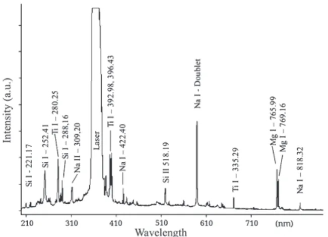
The experiments with beach sand were performed in order to examine the separation potentiality of our experimental VHWXS IRU YHU\ FRPSOH[ WDUJHWV 2UGLQDU\ EHDFK VDQG ZDV heat dried, pressed and sintered with the purpose of getting compact samples, which have a very complex composition and are inhomogeneous, i.e., the spectrum depends on the position the laser strikes the sample surface. It was not possible to build a synthetic spectrum because of the complexity and lack of information about the sample composition. We focused our attention to some peaks that are repetitive and very distinct from the background, by comparing their resonance wavelength with the NIST database. Several of the more intense observed lines were due to sodium and silicon, as shown in Fig. 5.
With regards to Mo samples, a Jobyn-Yvon spectrometer model TRIAX 550, with a 0.025 nm resolution (at 546 nm), was also used and plasma temperature was measured by means of the Boltzmann plot method (Amoruso et al., 1999). The plasma
temperature was about 0.9 eV, in agreement with results found in the literature for spectroscopic measurements (Noll, 2012;
Capitelli et al., 2004), but in contrast with the results obtained
in this work with Langmuir probe for other metallic samples.
ANALYSIS
In order to establish the magnitudes this work refers to, let us consider the experiments performed with tungsten targets and the CuHBr laser. The tungsten properties are: a
A = 0.493 for Ȝ= 511 nm; K = 1.74 W/(cm ºC), ț = 0.7 cm2/s , atomic mass M
W = 183.84 amu, cE = 0.133 J/(g ºC), LV = 4.48 kJ/g, and ȡW = 19.3 g/cm3. The laser parameters are provided in
Table 1. With these, T§.7KLVWHPSHUDWXUHLV
much higher than the tungsten boiling point of 5,930 K (Lide, 1996), and before the surface had achieved this temperature level, a fraction of the sample had been evaporated and ejected, starting the formation of the ablation plume. This evaluation was done without taking changes of the thermal parameters with temperature into consideration, and without considering the interaction of the laser beam with the ejected plume. However, the value is in the same order of magnitude as the translational temperature measured, both with the PVDF sensor and the electrostatic probe. It means that the laser pulse energy is in some way delivered to the ejected plume.
Using Eq. 3, the mass that is removed in one single pulse is estimated in m
E = 1.0 × 10
-7 g, which implies that, taking the
laser repetition rate of 16 kHz, the ejected mass rate is about 5 g/h. This evaluation requires some care, and some fraction of the removed material expands as clusters and/or droplets. The following calculations assume that all the removed material is PDGHRIDWRPVQHXWUDORULRQL]HG7KXVWKHREWDLQHG¿JXUHV must be faced as limit values, which are useful only to provide orders of magnitudes for the analyzed parameters.
Figure 5. A typical sand LIBS spectrum. The saturated peak around
355 nm is due to the scattering of the laser beam.
Figure 4. Comparison between measured (black line) and synthetic
If one takes an intermediate value for the expansion velocity (drift velocity) of v
D = 5 × 10
3 m/s, at the end of the
laser pulse the plume will expand a distance given by L
E = vDIJ §ȝP7KHSDUWLFOHGHQVLW\QXPEHURISDUWLFOHVSHUXQLW\ of volume) at the end of the laser pulse can estimate if it is assumed that all evaporated material had been expanded to a volume given by a hemisphere with radius L
E as in Eq. 5:
/ . .
n mm
L cm
2 3
1 2 9 10
w E E 3 19 3 # c r = -(5)
The mean free path is given by Eq. 6:
,
Lp n1 560nm
c
c v
= (6)
where:
ıC: the collisional cross section was estimated taking the tungsten atomic radius (1,41 Å) and thus ıC§EDUQ
Time between two successive collisions is given by Eq. 7:
v L ps 166 C T p c
x = (7),
where:
v
T: is the thermal velocity, calculated by taking the temperature of 100.000 K previously estimated.
2UUDWKHUGXULQJDWLPHSHULRGHTXLYDOHQWWRWKHODVHUSXOVH duration, more than 200 collisions between particles happen in the plume. Kools et al. (1992), using Monte Carlo calculations,
showed that only about four collisions are necessary for thermalization in the expanding plume. Thus, it is reasonable to consider that the plume expands similarly to a gas in equilibrium DWKLJKWHPSHUDWXUHDQGSUHVVXUHFRQ¿QHGWRDVPDOOYROXPH which is suddenly released to expand into vacuum.
The particle density decreases as the plume expands and, after some distance, the plume is not collisional anymore. To estimate this distance, it is considered that the particle density decays with the distance from the target surface according to Eq. 8,
( )
n z n LE Lz E 3
= ^ h` j (8)
and that the plume stops being collisional when the mean free path is in the same order of magnitude as the plume size L
P, thus substituting Eq. 8 into 6, one has Eq. 9:
. .
LNC n LE CLE 3 1mm 3
v
= ^ h = (9)
If one considers a 5 km/s expanding velocity, the time to expand until L
NC is in the order of 600 ns.
Therefore, for the experimental conditions presented in this work, after an expansion of about 3 mm, the plume is not collisional anymore and the interaction between particles from this point on is essentially electrostatic. Basically, two kinds of behavior are expected, for low densities the charged particles behave like free particles and for high densities they KDYHDFROOHFWLYHSODVPDEHKDYLRU7KH¿JXUHRIPHULWWKDW allows identifying the particles’ behavior is the Debye Length
L
Db, given by Eq. 10:
.
L
q N k T / Db e B e 2 0 1 2 f
=c m (10)
Taking the electron densities and temperatures from Table 3, the Debye length ranges from 0.5 to 2.6 mm in the present experimental conditions. Therefore, the plume typical dimensions are in the same order of magnitude as the Debye length and the particles’ behavior is in the transition between the individual particles and plasma behavior regimes. It suggests that the charged particles (ions mainly) can be separated by WKHDSSOLFDWLRQRIDQHOHFWULF¿HOGEHWZHHQHOHFWURGHVDSDUW from distances in the range of few millimeters and that the remaining plume fraction will be made of single atoms.
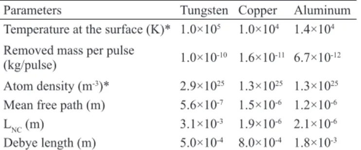
The same calculation was repeated for copper and aluminum, and the results, together with the values for WXQJVWHQDUHSUHVHQWHGLQ7DEOH7KH¿JXUHVDUHYHU\FORVH and the same comments made for tungsten are also applicable for copper and aluminum.
Table 4. Estimated plume parameters for tungsten, copper, and
aluminum, using the same calculation procedure described
in “Analysis”. The material constants were taken from Lide
(1996), laser parameters from the CVL laser in Table 1 and
plasma parameters from Table 3.
Parameters Tungsten Copper Aluminum Temperature at the surface (K)* 1.0×10⁵ 1.0×10⁴ 1.4×10⁴
Removed mass per pulse
(kg/pulse) 1.0×10
-10 1.6×10⁻¹¹ 6.7×10⁻¹²
Atom density (m-3)* 2.9×1025 1.3×1025 1.3×1025
Mean free path (m) 5.6×10⁻⁷ 1.5×10⁻⁶ 1.2×10⁻⁶
LNC (m) 3.1×10⁻³ 1.9×10⁻⁶ 2.1×10⁻⁶
Debye length (m) 5.0×10⁻⁴ 8.0×10⁻⁴ 1.8×10⁻³
CONCLUSIONS
In this work, several materials were evaluated in different experiments of laser ablation, using low energy (~ mJ per pulse), high repetition rate (~ tens of kHz) lasers in the visible and in the near ultraviolet, with pulse width in the range of tens of nanoseconds. The ablation experiments were in the thermal regime, with energy density in the range of tens of J/cm2 and intensities of about 109 W/cm2. The set of results for these experimental conditions leads to the following conclusions:
D VLJQL¿FDQW IUDFWLRQ RI WKH SOXPH JHQHUDWHG E\ ODVHU ablation is made of single atoms (neutral or ionized), even if complex targets are used;
WKHLRQVDQGQHXWUDODWRPVGHQVLW\DUHLQWKHVDPHRUGHURI magnitude;
IRUGLVWDQFHVJUHDWHUWKDQIHZPLOOLPHWHUVWKHSOXPHLVQR longer collisional;
DWGLVWDQFHVODUJHUWKDQFPIURPWKHWDUJHWWKH'HE\H length is such that the charged fraction of the plume can be VHSDUDWHGE\WKHDSSOLFDWLRQRIHOHFWURPDJQHWLF¿HOGV DEODWLRQUDWHVRIDERXWJKDUHSRVVLEOH
In short, it is possible, using laser ablation, to generate an atomic beam adequate for AVLIS purpose. This is possible even for very complex targets, such as ores. The main limitation is the small amount of material that is removed, limiting the method for the separation of small amounts of material. This is a severe limitation for the separation of materials that are needed in large amounts, such as uranium, however it is adequate for the separation of materials used in photonics or in magneto-optic sensors, which require small amounts of isotopes.
REFERENCES
Amoruso, S. et al., 1999, “Characterization of laser-ablation
SODVPDV´-RXUQDORI3K\VLFV%$WRPLF0ROHFXODUDQG2SWLFDO Physics, Vol. 32, No. 14, pp. R131-R172. doi:10.1088/0953-4075/32/14/201
Beams, J.W. and Haynes, F.B., 1936, “The Separation of Isotopes by Centrifuging”, Physical Review, Vol. 50, pp. 491-492. doi: 10.1103/PhysRev.50.491
Becker, E.W. et al., 1967, “Separation of the Isotopes of
Uranium by the Separation Nozzle Process”, Angewandte
Chemie International – Edition in English, Vol. 6, No. 6, pp 507-518. doi:10.1002/anie.196705071
%HQQHW*/³6SDFH1XFOHDU3RZHU2SHQLQJWKH)LQDO Frontier”, 4th International Energy Convertion Engineering Conference and Exhibit (IECEC), San Diego, California, USA, AIAA, pp. 2006-4191.
%RUQ 0 DQG :ROI ( ³3ULQFLSOHV RI 2SWLFV Electromagnetic Theory of Propagation: Interference and Diffraction of Light”, 7th ed., Cambridge University Press, 952p.
Calusaru, A. and Murgulescu, S., 1976, “Chemical and Ion Exchange Unit for a Cascade of Uranium Isotope Separation”, Naturwissenschaften, Vol. 63, No. 12, pp. 578-579. doi:10.1007/BF00622798
Capitelli, M. et al., 2004, “Laser-induced plasma expansion:
theoretical and experimental aspects”, Spectrochimica Acta: Part B, Vol. 59, No. 3, pp. 271-289. doi:10.1016/j. sab.2003.12.017
&KXQJ30³(OHFWULFSUREHVLQVWDWLRQDU\DQGÀRZLQJ plasmas: theory and applications”, Springer-Verlag, New York, 150p.
Del Bosco, E. et al., 1987, “Isotopic enrichment in a plasma
centrifuge”, Applied Physics Letters, Vol. 50, No. 24, pp. 1716. doi:10.1063/1.97725
Dolgolenko, D.A. and Muromkin, Y.A., 2009, “Plasma isotope separation based on ion cyclotron resonance”, Physics Uspekhi, Vol. 52, No. 4, pp. 345-357. doi:10.3367/ UFNe.0179.200904c.0369
'XOH\::³&22 Lasers: Effects and Applications”, Academic Press, New York, 427p.
Flicker, H. et al., 1964, “Construction of a promethium-147
atomic battery”, IEEE Transactions on Electron Devices, Vol. 11, No. 1, pp. 2-8. doi:10.1109/T-ED.1964.15271
Furry, W.H. et al.³2QWKHWKHRU\RILVRWRSHVHSDUDWLRQ
*LmR0$3et al., 2004, “PVDF sensor in laser ablation
H[SHULPHQWV´5HYLHZRI6FLHQWL¿F,QVWUXPHQWV9RO1R 12, pp. 5213-5215. doi:10.1063/1.1819556
Itoh, N. et al.³6PDOORSWLFDOPDJQHWLF¿HOGVHQVRUWKDW
XVHVUDUHHDUWKLURQJDUQHW¿OPVEDVHGRQWKH)DUDGD\HIIHFW´ $SSOLHG2SWLFV9RO1RSSGRL $2
Jensen, R.J. et al., 1982, “Separating isotopes with lasers”,
Los Alamos Science, Vol. 3, No. 1, pp. 02-33.
.DPDGD2et al., 2009, “Mixed rare earth iron garnet (TbY)
,*IRUPDJQHWLF¿HOGVHQVRUV´-RXUQDORI$SSOLHG3K\VLFV Vol. 61, No. 8, pp. 3268-3270. doi:10.1063/1.338877
.DWR'DQG/DPRQW5*³,VRWRSLFFKHPLFDOYDSRU deposition of fused silica and high-silica-content glasses for the SURGXFWLRQRIORZORVVRSWLFDOZDYHJXLGHV´$SSOLHG2SWLFV 9RO1RSSGRL$2
Kholpanov, L.P. et al., 1997, “Multicomponent isotope
separating cascade with losses”, Chemical Engineering DQG3URFHVVLQJ3URFHVVDQG,QWHQVL¿FDWLRQ9RO1R pp. 189-193. doi:10.1016/S0255-2701(96)04187-6
Kim, D.W. et al., 2001, “Separation of magnesium isotopes
by ion exchange chromatography”, Journal of Industrial and Engineering Chemistry, Vol. 7, No. 3, pp. 173-177.
Kools, J.C.S. et al. ³*DV ÀRZ G\QDPLFV LQ ODVHU
ablation deposition”, Journal of Applied Physics, Vol. 71, No. 9, pp. 4547-4556. doi:10.1063/1.350772
Koopman, D.W., 1971, “Langmuir probe and microwave measurements of the properties of streaming plasmas generated by focused laser pulses”, The Physics of Fluids, Vol. 14, No. 8, pp. 1707-1716. doi:10.1063/1.1693667
Lide, D.R., 1996, “CRC Handbook of Chemistry and Physics”, 76th ed., CRC Press, Boca Raton, USA, 2650p.
Louvet, P., 1995, “Device for isotope separation by ion cyclotron resonance”, Patent US005422481A.
Mack, E. and Arroe, H., 1956, “Isotope shift in atomic spectra”, Annual Review of Nuclear Science, Vol. 6, pp. 117-128. doi:10.1146/annurev.ns.06.120156.001001
Martynenko, Y.V., 2009, “Electromagnetic isotope separation method and its heritage”, Physics-Uspekhi, Vol. 52, No. 12, pp. 1266-1272. doi:10.3367/UFNe.0179.200912n.1354
1D\ORU5:DQG%DFNHU32³(QULFKPHQWFDOFXODWLRQV in gaseous diffusion: Large separation factor”, AIChE Journal, Vol. 1, pp. 95-99. doi:10.1002/aic.690010114
NIST 2012, “NIST Atomic Spectra Database Lines Form”, Retrieved in Mach 08, 2012, from http://physics.nist.gov/ PhysRefData/ASD/lines_form.html.
Noll, R., 2012, “Laser-induced breakdown spectroscopy”, Springer-Verlag, Heidelberg, 543p. doi:10.1007/978-3-642-20668-9
Paisner, J.A., 1988, “Atomic Vapor Laser Isotope Separation”, $SSOLHG 3K\VLFV % /DVHUV DQG 2SWLFV 9RO 1R pp. 253-260. doi:10.1007/BF00692883
Prasad, R.R. and Krishnan, M., 1987, “Theoretical and experimental study of rotation in a vacuum arc centrifuge”, Journal of Applied Physics, Vol. 61, No. 1, pp. 113-119. doi:10.1063/1.338976
Riley Jr., J.E., 1987, “The effects of lithium isotopic anomalies on lithium niobate”, Ferroelectrics, Vol. 75, No. 1, pp. 59-62. doi:10.1080/00150198708008209
Rutherford, W.M., 1986, “Separation of Zinc Isotopes by Liquid-Phase Thermal Diffusion”, Industrial & References Engineering Chemistry Process Design and Development, Vol. 25, No. 4, pp. 855-858. doi:10.1021/i200035a003
Schiller, S. et al., 1983, “Electron Beam Technology”, John
Wiley & Son, New York, Chichester, Brisbane, Toronto, Singapore, 508p.
Schwab, C. et al., 1998, “Laser techniques applied to isotope