Correspondence to: Branislav V. Bajkin, Dental Clinic of Vojvodina, Faculty of Medicine, University of Novi Sad, Hajduk Veljkova 12,
O R I G I N A L A R T I C L E S UDC: 616.314-089.87-005.1-08
DOI: 10.2298/VSP1412097B
Comparison of efficacy of local hemostatic modalities in
anticoagulated patients undergoing tooth extractions
Pore
ÿ
enje efikasnosti razli
þ
itih metoda lokalne hemostaze kod pacijenata na
oralnoj antikoagulantnoj terapiji posle ekstrakcije zuba
Branislav V. Bajkin, Sreüko D. Selakoviü, Siniša M. Mirkoviü, Ivan N. Šarþev, Ana J. Tadiü, Bojana R. Milekiü
Dental Clinic of Vojvodina, Faculty of Medicine, University of Novi Sad, Novi Sad, Serbia
Abstract
Background/Aim. Patients receiving long-term oral anti-coagulant therapy pose a clinical challenge during invasive dental procedures. The goal of this study was to compare different local hemostatic modalities after tooth extraction in patients receiving chronic Vitamin-K antagonist ther-apy. Methods. Totally 90 patients with International Normalized Ratio (INR) 3.0 requiring simple extraction of one or two teeth were randomized into three groups, 30 patients in each group. The patients with the mean INR value of 2.35 ± 0.37, in whom extraction wound was su-tured comprised the group A. In the group B with the mean INR of 2.43 ± 0.4, local hemostasis was achieved by placing absorbable gelatin sponges into the wound without suturing. The group C consisted of the patients with the mean INR of 2.36 ± 0.34 in whom neither gelatin sponge nor suturing were used for providing local hemostasis. Bleeding was registered as an event if other than initial hemostatic measure was needed or additional oral surgeon intervention required. Results. The obtainded results show that 1 (3.3%) patient in the group A, 2 (6.7%) pa-tients in the groups B and C manifested post-extraction bleeding. All cases of hemorrhage were easily solved with local hemostatic measures and all, except one case, were registered in the first two hours after the procedure until the dismissal. A difference between the groups was not statistically significant (Ʒ2 = .42, p > 0.05). Conclusion. In therapeutically anticoagulated patients tooth extractions can be safely performed without altering the dose of anti-coagulant medication if efficient local hemostasis is pro-vided. In most cases, in patients with INR 3.0 after ex-traction of one or two teeth postoperative bleeding can be controlled with local pressure, without any additional local hemostatic measures.
Key words:
tooth extraction; antiacoagulants; administration, oral; drug therapy; hemostasis.
Apstrakt
Uvod/Cilj. Pacijenti koji duže vremena primaju antikoagu-lantnu terapiju predstavljaju poseban izazov prilikom invaziv-nih dentalinvaziv-nih procedura.Cilj rada bio je da se uporede razliÿ -ite metode lokalne hemostaze kod pacijenata na oralnoj an-tikoagulatnoj terapiji nakon ekstrakcije zuba. Metode. Uk-upno 90 pacijenata na oralnoj antikoagulantnoj terapiji sa In-ternational Normalized Ratio (INR) vrednostima 3,0 kojima je bila potrebna ekstrakcija jednog ili dva zuba, bilo je podeljeno u tri grupe, po 30 ispitanika u svakoj grupi, zavisno od pri-menjenog metoda lokalne hemostaze. Grupu A ÿinili su pacijenti ÿija je ekstrakciona rana ušivena, a zabeležena proseÿna INR vrednost u ovoj grupi iznosila je 2,35 ± 0,37. U grupi B za postizanje lokalne hemostaze u ekstrakcionu ranu su postavljani resorptivni želatinski sunĀeri, bez ušivanja. Proseÿna INR vrednost u ovoj grupi iznosila je 2,43 ± 0,4. U grupi C (pacijenti sa proseÿnom INR vrednošýu 2,36 ± 0,34) posle ekstrakcije zuba nisu korišteni ni lokalni hemostatici, niti je rana ušivana. Produženo postekstrakcijsko krvarenje definisano je kao dogaĀaj u sluÿaju da poÿetne mere hemo-staze nisu bile dovoljne ili je bila potrebna dodatna oralnohi-rurška intervencija. Rezultati. Kod 1 (3,3%) pacijenta u grupi A i po 2 (6,7%) pacijenta u grupi B i grupi C javilo se produ-ženo postekstrakcijsko krvarenje. Nije utvrĀeno postojanje statistiÿki znaÿajne razlike u uÿestalosti krvarenja po grupama (Ʒ² = .42; p > 0.05). Svi pacijenti sa produženim krvarenjem bez poteškoýa zbrinuti su merama lokalne hemostaze i svi su, izuzev jednog, registrovani u prva dva ÿasa nakon interven-cije. Zakljuÿak. Kod pacijenata na oralnoj antikoagulantnoj terapiji moguýe je bezbedno izvršiti ekstrakciju zuba bez iz-mene terapijskog režima uz primenu adekvatnih mera lokalne hemostaze. Kod pacijenata sa INR 3,0 nakon jednostavnih ekstrakcija zuba kompresija rane gazom u veýini sluÿajeva dovoljna je mera za postizanje lokalne hemostaze.
Kljuÿne reÿi:
Introduction
Patients receiving long-term oral anticoagulant therapy (OAT) pose a clinical challenge during invasive dental pro-cedures. The important question is whether to continue, modify or interrupt OAT before dental treatment. Cessation or reduction of anticoagulant intake for several days prior to dental procedure may expose these patients to the risk of thromboembolism 1, especially in high risk patients, i.e. those with artificial heart valves and atrial fibrillation with related risk factors. An increasing number of authors state that tooth extraction in anticoagulated patients within the therapeutic International Normalized Ratio (INR) values (INR 4.0) can be safely done without changing OAT regi-men if using local hemostatic measures which stabilize or enhance clot formation at the surgical site 2–19. Hemostatic agents used for bleeding control in patients on OAT are: oxi-dized cellulose 2–7, absorbable gelatin sponges 5–8, absorbable collagen sponges 9, 19, fibrin glue 10, cyanoacrylate glue 11, platelet rich plasma gel 12 and topical thrombin 17. The im-portance of antifibrinolytic solutions for mouthwash in post-extractional bleeding prevention has also been noted in sev-eral articles 4, 20. None of these hemostatic agents has been proved to be superior compared to others 21–23.
The aim of this study was to compare some local hemo-static modalities in anticoagulated patients without interrup-tion of OAT, including a group of patients in whom local hemostatic agents were not used.
Methods
The study was initiated after it had been approved by local Ethics Committee and all study participants were pro-vided with a written informed consent.The study lasted for ten months and included patients on OAT who were referred to the Department of Oral Surgery, Dental Clinic of Vo-jvodina by their general dental or medical practitioners in the need for tooth extractions.
The study included patients on long-term OAT whose INR was 3.0 on the day of the procedure and in whom ex-traction of one or two teeth could be done using local anes-thesia without the need to raise a mucoperiosteal flap. The following patient categories were excluded from the study: patients with liver disease or bleeding disorders, patients taking medications that affect hemostasis, patients with se-vere bleeding after dental extractions even before starting OAT and patients who did not agree to participate in the in-vestigation.
During a patient’s first visit, retroalveolar or ortopan-thomographic x-ray and fulfilling of the standard form on the basis of a patient’s prior medical documentation were done. The underlying diagnosis which was the reason for starting OAT was noted, as well as the duration of OAT, type and dosage of oral anticoagulant used, recent INR values and every other medication a patient had been using.
The first 30 patients were randomized in the group A, next 30 patients into the group B and the remaining 30 pa-tients comprised the group C. The INR value was determined
for each patient on the day of the procedure. Antibiotic pro-phylaxis was given for patients at risk of endocarditis in ac-cordance with the American Heart Association guidelines 24.
Group A patients underwent suturing of the extractional wound with “figure of eight“ nonresorbable suture (black silk 3–0) without using of any local hemostatic agent. In group B absorbable gelatin sponge was used as local hemo-static agent without wound suturing. In the group C no addi-tional local hemostatic measures were used, except local pressure with gauze.
All extractions were done by the same surgeon, on an outpatient basis. Lidocaine hydrochloride 2% with 1/80,000 adrenaline was used as local anesthetic. Local infiltration method and intraligamentary anesthesia were mostly used, although regional blocks of the inferior alveolar and lingual nerve were also used 25. Extractions were as atraumatic as possible. Local pressure was applied in the patients of all the three groups afterwards, i.e. the patients were asked to hold sterile gauze in a firm bite for thirty minutes. The patients were observed for the next two hours. The first measure in case of post-extraction bleeding was a superficial gauze tam-ponade of the wound for ten minutes. This was repeated, if needed, twice at the most. It was planned, in cases of unsuc-cessful superficial gauze tamponade in the patients of the group A, to put a hemostatic agent, gelatin sponge, into the wound along with suturing, while in the same situation in the group B the wound would be sutured. In the group C the in-sertion of gelatine sponge was to be the first measure after which suturing of the wound would be done if necessary. Repeated hemorrhage in all the three groups of patients would be treated by insertion of a new hemostatic agent – oxidized regenerated cellulose into the wound. Should there be hemorrhage that could not be controlled with repeated lo-cal measure, the attending physician would be consulted, and if needed vitamin K or fresh frozen plasma would be admin-istered.
All the patients were advised to continue OAT after the procedure. Paracetamol was suggested for pain relief. Each patient was given a telephone number to contact the surgeon in case of hemorrhage.
All the subjects were examined thirty minutes, two hours, on the first, second and fifth day after the extraction, when sutures in the patients in the group A were removed. The hemorrhage was registered as an event if other than ini-tial local hemostatic measure was needed or an additional oral surgeon intervention was required. Hemorrhages that occurred in the first two hours after the procedure, i.e. during the observational period until the dismissal of the patient were characterized as “immediate bleeding”. Hemorrhages after this period were considered as “late bleeding”.
Data were analyzed using Ȥ2-test and analysis of vari-ance (ANOVA) as appropriate, and the probabilities of less than 0.05 were accepted as significant.
Results
ex-cluded, but were contacted by telephone to exclude postex-tractional hemorrhage. Two patients who were on combined oral anticoagulant and aspirin therapy were also excluded. Finally, the study included 90 patients, 30 in each group. In-dications for OAT are shown in the Table 1.
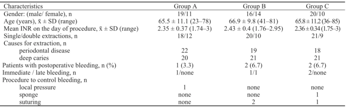
The mean INR values on the day of procedures were 2.35 ± 0.37 in the group A, 2.43 ± 0.4 in the group B and 2.36 ± 0.34 in the group C. The most common anticoagulant drug in all the three patient groups was acenocoumarol (28, 26 and 28 patients in the group A, B and C, respectively). All the other patients were taking warfarin. Basic characteristics of all the three groups of patients are shown in Table 2. The groups did not differ significantly in INR-values (p = 0.662), number of tooth extractions (p = 0.708), gender (p = 0.543) and age (p = 0.868).
Postoperative bleeding was noted in 1 (3.3%) patient in the group A, and 2 (6.7%) patients in the groups B and C (Table 2). Except one patient in the group B with “late bleeding”, which occurred several hours after the interven-tion and whose wound had to be sutured, all the other cases of postextractional bleeding were noted in the first two hours after the procedure and characterized as “immediate bleed-ing”. All cases of hemorrhage were easily solved only with
local hemostatic measures and none of the patients had seri-ous bleeding that would require systemic therapy. No statis-tically significant difference between these two groups of patients was found (Ȥ2 = .42; p= 0.811).
Discussion
Most authors suggest that the vast majority of oral sur-gical procedures in anticoagulated patients can be safely done without alteration of their regular OAT, thus avoiding the risk of thromboembolic complications 1–20. The need for efficient local hemostasis is emphasized. Local hemostatic measures usually mean the use of certain hemostatic agent such as oxidized regenerated cellulose, gelatine sponges, collagen sponges, fibrin glue and antifibrinolytic mouthwash.
Studies that compare different local hemostatic measures do not confirm advantages of certain agents 21–23. Nevertheless, having in mind limited possibilities in the use of some prepa-rations (such as fibrin glue since it costs a lot and carries the risk of viral transmission; tranexamic acid in the form of mouthwash is not available commercially in many countries and may have an effect only on the superficial clot and not on bleeding from the depth of the socket), there is still a need
Table 1 Indications for anticoagulant treatment
Indications Group A
(n)
Group B (n)
Group C (n)
Prosthetic valve replacement 11 4 7
Cardiac arrhythmia (atrial fibrillation) 6 14 11
Atrial fibrillation and valvular diseases 3 2 1
Atrial fibrillation and cerebrovascular accident 1 5 3
Deep vein thrombosis/pulmonary embolus 6 2 7
Ischemic heart disease 1 none 1
Cerebrovascular accident 1 2 none
Dilated cardiomyopathy none 1 none
Thrombophilia 1 none none
n – number of patients; group A – patients underwent suturing of the extractinal wound; group B – patient with absorbable gelatin sponge used for local hemostasis; group C – patients with no local hemostatic measures except local pressure with gauze.
Table 2 Characteristics of patients under coumarin treatment undergoing tooth extraction
Characteristics Group A Group B Group C
Gender: (male/ female), n Age (years), ʉ ± SD (range)
Mean INR on the day of procedure, ʉ ± SD (range) Single/double extractions, n
Causes for extraction, n periodontal disease deep caries
19/11 65.5 ± 11.1 (23–78) 2.35 ± 0.37 (1.74–3)
18/12
22 20
16/14 66.9 ± 9.8 (41–81) 2.43 ± 0.4 (1.76–2.95)
20/10
19 21
20/10 65.8 ± 11.2 (36–85) 2.36 ± 0.34 (1.75–3)
21/9
18 21 Patients with postoperative bleeding, n (%)
Immediate / late bleeding, n Procedure to control bleeding, n
local pressure sponge suturing
1 (3.3) 1/none
1 none none
2 (6.7) 1/1
none none 2
2 (6.7) 2/none
to find the safest method for local hemostasis in these pa-tients.
In most studies that dwell on tooth extractions in an-ticoagulated patients, the suturing of the extractional wound had been done 2–8. Ferrieri et al. 26 point out that only suturing of the wound with local application of anti-fibrinolytics in cases of hemorrhage is sufficient for suc-cessful local hemostasis. Recent studies, however, suggest that suturing of the extractional wound is not always nec-essary 9, 19, 27.
Up to now there is only one study by Campbell et al. 28 in which tooth extractions in anticoagulated patients were performed without additional local hemostatic measures. The authors found no difference in blood loss among groups of patients who continued, stopped and have never been on OAT during oral surgery. However, the number of patients who continued OAT was small and only 12 patients with INR < 3 were included in the study.
The results of our study show that minor oral surgical procedures, such as extraction of one or two teeth, could be safely done without alteration of OAT and without the use of any topical hemostatic agent. Wound suturing is as efficient in local hemostasis as the use of topical hemostatic agent, but it is not necessary measure in each patient. However in pa-tients on OAT in whom adequate primary local hemostasis
cannot be achieved, suturing is a procedure of a great im-portance.
This study has some potential drawbacks and limita-tions: its relatively small sample size; only oral surgery pro-cedures with low bleeding risk, simple extraction of one or two teeth, were performed; only patients with INR 3.0 were included although the current recommendation is that oral surgery can be safely done if INR values are 4.0. The reason for this is our intention to check safety of dental ex-tractions without the use of any local hemostatic agents.
Conclusion
In therapeutically anticoagulated patients tooth extrac-tions can be safely performed without altering the dose of anticoagulant medication, provided efficient local hemosta-sis. In most cases, in patients with INR 3.0 after extraction of one or two teeth postoperative bleeding can be controlled with local pressure, without any additional local hemostatic measures.
Acknowledgements
The authors thank the Center for Laboratory Medicine (Department of Hemostasis, Thrombosis and Hematology Diagnostics, Clinical Center of Vojvodina, Novi Sad, Serbia) for help and cooperation during this study.
R E F E R E N C E S
1. Wahl MJ. Dental surgery in anticoagulated patients. Arch In-tern Med 1998; 158(15): 1610î6.
2. Evans IL, Sayers MS, Gibbons AJ, Price G, Snooks H, Sugar AW. Can warfarin be continued during dental extraction? Results of a randomized controlled trial. Br J Oral Maxillofac Surg 2002; 40(3): 248î52.
3. Devani P, Lavery KM, Howell CJ. Dental extractions in patients on warfarin: Is alteration of anticoagulant regime necessary. Br J Oral Maxillofac Surg 1998; 36(2): 107î11.
4. Carter G, Goss A. Tranexamic acid mouthwash: A prospective randomized study of a 2-day regimen vs 5-day regimen to pre-vent postoperative bleeding in anticoagulated patients requir-ing dental extractions. Int J Oral Maxillofac Surg 2003; 32(5): 504î7.
5. Karaca I, Simûek S, Uøar D, Bozkaya S. Review of flap design in-fluence on the health of the periodontium after mandibular third molar surgery. Oral Surg Oral Med Oral Pathol Oral Ra-diol Endod 2007; 104(1): 18î23.
6. Zanon E, Martinenelli F, Bacci C, Cordioli G, Girolami A. Safety of dental extraction among consecutive patients on oral antico-agulant treatment managed using a specific dental management protocol. Blood Coagul Fibrinolisis 2003; 14(1): 27î30. 7. Salam S, Yusuf H, Milosevic A. Bleeding after dental extractions
in patients taking warfarin. Br J Oral Maxillofac Surg 2007; 45(6): 463î6.
8. Blinder D, Manor Y, Martinowitz U, Taicher S. Dental extractions in patients maintained on oral anticoagulant therapy: Compari-son of INR value with occurrence of postoperative bleeding. Int J Oral Maxillofac Surg 2001; 30(6): 518î21.
9. Bajkin BV, Popovic SL, Selakovic SD. Randomized, prospective trial comparing bridging therapy using low-molecular-weight heparin with maintenance of oral anticoagulation during ex-traction of teeth. J Oral Maxillofac Surg 2009; 67(5): 990î5.
10.Bodner L, Weinstein JM, Baumgarten AK. Efficacy of fibrin seal-ant in patients on various levels of oral seal-anticoagulseal-ant under-going oral surgery. Oral Surg Oral Med Oral Pathol Oral Ra-diol Endod 1998; 86(4): 421î4.
11. Al-Belasy FA, Amer MZ. Hemostatic effect of n-butyl-2-cyanoacrylate (histoacryl) glue in warfarin-treated patients under-going oral surgery. J Oral Maxillofac Surg 2003; 61(12): 1405î9. 12.Della Valle A, Sammartino G, Marenzi G, Tia M, Espedito di Lauro
A, Ferrari F, et al.Prevention of postoperative bleeding in an-ticoagulated patients undergoing oral surgery: use of platelet-rich plasma gel. J Oral Maxillofac Surg 2003; 61(11):1275î8. 13.Morimoto Y, Niwa H, Minematsu K. Risk factors affecting
post-operative hemorrhage after tooth extraction in patients re-ceiving oral antithrombotic therapy. J Oral Maxillofac Surg 2011; 69(6): 1550î6.
14.Rodríguez-Cabrera MA, Barona-Dorado C, Leco-Berrocal I, Gómez-Moreno G, Martínez-González JM. Extractions without eliminat-ing anticoagulant treatment: A literature review. Med Oral Patol Oral Cir Bucal 2011; 16(6): 800î4.
15.Jiménez Y, Poveda R, Gavaldá C, Margaix M, Sarrión G. An up-date on the management of anticoagulated patients pro-grammed for dental extractions and surgery. Med Oral Patol Oral Cir Bucal 2008; 13(3): 176î9.
16.Scully C, Wolf A. Oral surgery in patients on anticoagulant ther-apy. Oral Surg Oral Med Oral Pathol Oral Radiol Endod 2002; 94(1): 57î64.
17.Marjanoviý M. Use of thrombin powder after tooth extraction in patients receiving anticoagulant therapy. Vojnosanit Pregl 2002; 59(4): 389î92. (Serbian)
19.Bajkin BV, Bajkin IA, Petrovic BB. The effects of combined oral anticoagulant-aspirin therapy in patients undergoing tooth ex-tractions: a prospective study. J Am Dent Assoc 2012; 143(7): 771î6.
20.Sindet-Pedersen S, Ramstrom G, Bernvil S, Blomback M. Hemostatic effect of tranexamic acid mouthwash in anticoagulant-treated patients undergoing oral surgery. N Engl J Med 1989; 320(13): 840î3.
21.Blinder D, Manor Y, Martinowitz U, Taicher S, Hashomer T. Dental extractions in patients maintained on continued oral antico-agulant: Comparison of local hemostatic modalities. Oral Surg Oral Med Oral Pathol Oral Radiol Endod 1999; 88(2): 137î40.
22.Carter G, Goss A, Lloyd J, Tocchetti R. Tranexamic acid mouth-wash versus autologous fibrin glue in patients taking warfarin undergoing dental extractions: A randomized prospective clinical study. J Oral Maxillofac Surg 2003; 61(12): 1432î5. 23.Halfpenny W, Fraser JS, Adlam DM. Comparison of 2
hemo-static agents for the prevention of postextraction hemorrhage in patients on anticoagulants. Oral Surg Oral Med Oral Pathol Oral Radiol Endod 2001; 92(3): 257î9.
24.Wilson W, Taubert KA, Gewitz M, Lockhart PB, Baddour LM, Levison M, et al. Prevention of infective endocarditis: Guide-lines from the American Heart Association: A guideline from
the American Heart Association Rheumatic Fever, Endocardi-tis and Kawasaki Disease Committee, Council on Cardiovas-cular Disease in the Young, and the Council on Clinical Cardi-ology, Council on Cardiovascular Surgery and Anesthesia, and the Quality of Care and Outcomes Research Interdisciplinary Working Group. J Am Dent Assoc 2008; 139(1): 3î24. 25.Bajkin BV, Todorovic LM. Safety of local anaesthesia in dental
patients taking oral anticoagulants: is it still controversial. Br J Oral Maxillofac Surg 2012; 50(1): 65î8. PubMed PMID: 21130546
26.Ferrieri GB, Castiglioni S, Carmagnola D, Cargnel M, Strohmenger L, Abati S. Oral surgery in patients on anticoagulant treatment without therapy interruption. J Oral Maxillofac Surg 2007; 65(6): 1149î54.
27.Mubarak S, Ali N, Rass M, Sohail A, Robert A, Al-Zoman K, et al. Evaluation of dental extractions, suturing and INR on postoperative bleeding of patients maintained on oral anticoagulant therapy. Br Dent J 2007; 203(7): E15.
28.Campbell JH, Alvarado F, Murray RA. Anticoagulation and mi-nor oral surgery: Should the anticoagulation regimen be al-tered. J Oral Maxillofac Surg 2000; 58(2): 131î5.