ABSTRACT
Biocompatibility of RealSeal, its primer and AH
Plus implanted in subcutaneous connective tissue
of rats
Fabiana Soares GRECCA1, Patrícia Maria Poli KOPPER1, Régis Burmeister dos SANTOS1, Anna Christina FOSSATI2, Vinicius Coelho CARRARD1, Gerson Arison Xavier ACASIGUA3, José Antônio Poli de FIGUEIREDO4
1- DDS, MSc, PhD, Assistant Professor, Department of Conservative Dentistry, Dental School, Federal University of Rio Grande do Sul, Porto Alegre, RS, Brazil. 2- DDS, MSc, PhD, Associate Professor, Department of Oral Biology, Dental School, Federal University of Rio Grande do Sul, Porto Alegre, RS, Brazil 3- DDS, MSc student, Department of Conservative Dentistry, Dental School, Federal University of Rio Grande do Sul, Porto Alegre, RS, Brazil. !"
Corresponding address: Fabiana Soares Grecca - Avenida Ramiro Barcelos, 2492 - 90035-003 - Porto Alegre, RS - Brazil - Phone/Fax: +55-51-3308-5191 - e-mail: [email protected]
5HFHLYHG0D\0RGL¿FDWLRQ-XO\$FFHSWHG$XJXVW
O
bjective: This study tested rat connective tissue response to RealSeal, RealSeal primer or AH Plus after 7, 15, 30, 60 and 90 days of implantation. Material and methods: Thirty Wistar rats had subcutaneous sockets created on their back and received four implants each of polyethylene tubes containing one of the materials tested according to the groups: AH (AH Plus Sealer); RS (RealSeal Sealer); RP (RealSeal Primer); CG (control group – empty tube). After histological processing, sections were analyzed to identify the presence of neutrophils, lymphocytes and plasma cells, eosinophils, macrophages and giant cells, as ! "#$%& of the control group were observed at 14 and 60 days in AH group, and at 90 days in RS group (p<0.05). There were no differences in terms of presence of macrophages, giant '*+ -days than after any other period (p=0.031). RP group scored higher for lymphoplasmacytic /28>?@>>2DE%F study was to demonstrate that issues involved with tissue tolerance of a Resilon-containing sealer, RealSeal Sealer, cannot be attributed to its primer content.Key words:J $
Endodontics.
INTRODUCTION
$ ' treatment success. Materials used during obturation should meet a number of criteria so that success can be achieved. Of these, biocompatibility is a
can be placed in close contact with periapical tissues.
The implantation of materials in subcutaneous tissues of rats has been used as a method to study biocompatibility4,11,16. The material under study
may be placed in dentin3,5, silicone6,16,17FL7,11
or polyethylene9 tubes. When animal testing is
applied, material implant in polyethylene tubes has been described as gold standard. Figueiredo, et al.2
(2001) did not observe tissue reaction differences compared with sealer sub-mucous injection, but polyethylene tubes helped control the amount of sealer in contact with the tissues.
Resilon). Previous studies showed lower cytotoxicity levels of Resilon-containing sealers when compared with commonly used sealers13,15. The fact that Resilon
has low setting time in anaerobic environment10
may account to a better tissue response. Also, when associated with good coronal restoration, it seems to display good tissue reaction8. However,
the literature is scarce as to the biocompatibility of Resilon. Onay, Ozdemir and Ungor11 (2007) tested
$ [ \ subcutaneous tissue response to this material. However, they have not tested the primer, which is potentially an irritant to the tissues.
Resilon is not the only available resin-containing sealer. A widely used resinous sealer, AH Plus (Dentsply-Maillefer, Tulsa, OK, USA), which is commonly used in conjunction with gutta-percha, has been used as a control for comparison in many tests12,14.
To test the hypothesis that RealSeal and its primer are biocompatible, this study evaluated tissue response to AH Plus, RealSeal or RealSeal primer in polyethylene tubes implanted in subcutaneous connective tissue of rats for 7, 15, 30, 60 and 90 days.
MATERIAL AND METHODS
This study was approved by the Research Ethics Committee of the Dental School of the Federal University of Rio Grande do Sul (UFRGS), Brazil.
Thirty Wistar male rats (Rattus novergicus albinus) weighing 180 to 220 g were obtained from the UFRGS animal care facility. During the study, the animals were kept in routinely cleaned cages at controlled temperature and received water, dry Nuvelab CR1 (Nuvital, Curitiba, PR, Brazil) and Labina (Purina, Campinas, SP, Brazil) chows.
One hundred and twenty polyethylene (nontoxic Scalp Vein 19G) test tubes (1.3 mm inner diameter X 5 mm long) were manufactured. Thirty tubes (AH D '* + ?{ Maillefer); 30 (RS group), with RealSeal Sealer (SybronEndo); 30 (RP group) with RealSeal Primer (SybronEndo) and the other 30 (CG - control group) were left empty.
Following preparation of the tubes, the animals received general anesthesia by intramusctular injection of 0.008 mL/100 g ketamine (Francotar™,
Virbac do Brasil Indústria e Comércio Ltda., Roseira, SP, Brazil) and 0.004 mL/100 g xylazine chloride 2%
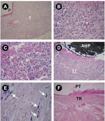
Figure 2-# $$%"& '# ()$*%)*67
(Virbaxyl®, Virbac do Brasil Indústria e Comércio
Ltda.). After that, the dorsum of the animals was manually shaved and scrubbed with gauze soaked in 3% alcohol-iodine (Quinta Essência Cosméticos e Medicamento Ltda., Porto Alegre, RS, Brazil).
Four incisions of about 1 cm long and 2 cm distant /" (Free-Bac, Embramac Empresa Brasileira de Material Cirúrgico Ltda., Itapira, SP, Brazil) and handle. After each incision, subcutaneous tissue was dissected
?{L®, SS White
Artigos Dentários Ltda., Rio de Janeiro, RJ, Brazil). The surgical pockets were produced on the dorsum of each animal.
A test tube was inserted in each pocket using surgical forceps (Duflex®, SS White Artigos
Dentários Ltda., Rio de Janeiro, RJ, Brazil). Each animal received one tube from each experimental was used to determine in which pocket the test tube would be inserted, and ensured that the different types of test tubes were inserted in all different positions. After that, the incisions were sutured with 4-0 mononylon stitch (Somerville Ltda., Jabotão dos Guarapes, PE, Brazil).
Six rats were killed at 7, 15, 30, 60, and 90 days postoperatively. The animals were anesthetized again as described above and killed by cervical
Event Time Group p
AH RS RP CG
Neutrophils 7 days 11.10 12.00 9.30 7.00 0.390
14 days 9.50 12.83 9.50 9.50 0.177
30 days 10.00 10.00 11.67 10.00 0.506
60 days 13.00 14.17 10.50 10.50 0.282
90 days 8.00 8.00 10.00 8.00 0.392
Eosinophils 7 days 9.80 8.00 12.00 10.25 0.470
14 days 9.00 10.58 11.38 11.20 0.742
30 days 13.75 9.92 8.50 11.13 0.250
60 days 12.60 15.50 10.00 10.00 0.104
90 days 8.50 8.50 8.50 8.50 1.000
LPI 7 days 14.30 9.20 8.50 7.50 0.160
14 days 14.50A 10.00AB 12.25AB 5.70B 0.049
30 days 15.38 9.50 9.00 9.38 0.236
60 days 17.00A 14.75AB 9.67AB 7.42B 0.030
90 days 10.50AB 12.40A 6.50AB 5.40B 0.032
Macr.+ Giant 7 days 12.20 11.90 7.20 8.38 0.354
14 days 6.80 12.00 12.50 10.80 0.385
30 days 10.38 8.83 13.58 8.50 0.423
60 days 11.10 15.75 11.00 10.00 0.384
90 days 7.50 12.70 5.00 7.50 0.074
Fibrosis 7 days 11.50 7.60 11.50 9.25 0.241
14 days 12.00 12.00 9.50 8.00 0.215
30 days 13.50 10.17 11.83 6.00 0.121
60 days 14.70 13.17 11.25 9.33 0.446
90 days 11.00 9.40 7.00 7.80 0.591
Abscesses 7 days 13.80 8.00 9.80 8.00 0.079
14 days 10.50 10.50 10.50 10.50 1.000
30 days 10.50 10.50 10.50 10.50 1.000
60 days 12.00 12.00 12.00 12.00 1.000
90 days 8.50 8.50 8.50 8.50 1.000
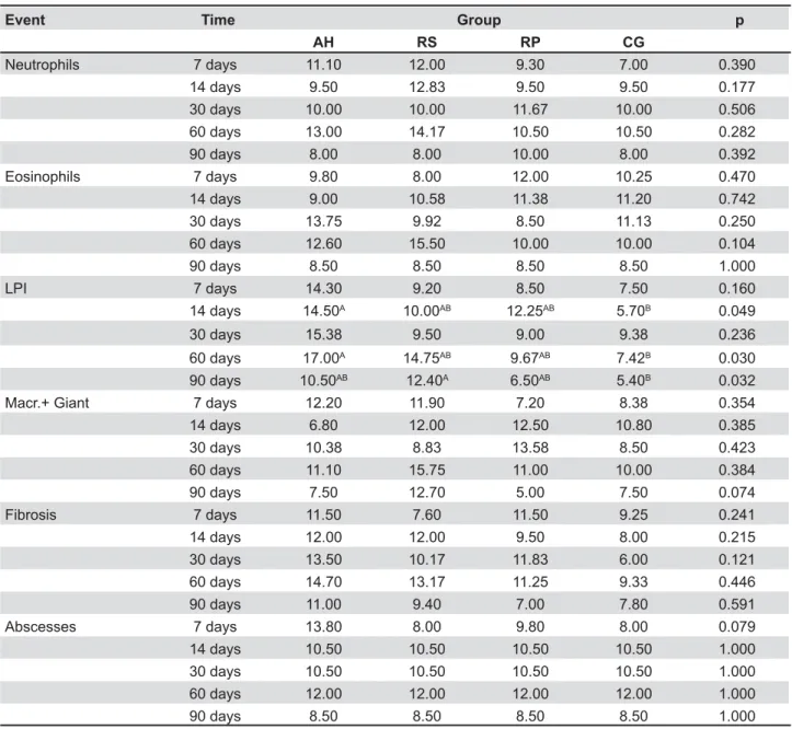
Table 1- Mean scores attributed to the AH Plus Sealer (AH), RealSeal Sealer (RS), RealSeal Primer (RP), and Control (CG) groups, after experimental periods of 7, 14, 30, 60 and 90 days, for the six events assessed
(]%%%$ $^'_(%
dislocation. Immediately after that, excisional biopsies of each implant area were obtained which were immersed in 10% buffered formaldehyde for 48 h.
The samples were embedded in paraffin. A microtome (Leica RM 2025, Nussloch, Baden-Württemberg, Germany) was used to section the blocks to reach the tube. The tube was removed
?{L®, SS White Artigos
Dentários Ltda.), and the block was immersed again U' and 5 semi-serial sections 5- to 6-μm-thick were obtained and then stained with Harris hematoxylin and alcoholic eosin (HE). Results were analyzed by a blinded examiner using a light microscope (BX41TF, D />> >> 2>> The examiner was calibrated before data analysis (kappa=0.6).
For each study sample, only the section that was most representative of the histological condition was chosen. Adjacent tissue in at least one of the tube ends was visualized in every section chosen.
E ! following scores: 1- absent; 2- mild (sparse cells or very small groups of cells); 3- moderate (cells D 2 ?D
following scores: 1- absent; 2- thin layer of collagen
Abscesses, characterized by the presence of dead neutrophils (pus) in a large clearly stained area, % 1- absent; 2- abscess in contact with the area that contained the material; 3- abscess also in areas distant from the area that contained the material.
The nonparametric Kruskal-Wallis test was used, and the multiple comparisons test, to determine ! set at D=0.05.
RESULTS
Some specimens were lost during histological processing, resulting in the following sample distribution according to the experimental periods and groups: 7 days – AH (n=5), RS (n=5), RP (n=5), CG (n=4); 14 days – AH (n=5), RS (n=6), RP (n=4), CG (n=5); 30 days – AH (n=6), RS (n=4), RP (n=4), CG (n=6); 60 days – AH (n=5), RS (n=6), RP (n=6), CG (n=6); and 90 days – AH (n=4), RS (n=5), RP (n=4), CG (n=5).
Figure 1 and Table 1 illustrate the behavior of the groups in terms of the events assessed, at each evaluation period. The groups did not differ significantly from each other in terms of presence of macrophages and giant cells,
any of the different experimental periods (p>0.05). & higher than those of the control group were observed at 14 and 60 days in AH group and at 90 days in RS group.
Comparing the results for each material after different experimental periods, no significant differences were observed in relation to the presence of macrophages and giant cells, eosinophils, ?>>"D'* higher for abscesses at 7 days than after any other period (p=0.031). RP group scored higher for /28> days (p=0.04).
DISCUSSION
The objective of this study was to conduct an in vivo experiment to contribute with regards to the biocompatibility of RealSeal Sealer, comparing it with its own primer, and another resinous sealer (AH Plus).
Tubes containing test materials implanted into the subcutaneous tissue of experimental animals have been employed to test biocompatibility3-7,9,11,16-17.
Since this method brings the test substances into contact with connective tissue, it simulates what occurs in the periapical region after obturation of root canals.
Analysis of the results demonstrated that the median score for neutrophils and eosinophils was 1 in all groups, indicating that these cells were absent in the tissue close to the materials. This fact suggests that contact with AH Plus Sealer, RealSeal Sealer and RealSeal Sealer primer are all well-tolerated by the body.
Abscesses were not observed in the primer, RealSeal Sealer or control groups. At 7 days, AH Plus Sealer group exhibited abscesses in contact with the material, and the score after 7 days was Based on this observation, it can be stated that AH Plus Sealer was more aggressive, during the initial period of contact with connective tissue than the other materials.
The results of this investigation are comparable
14 (2006) and Onay,
Ozdemir and Ungor11 (2007), who concluded that
Epiphany Sealer, an endodontic sealer with the same chemical composition as that of RealSeal Sealer, is biocompatible. Batista, et al.1 (2007) also
CONCLUSION
The main contribution of this study was to demonstrate that issues involved with tissue tolerance of a Resilon-containing sealer, RealSeal Sealer, cannot be attributed to its Primer content. Further studies should be conducted to assess long-term tissue response of these materials, since ageing has not been tested from the biocompatibility point of view.
REFERENCES
1- Batista RF, Hidalgo MM, Hernandes L, Consolaro A, Velloso TR, Cuman RK, et al. Microscopic analysis of subcutaneous reactions to endodontic sealer implants in rats. J Biomed Mater Res A. 2007;81(1):171-7.
2- Figueiredo JA, Pesce HF, Gioso MA, Figueiredo MA. The histological effects of four endodontic sealers implanted in the oral mucosa: submucous injection versus implant in polyethylene tubes. Int Endod J. 2001;34(5):377-85.
3- Holland R, Souza V, Nery MJ, Bernabé FE, Filho JA, Junior ED, et al. Calcium salts deposition in rat connective tissue after the implantation of calcium hydroxide-containing sealers. J Endod. 2002;28(3):173-6.
4- Holland R, Souza V, Nery MJ, Faraco IM Jr, Bernabé PF, Otoboni JA Filho, et al. Reaction of rat connective tissue to implanted dentin + calcium hydroxide. Braz Dent J. 2001;12(1):3-8.
5- Holland R, Souza V, Nery MJ, Otoboni Filho JA, Bernabé PF, Dezan E Jr. Reaction of rat connective tissue to implanted dentin
J Endod. 1999;25(3):161-6.
6- Kaplan AE, Ormaechea MF, Picca M, Canzobre MC, Ubios AM. Rheological properties and biocompatibility of endodontic sealers. Int Endod J. 2003;36(8):527-32.
7- Kolokouris I, Economides N, Beltes P, Vlemmas I. In vivo
comparison of the biocompatibility of two root canal sealers implanted into the subcutaneous connective tissue of rats. J Endod. 1998;24(2):82-5.
8- Leonardo MR, Barnett F, Debelian GJ, Pontes Lima RK, Bezerra ! &' $ ! \ without coronal restoration: a histopathological evaluation. J Endod. 2007;33(11):1299-303.
9- Molloy D, Goldman M, White RR, Kabani S. Comparative tissue tolerance of a new endodontic sealer. Oral Surg Oral Med Oral Pathol. 1992;73(4):490-3.
10- Nielsen B, Beeler W, Vy C, Baumgartner J. Setting times of Resilon and other sealers in aerobic and anaerobic environments. J Endod. 2003;32(2):130-2.
11- Onay EO, Ungor M, Ozdemir BH. In vivo evaluation of the biocompatibility of a new resin-based obturation system. Oral Surg Oral Med Oral Pathol Oral Radiol Endod. 2007;104(3):e60-6. 12- Scarparo RK, Grecca FS, Fachin EV. Analysis of tissue reactions to methacrylate resin-based, epoxy resin-based, and zinc oxide-eugenol endodontic sealers. J Endod. 2009;35(2):229-32. 13- Scotti R, Tiozzo R, Parisi C, Croce MA, Baldissara P. J !ex vivo. Int Endod J. 2008;41(8):651-7.
14- Sousa CJA, Montes CRM, Pascon EA, Loyola AM, Versiani MA. Comparison of the intraosseous biocompatibility of AH Plus, EndoREZ, and Epiphany root canal sealers. J Endod. 2006;32(7):656-62.
15- Susini G, About I, Tran-Hung L, Camps J. Cytotoxicity of Epiphany and Resilon with a root model. Int Endod J. 2006;39(12):940-4.
16- Zmener O, Guglielmotti MB, Cabrini RL. Biocompatibility of two calcium hydroxide-based endodontic sealers: a quantitative study in the subcutaneous connective tissue of the rat. J Endod. 1988;14(5):229-35.