3 2 0 Arq Bras Oftalmol. 2012;75(5):320-3
Artigo Original |
Original articleABSTRACT
Purpose: To evaluate the reproducibility of peripapillary retinal nerve iber layer (RNFL) thickness measurements in normal eyes and eyes with glaucoma using spectral domain optical coherence tomography (SDOCT).
Methods: One eye of 79 normal and 72 glaucoma patients was analyzed. All patients underwent a complete ophthalmological examination, including visual acuity testing; intraocular pressure, slit-lamp examination, indirect ophthalmos-copy; and the glaucoma group underwent achromatic perimetry with the 24-2 SITA Fast Humphrey Field Analyzer. All patients’ eyes were scanned using the spectral domain optical coherence tomography - Spectralis®
and one of them was cho sen randomly. Three con secutive circular B-scan centered at the optic disc were performed in one visit.
Results: The intraclass correlation coefficient (ICC), coefficient of variation and test-retest variability for the mean retinal nerve iber layer thickness were respec-tively: 0.94, 2.56% and 4.85 µm for the normal group and 0.93, 4.65% and 6.61 µm for the glaucomatous group. The intraclass correlation coeicient for retinal nerve iber layer thickness in all quadrants were all excellent in both groups, with the superior quadrant having the highest ICCs (0.964) in glaucomatous eyes and nasal quadrant measurements having the lowest (0.800), but still excellent in eyes without glaucoma. The coeicient of variation was between 2.56% - 8.74% and between 4.65% - 11.44% in normal and glaucomatous group respectively. The test-retest variability was between 4.85 µm and 11.51 µm in the normal group and between 6.61 µm and 14.24 µm in the glaucomatous group. The measurements in glauco-matous eyes were more variable than normal eyes.
Conclusions: Spectral domain optical coherence tomography showed excellent reproducibility with regard to retinal nerve iber layer thickness measurements in normal and glaucomatous eyes.
Keywords: Diagnostic techniques, ophthalmological; Glaucoma/diagnosis; Tomo-graphy, optical coherence/methods; Retinal ganglion cells; Optic nerve/pathology; Nerve ibers; Reproducibility of results
RESUMO
Objetivo: Avaliar a reprodutibilidade da medida da espessura da camada de fibras nervosas da retina (CFNR) em olhos sem e com glaucoma utilizando-se tomografia de coerência óptica de domínio espectral (spectral domain OCT - SDOCT).
Métodos: Foram analisados apenas um olho de 79 pacientes normais e 72 com glau-coma. Todos os pacientes realizaram um exame oftalmológico completo, incluindo acuidade visual, pressão intraocular, biomicroscopia, oftalmoscopia indireta e, para o grupo com glaucoma, perimetria acromática 24-2 SITA Fast Humphrey Field Ana ly zer. Foram realizados em todos os olhos e em apenas uma visita, três B-scans circulares centrados no disco óptico utilizando-se o SDOCT - Spectralis®
.
Resultados: O coeficiente de correlação intraclasse (ICC), coeficiente de variação e va -riabilidade teste-reteste para a média de espessura da camada de fibras nervosas da retina foram respectivamente: 0,94, 2,56% e 4,85 µm para o grupo sem glaucoma e 0,93, 4,65% e 6,61 µm para o grupo glaucomatoso. O coeficiente de correlação intraclasse foi excelente em ambos os grupos em todos os quadrantes, com o quadrante superior sendo o maior (0,964) no grupo glaucomatoso e o nasal sendo o menor (0,800), mas ainda excelente, em olhos sem glaucoma. O coeficiente de variação foi entre 2,56% - 8,74% e entre 4,65% - 11,44%, nos grupos sem e com glaucoma, respectivamente. A variabilidade teste-reteste variou de 4,85 µm e 11,51 µm no grupo sem glaucoma; e entre 6,61 µm e 14.24 µm no com glaucoma. Olhos com glaucoma apresentaram-se mais variáveis que os sem glaucoma.
Conclusão: A tomografia de coerência óptica “spectral domain” apresentou excelente reprodutibilidade da espessura da camada de fibras nervosas da retina em pacientes sem e com glaucoma.
Descritores: Técnicas de diagnóstico oftalmológico; Glaucoma/diagnostico; Tomografia de coerência óptica/métodos; Células ganglionares da retina; Nervo óptico/patologia; Fibras nervosas; Reprodutibilidade dos testes
INTRODUCTION
Glaucoma is an optic neuropathy, multifactorial, characterized by accelerated death of the retinal ganglion cells, subsequent axonal loss, and visual ield impairment(1).
Detected defects in the retinal nerve iber layer (RNFL) years before arising alterations in the visual ield suggests that anatomical alterations of the RNFL can identify progression of the glaucoma before it causes functional loss in the visual ield(2).
Methods for reliably establishing glaucomatous nerve atrophy are limited. Optical coherence tomography (OCT) is a noninvasive, high-resolution imaging technique that allows in vivo measurements of RNFL in cross section. High-resolution imaging of retinal structure is clinically relevant for the diagnosis of glaucoma(3).
Until recently, the OCT commercially available was the time do-main OCT (TDOCT), which provides an axial resolution of 10 μm, and cross sectional retinal images consisting of 512 A-scans can be acquired
Reproducibility of peripapillary retinal nerve fiber layer thickness measurements
using Spectral Domain OCT in Brazilian patients
Reprodutibilidade da espessura da camada de ibras nervosas da retina utilizando-se o
Spectral Domain OCT em pacientes brasileiros
Daniela araújo Toscano1, Marcos PereiraDe Ávila2, Maria regina caTai chaliTa1
Submitted for publication: October 4, 2011 Accepted for publication: August 31, 2012
Study carried out at Department of Ophthalmology, Universidade de Brasília - UnB.
1 Physician, Department of Ophthalmology, Universidade de Brasília - UnB - Brasília (DF), Brazil. 2 Professor, Department of Ophthalmology, Universidade Federal de Goiás - UFG - Goiânia (GO),
Brazil.
Funding: No specific financial support was available for this study.
Disclosure of potential conflicts of interest: D.A.Toscano, None; M.P.Ávila, None; M.R.C.Chalita, None.
Correspondence address: Daniela Araújo Toscano. Av. Ingá, 250, Apto. 302 - João Pessoa (PB) 58038-250 - Brazil - E-mail: [email protected]
Toscano DA, et al.
3 2 1
Arq Bras Oftalmol. 2012;75(5):320-3 in 1.28 seconds. The main disadvantage of TDOCT technology is the
limited resolution and slow acquisition time. With the spectral do-main OCT (SDOCT) or Fourier dodo-main, the echo time delays of light are measured by acquiring the interference spectrum of the light signal and converted to depth information by Fourier transform(4).
The detection method using a spectrometer allowed a conside-rable increase in imaging speed and resolution without compromi-sing image quality(5). The increase in imaging speed minimizes
mo-tion artifacts. Resolumo-tion is up to 5 times higher, and imaging speed is 60 times faster than in conventional time domain OCT(4).
Estimate reproducibility of RNFL thickness using OCT is essential for diagnostic precision and in particular, describes the smallest changes detectable for identifying and monitoring the progression of glaucoma as well as indicates therapeutic interventions(6,7).
Quantifying the reproducibility of the RNFL thickness measure-ments using Spectralis® spectral domain OCT is an important step in evaluating the potential usefulness of this device for the diagnosis of glaucoma and for determining glaucomatous progression.
To our knowledge the present study is the irst to report on the reproducibility and test-retest variability of RNFL thickness measure-ments using Spectralis® SD-OCT device in Brazilian population.
The purpose of this study was to evaluate the reproducibility of peripapillary retinal nerve iber layer (RNFL) thickness measurements in normal and glaucomatous eyes using spectral domain optical coherence tomography (Spectralis®) in Brazilian population.
METHODS
This prospective observational cross-sectional study was approved by the Institutional Review Board / Ethics Committee of the University of Brasília, Brasília, Brazil. All participants in this study gave their written informed consent. All normal and glaucoma participants were enrolled in this study through the glaucoma and cataract service of Brasília Center of Vision (CBV, Brasília, Brazil) between August and December 2010.
A total of 151 eyes from 79 normal patients and 72 patients with mo-derate to advanced glaucoma were analyzed. All subjects underwent a complete ophthalmological examination, including medical and family history; visual acuity testing with refraction; intraocular pressure mea-surements using Goldmann applanation tonometry (GAT), a complete slit-lamp examination, including indirect ophthalmoscopy. Patients from the glaucoma group underwent achromatic perimetry using 24-2 SITA FAST Humphrey Field Analyzer (Humphrey- Zeiss Systems, Dublin CA).
Inclusion criteria for both groups were: age more than or equal to 40 years, spherical refractive error less than or equal to 5 diopters (D), cylindrical refractive error less or equal to 3D.
Inclusion criteria for normal subjects were: best-corrected visual acuity of 20/60 or better; normal slit-lamp examination; intraocular pres-sure of 21 mmHg or less; normal appearing optic nerve heads; and no history of ocular surgery or laser treatments.
Glaucoma patients were deined on the basis of having either: 1. An abnormal Humphrey Field Analyzer, deined as having MD
less than -12dB; Less than 50% of the points are depressed bellow the 5% level and less than 20 points are depressed bellow the 1% level on the patterns deviation plot; No points in the central 5o can have a sensitivity of 0 dB; Only one hemiield may
have a point with sensitivity of < 15 dB within 5o of ixation(8) and
2. Glaucoma optic disc change deined as a cup-to-disc ratio greater than or equal to 0.6, cup-to-disc ratio asymmetry be-tween the eyes greater than or equal to 0.2, disc rim thinning, notching, localized pallor or nerve iber layer defect (9).
All patients from the glaucoma group had primary openangle glau -coma or chronic angle-closure glau-coma, being excluded any other kind of glaucoma.
Exclusion criteria for both groups included other intraocular diseases, as well as diseases that afect the visual ield, like pituitary lesion, diabe tes, retinal conditions or secondary cause of intraocular pressure increase.
Patients were excluded in the glaucoma group if they had unre-liable automated perimetry results: ixation loss, false positives or false negatives more than 33%. Visual ields were carried out at least twice(9).
OCT
MEASUREMENTSAll patients’ eyes were scanned using the commercially available SDOCT Spectralis® HRA (Heidelberg Retina Angiograph) + OCT (Hei-delberg Engineering). This instrument uses a wavelength of 820 nm in the near infrared spectrum in the SLO (scanning laser ophthalmos-copy) mode. The light source of the SDOCT is a super luminescent diode with a wavelength of 870 nm. Infrared images and OCT scans (40,000 A-Scan/sec) of the dual laser scanning systems are acquired simultaneously(10). Three consecutive circular B-scan (3.4-mm
diame-ter, 768 A-scans) centered at the optic disc were performed in one visit by the same operator. The scanning circle was centered manually on the optic disc irst, although the participant was looking at the internal ixation light. The RNFL borders could be clearly identiied and were marked automatically by the segmentation software. Ima ges were judged to be of suicient quality on the basis of subjecti -ve operator evaluation. Within each scan session, the instrument alignment and controls were not changed, unless as part of image acquisition process. The subject was repositioned between the scan measurements only when necessary. There was no speciic attempt to reposition the subjects between scans done during the same ses-sion. Images were acquired through undilated pupils.
The Spectralis® reports the average RNFL thickness in the supe-rior, temporal, infesupe-rior, nasal and overall (mean). A RNFL thickness graph includes the colored normative database range.
Eyes were placed into 1 of 3 categories that indicate comparison versus normative database. Green: within normal limits, with values inside the 95% normal range. Yellow: borderline, with values outsi-de 95% but within 99% conioutsi-dence interval of normal distribution (0.01<p<0.05). Red: outside normal limits, with values outside 99% conidence interval of normal distribution.
S
TATISTICALANALYSISIntraclass correlation coeicient (ICC), coeicient of variation (COV), and test-retest variability (TRV) were analyzed to determine the reproducibility of SDOCT with regards to RNFL thickness mea-surements.
Number of participants were calculated for the 95% lower con-idence interval (CI) of ICC 0.8 not to be lower than 0.75, a generally accepted lower value of good reproducibility. The average of ICC, COV and TRV were calculated for the RNFL thickness in each quadrant and overall.
Coeicient of variation was calculated using the standard devia-tion divided by the mean thickness, expressed as a percentage. The test-retest variability in RNFL thickness, measured in micrometers, was calculated as two times the SD of the two repeated measu-rements for each measure of RNFL thickness.
RESULTS
One hundred ifty one subjects were enrolled in this study, 79 without glaucoma and 72 with moderate to advanced glaucoma. Mean age was 58 (11) years old (range, 40-86) and 68 (10) years old (range, 44-97) for normal and glaucoma groups, respectively. Table 1 shows demographic characteristics of the study population.
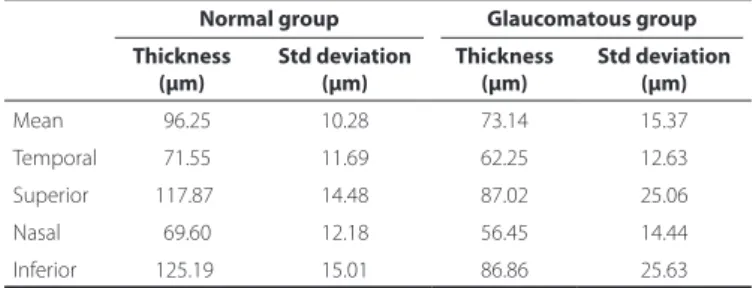
Table 2 summarizes the mean RNFL thickness in all quadrants and overall for normal and glaucomatous groups. The inferior and superior quadrant were the thickest in the normal group and glauco-matous group, respectively. The nasal quadrant was the thinnest in both groups.
Reproducibility of peripapillary retinal nerve fiber layer thickness measurements using Spectral Domain OCT in Brazilian patients
3 2 2 Arq Bras Oftalmol. 2012;75(5):320-3
ICC, COV and TRV for both groups are presented in tables 3 and 4, respectively. The lowest ICC was 0.800 in the nasal quadrant for the normal group and 0.827 in the temporal quadrant for the glaucoma-tous group. The highest ICC was 0.944 for the mean RNFL in the normal group and 0.964 for the superior quadrant in the glaucomatous group. Even the lower 95 % CIs were greater 0.70, indicating excellent repro-ducibility of all measurements. The overall RNFL had the lowest COV and the nasal quadrant the highest COV in both groups. TRV showed the lowest for the overall RNFL thickness, 4,85 µm in the normal group and 6.61 µm in the glaucomatous group. The highest TRV was obser-ved in the nasal quadrant for the normal group (11.51 µm) and in the inferior quadrant for the glaucomatous group (14.24 µm). RNFL mea-surements in glaucomatous eyes were more variable than normal eyes.
DISCUSSION
In glaucoma patients the OCT is an important instrument for diagnosis and to follow the progression of the disease. Assessing the reproducibility of the RNFL thickness using OCT is of paramount importance for its use in clinical practice of glaucoma.
This study showed excellent reproducibility of RNFL thickness measurements with Spectralis® in patients without and with mode-rate to advanced glaucoma (generally deined as 0.75 - 1.00). The lowest ICCs were observed in the nasal quadrant for the patients without glaucoma (0.800) and in the temporal quadrant (0.827) for patients with glaucoma. Even the lower 95% CIs were greater 0.70, indicating excellent reproducibility of all measurements.
Comparing two diferent SD OCTs (CirrusTM and Spectralis®), ex-cellent repeatability of RNFL thickness measurement was observed in normal participants for both devices. Using Spectralis® in undilated pupils the average of TRV was 4.95, COV 1.7% and ICC 0.971(11). In our
study the TRV was 4.85, COV 2.56% and ICC 0.944. These diferences can be explained because the number and the age of the participants in our study were higher.
Many other studies reported the reproducibility of RNFL thickness using diferent kinds of OCT. Blumenthal et al., in 1999 studied 10 eyes with glaucoma and 10 normal eyes and reported, using the commercially available OCT, that glaucoma patients were found to be signiicantly more variable than normal subjects (P=0.03). The coeicient of variation for the mean RNFL thickness was signiicantly smaller (P=0.02) in normal eyes (6.9%) than in glaucomatous eyes (11.8%). The coeicient of variation was larger in the temporal and nasal quadrants than in the superior and inferior quadrants, as found in our study(12) .
Studies with StratusTM OCT showed good RNFL thickness
repro-ducibility. Peripapillary RNFL thickness of 51 subjects with glaucoma was measured using the Standard and Fast scan protocols of StratusTM
OCT 3 times on the same day to determine intrasession variability and on 5 diferent days within a 2-month period to determine intersession variability. It was observed the RNFL thickness test-retest variability intrasession ranged between 5.2 µm and 17.1 µm (mean 5.2 µm) using standard protocol and between 5 µm and 16.7 µm (mean 5.0 µm) for fast protocol, for clock hours. The test-retest variability of the tempo-ral quadrant and of most tempotempo-ral clock hours seemed to be less than that of other quadrants orlocations, and theFast scanning protocol, ICC and COV tended also to be worst for the nasal quadrant, similar to our results. The best ICC was 0.98 for both protocols. Intraclass correlation coeicients were essentially all excellent with the mean Standard RNFL and Fast RNFL values having the highest of 0.98(13).
Still assessing the reproducibility of the StratusTM, Budenz et al.,
performed 3 peripapillary circulars scans in 147 subjects normal and with glaucoma. The ICC was excellent for both Standard (mean 0.97) and Fast (mean 0.95) RNFL measurements. TRV of the quadrants mea-surements ranged from 3.5 µm to 13.0 µm in normal subjects and 5.2 µm to 13.8 µm in glaucomatous eyes analyzing standard and fast protocols, and four quadrants(14). Reproducibility of StratusTM
OCT as also studied in 10 normal subjects that were scanned three consecutive times with each of the following: macular scans, RNFL scans, and ONH scans before dilation and three additional times for each scan type after dilation. Similarly, all the subjects were scanned on two additional days within 5 months and the inter and intravisit reproducibility was calculated. The ICCs for the RNFL quadrants were higher after dilation, ranging between 71% and 84%, with the exception of the superior quadrant (ICC was 79% before and 75% after dilation). StratusTM OCT demonstrated reproducible
measure-ments for NFL thickness, macular thickness, and optic nerve head parameters(15).
The reproducibility of macula thickness using spectral domain OCT (Spectralis®) was studied in 41 normal eyes. Intravisit reprodu-cibility was analyzed by performing 3 scans in the macula. It was
Table 1: Demographic characteristics
Groups Number
Gender
Age Male Female
Normal group 79 49.37% 50.63% 58 (range 40-86) Glaucomatous group 72 31.94% 68.06 % 68 (range 44-97)
Table 2. RFNL Thickness in normal and glaucomatous groups
Normal group Glaucomatous group
Thickness (µm)
Std deviation (µm)
Thickness (µm)
Std deviation (µm)
Mean 096.25 10.28 73.14 15.37
Temporal 071.55 11.69 62.25 12.63
Superior 117.87 14.48 87.02 25.06
Nasal 069.60 12.18 56.45 14.44
Inferior 125.19 15.01 86.86 25.63
Std= standard
Table 4. Intraclass correlation coeicient, coeicient of variation and test-retest variability in glaucomatous eyes
ICC COV (%) TRV (µm)
Mean 0.937 (0.908) 04.65 06.61
Temporal 0.827 (0.757) 08.44 09.95
Superior 0.964 (0.947) 07.23 11.15
Nasal 0.877 (0.824) 11.44 11.78
Inferior 0.855 (0.795) 08.60 14.24
ICC= intraclass correlation coeicient, with lower 95% CI (conidence interval) in paren-theses; COV= coeicient of variation; TRV= test-retest variability
Table 3. Intraclass correlation coeicient, coeicient of variation and test-retest variability in normal eyes
ICC COV (%) TRV (µm)
Mean 0.944 (0.921) 2.56 04.85
Temporal 0.895 (0.852) 5.93 08.41
Superior 0.928 (0.898) 4.09 09.49
Nasal 0.800 (0.726) 8.74 11.51
Inferior 0.931 (0.902) 3.80 09.33
Toscano DA, et al.
3 2 3
Arq Bras Oftalmol. 2012;75(5):320-3 observed excellent reproducibility of retinal thickness measurements
with mean diference among measurements of about 1 µm(6).
Other SDOCT were also analyzed as RTVue which the RNFL thickness reproducibility showed the ICC ranged between 0.91 and 0.97 for the healthy group (60 eyes) and 0.86 and 0.97 for the glaucomatous group (76 eyes)(7). Using the SD SLO/OCT OTI the lowest ICC was 0.961
at 3 o’clock in the normal group (98 eyes) and 0.951 at 4 o’clock in the glaucoma group (79 eyes)(9).
Bendschneider et al., analyzed the RNFL thickness in 170 healthy patients using the SDOCT Spectralis® and observed that the RNFL thickness was signiicantly associated with age (1.9 µm decline in mean total RNFL thickness per age decade), axial length (total RNFL decrease of -4.79 mm per every increasing 1-mm-axial length) and optic disc area (with a total RNFL increase of 6.28 mm for every 1-mm2-increase in disc area). The total RNFL thickness in the study
population was 97.2 ± 9.7 µm, which is comparable to our study (96.25 µm ± 10.28)(10).
Recently, Wu et al., reported the reproducibility of Spectralis in 45 normal patients and 33 glaucoma patients. Their ICCs ranged from 0.977 (temporal) to 0.990 (global and inferior-nasal sector) in normal eyes, and from 0.983 (temporal) to 0.997 (inferior quadrant) in glaucomatous eyes. CVs ranged from 1.45% (overall global) to 2.59% (temporal quadrant) for normal participants and ranged from 1.74% (overall global) to 3.22% (temporal quadrant) for glaucoma patients(16). There were some diferences between this study and the
present study: they analyzed the RNFL thickness using more than four quadrants (overall global - 360 degrees), for 4 quadrants (supe-rior, infe(supe-rior, nasal, and temporal), and then for 4 additional sectors: superior-temporal (TS, 45 to 90 degrees), superior-nasal (NS, 90 to 135 degrees), nasal (NI, 225 to 270 degrees), and inferior--temporal (TI, 270 to 315 degrees); the number of participants was smaller than ours; the subjects had diferent types of glaucoma, in cluding pseudoexfoliation glaucoma; and they used the TruTrack image alignment software (i.e., the eye tracking system). Probably because of all these facts theirs ICC and COV were better than the present study, especially because of the tracking system.
Evaluating the impact of Spectralis® self-acting eye tracking system and retest software on the reproducibility of RNFL thickness measurements in glaucomatous and healthy eyes it was observed that the reproducibility can be improved by using the eye tracker, and this gain was signiicantly higher in glaucomatous than in heal-thy eyes. In healheal-thy subjects, COVs for RNFL thickness measurements without using the system ranged from 3.5% to 7.4% and with the eye tracker and retest protocol ranged from 1.0% to 2.5%. In glaucoma patients measurements without the eye tracker ranged from 5.8% to 10.5% and with the system ranged from 1.6% to 3.8%. The impro-vement in reproducibility was signiicantly higher in glaucomatous than in healthy eyes(17).Our study showed COV ranged from 4.65%
to 11.44% in the glaucomatous group similar to the previous study without using the eye tracker system. Despite not using the eye tracking system, our study showed an excellent reproducibility of RNFL thickness in normal and glaucomatous eyes. It is necessary to emphasize that the patients included in the present study had
moderate to advanced glaucoma with diicult to ixation on one or both eyes and it may have contributed to the high COV.
CONCLUSION
The Spectralis® spectral domain OCT equipped showed excellent reproducibility with regards to RNFL thickness measurements in normal and in moderate to advanced glaucoma patients.
REFERENCES
1. Sehi M, Grewal DS, Sheets CW, Greenield DS. Diagnostic ability of Fourier-domain vs time-domain optical coherence tomography for glaucoma detection. Am J Oph-thalmol. 2009;148(4):597-605.
2. Sommer A, Miller NR, Pollack I, Maumenee AE, George T. The nerve iber layer in the diagnosis of glaucoma. Arch Ophthalmol. 1977;95(12):2149-56.
3. Huang D, Swanson EA, Lin CP, Schuman JS, Stinson WG, Chang W, et al. Optical coherence tomography. Science. 1991;254(5035):1178-81.
4. Chen TC, Cense B, Pierce MC, Nassif N, Park BH, Yun SH, et al. Spectral domain optical coherence tomography: ultra-high speed, ultra-high resolution ophthalmic imaging. Arch Ophthalmol. 2005;123(12): 1715-20.
5. Nassif N, Cense B, Park B, Pierce M, Yun S, Bouma B, et al. In vivo high-resolution video-rate spectral-domain optical coherence tomography of the human retina and optic nerve. Opt Express. 2004;12(3):367-76.
6. Menke MN, Dabov S, Knecht P, Sturm V. Reproducibility of retinal thickness mea-surements in healthy subjects using spectralis optical coherence tomography. Am J Oph thalmol. 2009;147(3):467-72.
7. Gonzalez-Garcia AO, Vizzeri G, Bowd C, Medeiros FA, Zangwill LM, Weinreb RN. Repro-ducibility of RTVue retinal nerve iber layer thickness and optic disc measurements and agreement with Stratus optical coherence tomography measurements. Am J Ophthalmol. 2009;147(6):1067-74, 1074 e1.
8. Leung CK, Ye C, Weinreb RN, Cheung CY, Qiu Q, Liu S, et al. Retinal nerve iber layer imaging with spectral-domain optical coherence tomography a study on diagnostic agreement with Heidelberg Retinal Tomograph. Ophthalmology. 2010;117(2):267-74. 9. Lee SH, Kim SH, Kim TW, Park KH, Kim DM. Reproducibility of retinal nerve iber thick-ness measurements using the test-retest function of spectral OCT/SLO in normal and glaucomatous eyes. J Glaucoma. 2010;19(9):637-42.
10. Bendschneider D, Tornow RP, Horn FK, Laemmer R, Roessler CW, Juenemann AG, et al. Retinal Nerve Fiber Layer Thickness in Normals Measured by Spectral Domain OCT. J Glaucoma. 2010;19(7):475-82.
11. Tan BB, Natividad M, Chua KC. Comparison of retinal nerve iber layer measurement between 2 spectral domain OCT instruments. J Glaucoma. 2012;21(4):266-73. 12. Blumenthal EZ, Williams JM, Weinreb RN, Girkin CA, Berry CC, Zangwill LM.
Reprodu-cibility of nerve iber layer thickness measurements by use of optical coherence to mography. Ophthalmology. 2000;107(12):2278-82.
13. Budenz DL, Fredette MJ, Feuer WJ, Anderson DR. Reproducibility of peripapillary retinal nerve iber thickness measurements with stratus OCT in glaucomatous eyes. Ophthalmology. 2008;115(4):661-6. e4.
14. Budenz DL, Chang RT, Huang X, Knighton RW, Tielsch JM. Reproducibility of retinal nerve iber thickness measurements using the stratus OCT in normal and glaucoma-tous eyes. Invest Ophthalmol Vis Sci. 2005;46(7):2440-3.
15. Paunescu LA, Schuman JS, Price LL, Stark PC, Beaton S, Ishikawa H, et al. Reproducibi-lity of nerve iber thickness, macular thickness, and optic nerve head measurements using StratusOCT. Invest Ophthalmol Vis Sci. 2004;45(6):1716-24.
16. Wu H, de Boer JF, Chen TC. Reproducibility of retinal nerve iber layer thickness mea surements using spectral domain optical coherence tomography. J Glaucoma. 2011;20(8):470-6.