Arq Neuropsiquiatr 2009;67(3-A):719-723 Clinical / Scientiic note
Contribution of the diffuSion-weighted
Mri in the diagnoSiS and follow-up of
enCephalopathy CauSed by Maple Syrup
urine diSeaSe in a full-terM newborn
José Roberto Lopes Ferraz-Filho
1, Valdeci Hélio Floriano
2, Marcelo Bianco Quirici
3,
Regina Pires de Albuquerque
4, Antônio Soares Souza
5ContribuiÇÃo da SeQÜÊnCia difuSÃo da Mr no diagnÓStiCo e aCoMpanhaMento da enCefalopatia por doenÇa da urina eM Xarope de bordo eM neonato a terMo
FAMERP Medical School, São José do Rio Preto, SP, Brazil: 1MD, Department of Radiology, Doctoral candidate Health Sciences Program FAMERP
Medi-cal School; 2MD, Department of Radiology, Fellow in Neuroradiology and Master’s candidate Health Sciences Program of the FAMERP Medical School; 3MD, Department of Radiology, Fellow in Neuroradiology of the FAMERP Medical School; 4MD, Department of Pediatrics, Assistant Professor of Child
Neurology of the FAMERP Medical School; 5MD, PhD, Head of the Department of Radiology, FAMERP Medical School.
Received 3 December 2008, received in inal form 8 April 2009. Accepted 1 June 2009.
Dr. José Roberto Lopes Ferraz-Filho – Av. Brigadeiro Faria Lima 5544 - 15090-000 São José do Rio Preto SP - Brasil. E-mail: [email protected] Maple syrup urine disease (MSUD) or leucinosis is
caused by a deiciency of the catalytic components of the
α-ketoacid-dehydrogenase complex, which is responsible for the catabolism of branched-chain amino acids (leucine, isoleucine, and valine)1,2. It is an inherited genetic disease with an autosomal recessive pattern affecting approxi-mately 1 out of 120,000–500,000 infants worldwide3,4. Diagnosis is made clinically based on the peculiar ma-ple syrup odor or sugar burnt of the urine, encephalop-athy, increased levels of branched-chain amino acids in the plasma and urine, and the presence of α-hydroxyacid and branched-chain α-ketoacids in urine. The presence of plasma L-alloisoleucine and urinary α-hydroxyisovalerate are pathognomonic for MSUD2. According to the litera-ture, ive forms of MSUD have been described: classic, in-termediate, intermittent, thiamine-responsive, and dihy-drolipoyl dehydrogenase-deicient. The commonest and severest form of the disease is the classic type, which is characterized by a neonatal onset of encephalopathy2. Magnetic resonance imaging (MRI) studies in the acute phase of classic MSUD are characterized by diffuse ede-ma corresponding to both myelinated and unmyelinated areas of the brain3-6.
The purpose of this case report is to show convention-al MRI and diffusion-weighted imaging (DWI) indings of the different evolutionary phases in MSUD of a newborn that evolved with brain white matter lesions.
CaSe
A full-term male infant born from an uneventful pregnan-cy and delivery, with a birth weight of 3.245g and Apgar scores
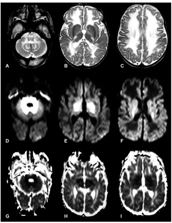
of 9/10 (at 1 and 5 min, respectively), was hospitalized because of sucking dificulties, weak cry, and lethargy. At 10 days of life, the baby had episodes of seizures, bradycardia, and apnea, lead-ing to coma. Biochemical examinations showed hypoglycemia and metabolic ketoacidosis. Brain MRI at 10 days of life showed hypersignal lesions on DWI and corresponding hyposignals on ADC maps throughout the white matter of the brainstem, cere-bella and internal capsules (Fig 1).
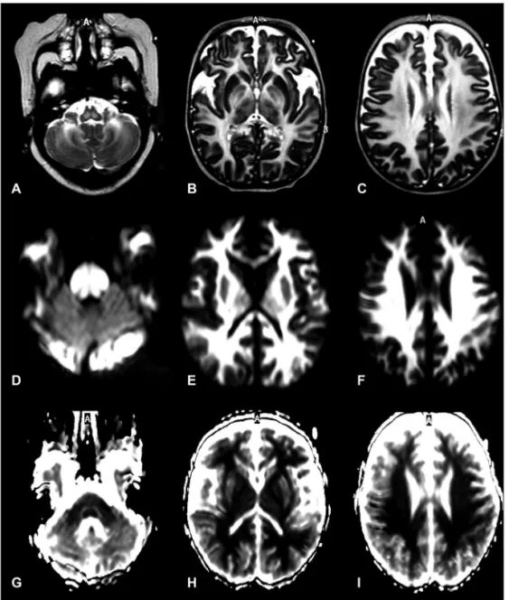
At 20 days of life the clinical condition of the baby became critical and maple syrup odor or sugar burnt was noted in the urine. A repeat MRI at this time showed increasing myelinating white matter lesions and new hypersignal lesions on T2-weight-ed sequences and hyposignal on the diffusion-weightT2-weight-ed imag-es located in the unmyelinated white matter of the frontal, pa-rietal and temporal lobes (Fig 2).
At 25 days of age, chromatography showed an increase of the branched-chain amino acids in the plasma with elevated
lev-els of leucine = 2,982 µmol/L (normal values: 48–160 µmol/L);
isoleucine = 648 µmol/L (normal values: 26–91 µmol/L) and
valine = 677 µmol/L (normal values: 86–190 µmol/L). The baby
was treated with a speciic aproteic diet and 48-hours of dialy-sis, which dramatically improved the clinical status. Treatment was continued only with a special nasogastric diet.
Repeat chromatography at 5 months of age showed persis-tently elevated plasma branched-chain amino acids levels with
leucine = 764 µmol/L (normal values: 47–155 µmol/L),
isoleu-cine = 91 µmol/L (normal values: 31–86 µmol/L) and valine = 65
µmol/L (normal values: 64–294 µmol/L).
hy-posignal on ADC maps (Fig 3). Chromatography of the amino ac-ids at this time continued to show increased levels of
isoleu-cine = 767 µmol/L (normal values: 31–86 µmol/L) and valine =
1,128 µmol/L (normal values: 64–294 µmol/L) but a decrease in
the leucine level = 25 µmol/L (normal values: 47–155 µmol/L)
for the age.
This study was approved by the Ethics Research Commit-tee of the Institution.
diSCuSSion
The present case report shows the indings of brain MRI DWI indings in different phases of classic MSUD in a full-term newborn with an unfavorable clinical outcome.
Conventional MRI during the acute metabolic decom-pensation phase of the disease is characterized by
hyper-signals on T2-weighted sequences and the contrary pat-tern in the signal on DWI and ADC maps, which occur both in myelinated and unmyelinated white matter, suggestive of different underlying pathogenetic mechanisms1,3,6.
Myelinated white matter changes during the acute phase of MSUD are characterized by a decreased value of ADC that may be a result of neurotransmitter disor-ders, such as an increase in glutamate, an impaired energy metabolism associated with increases in brain lactate and decreased synthesis of lipid and proteolipid proteins7-9. On the other hand, an increase in ADC intensity in unmy-elinated white matter may be due to blood-brain barrier alterations (vasogenic-edema)10.
In our case, during the acute phase of MSUD the brain-stem myelinated areas and the internal capsules were
Arq Neuropsiquiatr 2009;67(3-A) Maple syrup encephalopathy DW/MRIFerraz-Filho et al.
characterized by hypersignal on DWI and hyposignal on ADC maps, which suggests the presence of intramyelin-ic cytotoxintramyelin-ic edema. At this time, an increase in branched-chain aromatic amino acids in plasma (leucine, isoleucine, and valine) was observed coinciding with the appearance of cerebral edema and the consequent clinical aggravation characterized by seizures, bradycardia and apnea leading to coma.
Subsequently, the lesions in brain regions that are my-elinated early improved but new lesions appeared in the periventricular and subcortical white matter after the irst year of life. The new lesions had hyposignals on ADC maps compatible with cytotoxic edema. These new le-sions showed a rapid and progressive disease course
fol-lowing the normal brain myelinization, which was concur-rent with clinical deterioration associated with anemia, urinary tract infection and a persistent increase in isoleu-cine and valine amino acids.
The unfavorable outcome in the present report may be justiied because the diagnosis and speciic treatment were delayed; there was no improvement in the imaging pattern and amino acid plasma levels in follow-up study.
Ha et al.1 described a case of MSUD in which cytotox-ic edema regressed parallel to the increasing ADC value, however vasogenic-interstitial edema evolved to brain at-rophy with worsening of the ADC value. The patient also had increased leucine plasma levels at follow-up.
On the other hand, normalization of signal
es of DWI and ADC maps on MRI follow-up have been show in patients with decreases of amino acid plasma lev-els and corresponding clinical improvement, which is at-tributable to both the duration of encephalopathy in the acute phase of MSUD and the success of early speciic treatment3,5,6,11.
The unfavorable outcome in children with MSUD is consistently bad when the disease is diagnosed after 14 days1,12. According to Morton et al.12 the prolonged dei-ciencies of one or more of the amino acids caused by ex-cessive dietary restriction causes anemia and immunode-iciency, as well as dysmyelination, poor head growth and overall developmental delays. These authors also men-tioned that the brain edema and hyponatremia were as-sociated with hypersignals on T2-weighted MRI through-out the deep gray matter, different to what was observed
in our case in which the serum sodium levels remained normal.
Diffusion-weighted MRI can demonstrate the involve-ment of myelinated white matter in newborns in the acute phase of MSUD. Follow-up DWI associated with amino acid plasma level measurements may be of predictive val-ue for the clinical outcome and the eficacy of treatment.
referenCeS
1. Ha JS, Kim TK, Eun BL, et al. Maple syrup urine encephalopathy: a fol-low-up study in the acute stage using diffusion-weighted MRI. Pediatr Radiol 2004;34:163-166.
2. Sakai M, Inoue Y, Oba H, et al. Age dependence of
diffusion-weight-ed magnetic resonance imaging indings in maple syrup urine disease
encephalopathy. J Comput Assist Tomogr 2005;29:524-427.
3. Cavalleri F, Berardi A, Burlina AB, Ferrari F, Mavilla L. Diffusion-weighted MRI of maple syrup urine disease encephalopathy. Neuro-radiology 2002;44:499-502.
Arq Neuropsiquiatr 2009;67(3-A) Maple syrup encephalopathy DW/MRIFerraz-Filho et al.
4. Parmar H, Sitoh YY, Ho L. Maple syrup urine disease:
diffusion-weight-ed and diffusion-tensor magnetic resonance imaging indings. J Com -put Asssist Tomogr 2004;28:93-97.
5. Jan W, Zimmerman RA, Wang ZJ, Berry GT, Kaplan PB, Kaye EM. MR dif-fusion imaging and MR spectroscopy of maple syrup urine disease dur-ing acute metabolic decompensation. Neuroradiology 2003;45:393-399. 6. Righini A, Ramenghi LA, Parini R, Triulzi F, Mosca F. Water apparent
diffusion coeficient and T2 changes in the acute stage of maple syrup
urine disease: evidence of intramyelinic and vasogenic-interstitial ede-ma. J Neuroimaging 2003;13:162-165.
7. Tavares RG, Santos CE, Tasca CI, Wajner M, Souza DO, Dutra-Filho CS. Inhibition of glutamate uptake into synaptic vesicles of rat brain by the metabolites accumulating in maple syrup urine disease. J Neurol Sci 2000;181:44-49.
8. Felber SR, Sperl W, Chemelli A, Murr C, Wendel U. Maple syrup urine
disease: metabolic decompensation monitored by proton magnetic res-onance imaging and spectroscopy. Ann Neurol 1993;33:396-401. 9. Jouvet P, Rustin P, Taylor DL, et al. Branched chain amino acids induce
apoptosis in neural cells without mitochondrial membrane depolarization or cytochrome C release: implications for neurological impairment asso-ciated with maple syrup urine disease. Mol Biol Cell 2000;11:1919-1932. 10. Riviello JJ Jr, Rezvani I, DiGeorge AM, Foley CM. Cerebral edema
causing death in children with maple syrup urine disease. J Pediatr 1991;119:42-45.
11. Sener RN. Maple syrup urine disease: Maple syrup urine disease:
dif-fusion MRI, and proton MR spectroscopy indings. Comput Med Im -aging Graph 2007;31:106-110.
![Fig 1. Axial - T2-weighted [A–C], diffusion-weighted [D–F], ADC maps [G–I]: in the acute metabolic decompensation shows hyperintense lesions on the diffusion-weighted images and hypointense on ADC maps in the brainstem, basal ganglia, thalami and white m](https://thumb-eu.123doks.com/thumbv2/123dok_br/15432341.594924/2.955.177.735.91.778/weighted-diffusion-metabolic-decompensation-hyperintense-diffusion-hypointense-brainstem.webp)