Letters to the Editor
Radiol Bras. 2016 Jan/Fev;49(1):56–64
58
http://dx.doi.org/10.1590/0100-3984.2014.0057 infarction(5,6), spontaneous or traumatic rupture, congestive heart
failure, and Kasabach-Merritt syndrome(2,6,7).
A correct diagnosis of the pedunculated lesion may be diffi-cult, despite the typical radiological presentation, because of the limitation in define the origin of the mass, since a thin pedicle may be almost undetectable at images(1,4,5).
The most used modalities of imaging in diagnosis include US, CT and MRI(1–4,6,8). At US, the image is typically hyperechoic, homogeneous, with well defined margins; and, in cases of giant lesions, central heterogeneity may be present(8). At CT, with a certain frequency, giant hemangiomas do not present with the typical pattern of hypoattenuating lesion with centripetal enhance-ment and homogenization at delayed sections, due to the pres-ence of avascular areas of necrosis, fibrosis or hemorrhage(3,8). MRI is the most sensitive and specific (> 90%) diagnostic method(4,6). The lesions are well defined, homogeneous, with low signal intensity at T1-weighted sequences, and high signal in-tensity at T2-weighted sequences.
Biopsy is not recommended in such cases, due to the risk of hemorrhage(6).
There are reports in the literature describing pedunculated hemangiomas as gastric, adrenal tumor(1,4), retroperitoneal mass(1), other pedunculated liver tumors such as hepatocellular carcinoma, mesenchymal hamartoma, focal nodular hyperplasia or adenoma(4).
Surgical treatment is reserved for cases of giant or symp-tomatic lesions, uncertain diagnosis, lesions with complica-tions(1,2,4–7), and for cases of pedunculated hemangiomas due to their tendency to torsion(5,6).
REFERENCES
1. Ha CD, Kubomoto SM, Whetstone BM, et al. Pedunculated hepatic he-mangiomas often misdiagnosed despite their typical findings. The Open Surgery Journal. 2013;7:1–5.
2. Moon HK, Kim HS, Heo GM, et al. A case of pedunculated hepatic he-mangioma mimicking submucosal tumor of the stomach. Korean J Hepatol. 2011;17:66–70.
3. Choi BI, Han MC, Park JH, et al. Giant cavernous hemangioma of the liver: CT and MR imaging in 10 cases. AJR Am J Roentgenol. 1989;152: 1221–6.
4. Liang RJ, Chen CH, Chang YC, et al. Pedunculated hepatic heman-gioma: report of two cases. J Formos Med Assoc. 2002;101:437–41. 5. Ersoz F, Ozcan O, Toros AB, et al. Torsion of a giant pedunculated liver
hemangioma mimicking acute appendicitis: a case report. World J Emerg Surg. 2010;5:2.
6. Guenot C, Haller C, Rosso R. Hémangiome caverneux pédiculé géant du foie: à propôs d’un cas et revue de la littérature. Gastroenterol Clin Biol. 2004;28:807–10.
7. Acharya M, Panagiotopoulos N, Bhaskaran P, et al. Laparoscopic resec-tion of a giant exophytic liver haemangioma with the laparoscopic Habib 4× radiofrequency device. World J Gastrointest Surg. 2012;4:199–202. 8. D’Ippolito G, Appezzato LF, Ribeiro ACR, et al. Apresentações incomuns do hemangioma hepático: ensaio iconográfico. Radiol Bras. 2006;39:219– 25.
Paula de Castro Menezes Candido1, Izabela Machado Flores Pereira1, Breno Assunção Matos1, Mario Henrique Giordano Fontes1, Teófilo Eduardo de Abreu Pires1, Petrônio Rabelo Costa1
1. Hospital Felício Rocho, Belo Horizonte, MG, Brazil. Mailing Address: Dra. Paula de Castro Menezes Candido. Hospital Felício Rocho – Setor de Radiologia. Avenida do Contorno, 9530, Barro Preto. Belo Horizonte, MG, Brazil, 30110-934. E-mail: [email protected].
Posterior reversible encephalopathy syndrome following immunoglobulin therapy in a patient with Miller-Fisher syndrome
Síndrome encefálica reversível posterior em paciente com síndrome de Miller-Fisher pós-tratamento com imunoglobulina
Dear Editor,
A 54-year-old female patient presenting with ophthalmopa-resis, ataxia and areflexia for one week. The patient denied fever, muscle weakness, and did not report any previous comorbidity. At physical examination, she was normotensive, oriented, with bilat-eral flexor cutaneous-plantar reflex and preserved superficial/deep sensitivity. Human immunodeficiency virus, Epstein-Barr virus, cytomegalovirus, HTLV-1 and VDRL serologies were negative. Considering such findings, the hypothesis of Miller-Fisher
syn-drome was raised, and liquor cerebrospinalis analysis demonstrated hyperproteinorachia, confirming the diagnosis.
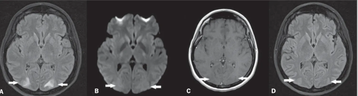
Within 24–48 hours after immunoglobulin therapy initiation, the patient presented with intense headache followed by tonic-clonic seizures and later decreased level of consciousness, with no asso-ciation with hypertensive peaks. Magnetic resonance imaging (MRI) (Figure 1A,B,C) showed sparse hyperintense areas in the white substance, bilaterally on T2-weighted and FLAIR sequences, predominantly in the parieto-occipital regions, without diffusion restriction and without gadolinium enhancement, demonstrat-ing an imagdemonstrat-ing pattern suggestive of posterior reversible encepha-lopathy syndrome (PRES). After the therapy suspension and adop-tion of support measures, the patient progressed satisfactorily, with no sequelae and reversion of the MRI findings (Figure 1D).
Figure 1.A: Axial MRI FLAIR sequence demonstrating hyperintensity in the occipital lobes white substance bilaterally and symmetrically (arrows). B: Axial diffusion-weighted MRI does not demonstrate any alterations (arrows). C: Contrast-enhanced T1-weighted sequence revealing absence of gadolinium-enhanced areas (arrows). D: Axial FLAIR sequence acquired after four weeks demonstrating resolution of the alterations in the occipital lobes white substance (arrows).
Letters to the Editor
Radiol Bras. 2016 Jan/Fev;49(1):56–64
59
http://dx.doi.org/10.1590/0100-3984.2015.0129 The Brazilian radiological literature has recently highlighted
the relevant role played by MRI in the improvement of the diag-nosis of central nervous system conditions(1–5).
PRES is a clinical-radiological entity of varied etiology, gen-erally occurring in the setting of severe arterial hypertension. In some cases, however, it may be associated with immunosuppres-sive therapy, and is rarely described in the literature after the use of immunoglobulin(6–12). Its physiopathogenesis is characterized by the presence of endothelial lesion and dysfunction of cerebral autoregulation mechanisms, leading to hypoperfusion and vasogenic edema(7–12). The clinical manifestations present acute/ subacute onset characterized by headache, decreased level of consciousness, visual alterations, tonic-clonic seizures and focal neurological signs. The symptoms are progressive. Complete re-gression is achieved provided the syndrome is appropriately treated; otherwise irreversible damages may occur(6–11).
MRI findings are quite suggestive and characterized by hyperintense areas on T2-weighted and FLAIR sequences, in general affecting the white substance bilaterally and symmetri-cally, with predilection for the parieto-occipital region. It may also affect the frontal lobes, internal and external capsules, cerebel-lum and encephalic trunk(7–9). At early stages of the condition, diffusion MRI does not demonstrate any abnormalities, but inap-propriate management may result in irreversible damages pre-sented as diffusion restriction corresponding to cytotoxic edema. Recent studies by means of retrospective analysis, utilizing MRI and laboratory data, have demonstrated the association be-tween PRES and albumin serum levels. There are evidences that significantly decreased albumin serum levels lead to a higher risk to develop vasogenic-type edema(12). This is due to the fact that, in conditions with endothelial damages caused by inflammatory processes, the decrease in the colloidosmotic pressure, directly re-lated to the albumin levels, may facilitate the development of vasogenic edema. Thus, the early administration of human se-rum albumin might prevent ischemic damages and reduce pos-sible sequelae(12).
Finally, despite being rare after administration of immuno-globulin, PRES should be considered in cases where typical MRI findings are present. One should not wait until the onset of a hy-pertensive episode to take such a diagnostic possibility into con-sideration.
REFERENCES
1. Bimbato EM, Carvalho AG, Reis F. Toxic and metabolic encephalopa-thies: iconographic essay. Radiol Bras. 2015;48:121–5.
2. Castro FD, Reis F, Guerra JGG. Intraventricular mass lesions at mag-netic resonance imaging: iconographic essay – part 1. Radiol Bras. 2014; 47:176–81.
3. Ono SE, Carvalho Neto A, Gasparetto EL, et al. X-linked adrenoleukodys-trophy: correlation between Loes score and diffusion tensor imaging parameters. Radiol Bras. 2014;47:342–9.
4. Alfenas R, Niemeyer B, Bahia PRV, et al. Parry-Romberg syndrome: findings in advanced magnetic resonance imaging sequences – case report. Radiol Bras. 2014;47:186–8.
5. Barbosa JHO, Santos AC, Salmon CEG. Susceptibility weighted imag-ing: differentiating between calcification and hemosiderin. Radiol Bras. 2015;48:93–100.
6. Stetefeld HR, Lehmann HC, Fink GR, et al. Posterior reversible en-cephalopathy syndrome and stroke after intravenous immunoglobulin treatment in Miller-Fisher syndrome/Bickerstaff brain stem encephali-tis overlap syndrome. J Stroke Cerebrovasc Dis. 2014;23:e423–5. 7. McKinney AM, Short J, Truwit CL, et al. Posterior reversible
encepha-lopathy syndrome: incidence of atypical regions of involvement and im-aging findings. AJR Am J Roentgenol. 2007;189:904–12.
8. Pereira PR, Pinho J, Rodrigues M, et al. Clinical, imagiological and etiological spectrum of posterior reversible encephalopathy syndrome. Arq Neuropsiquiatr. 2015;73:36–40.
9. Bartysnki WS, Boardman JF. Distinct imaging patterns and lesion dis-tribution in posterior reversible encephalopathy syndrome. AJNR Am J Neuroradiol. 2007;28:1320–7.
10. Bartynski WS. Posterior reversible encephalopathy syndrome, part 1: fundamental imaging and clinical features. AJNR Am J Neuroradiol. 2008;29:1036–42.
11. Wada A, Yoshida R, Oda K, et al. Acute encephalopathy associated with intravenous immunoglobulin therapy. AJNR Am J Neuroradiol. 2005; 26:2311–5.
12. Pirker A, Kramer L, Voller B, et al. Type of edema in posterior reversible encephalopathy syndrome depends on serum albumin levels: an MR im-aging study in 28 patients. AJNR Am J Neuroradiol. 2011;32:527–31.
Bruno Niemeyer de Freitas Ribeiro1, Tiago Medina Salata2, Rafael Silveira Borges2, Edson Marchiori3
1. Instituto Estadual do Cérebro Paulo Niemeyer, Rio de Janeiro, RJ, Bra-zil. 2. Hospital Casa de Portugal / 3D Diagnóstico por Imagem, Rio de Janeiro, RJ, Brazil. 3. Universidade Federal do Rio de Janeiro (UFRJ), Rio de Janeiro, RJ, Brazil. Mailing Address: Dr. Bruno Niemeyer de Freitas Ribeiro. Instituto Estadual do Cérebro Paulo Niemeyer – Serviço de Ra-diologia. Rua do Rezende, 156, Centro. Rio de Janeiro, RJ, Brazil, 20231-092. E-mail: [email protected].
Pulmonary paracoccidioidomycosis showing reversed halo sign with nodular/coarse contour
Paracoccidioidomicose pulmonar exibindo sinal do halo invertido com margens nodulares/rugosas
Dear Editor,
A 63-year-old man, living and working in urban area since his childhood, and smoking for 30 years. In 2012 he underwent investigation for chronic cough. At that same time, he reported gingival lesion. For a long time, he had the habit of weekly visit-ing rural areas for leisure and amateur fishvisit-ing. The patient de-nied history of fever, weight loss or comorbidities. Blood counts since 2009 without any abnormalities.
Chest computed tomography (CT) in December 30, 2013 showed focal pulmonary ground glass opacities predominantly in the middle fields, some of them completely or partially surrounded by a thin and coarse consolidation ring representing the “reversed halo sign”. Other findings include some areas with subtle inter-lobular septa thickening (Figures 1A, 1B and 1C).
The gingival lesion, characterized by granular, erythematous ulceration with fine blood-red dots, compatible with a “mulberry-like” appearance, was biopsied.
Biopsy result: eosinophilic epithelial cells of the squamous and spinous layers, giant, “foreign-body” type cells containing iso-lated and clustered spherical fungi with double and birefringent membranes, in association with inflammatory cells. The cytologi-cal diagnosis confirmed the presence of Paracoccidioides brasi-liensis (Figure 1D).
On February 2, 2015, post-itraconazol therapy chest CT dem-onstrated rare areas of hypoattenuation associated with fibrocicatricial septal thickening.