National University of La Plata and University of Algarve
DECIPHERING THE EFFECTS OF METAL-BASED DRUGS ON CELL SIGNALING
PATHWAYS IN CANCER CELLS
Kateryna Babych
Dissertação / Dissertation
Mestrado Erasmus Mundus em Inovação Química e Regulamentação / (Erasmus Mundus Master in Chemical Innovation and Regulation
Trabalho efetuado sob a orientação de/ Work supervised by: Prof. Ignacio E. León and Prof. Vera Linda Ribeiro Marques
Faculdade de Ciências e Tecnologia 2019
DECLARATION OF AUTHORSHIP AND COPYRIGHT
I declare that I am the author of this work, which is original. The work cites other authors and works, which are adequately referred in the text and are listed in the bibliography.
_____________ _______________________
(date) (name)
Copyright: Kateryna Babych. The Universidad Nacional de La Plata (National University of La Plata) and Universidade do Algarve (University of Algarve) have the right to keep and publicize this work through printed copies in paper of digital form, or any other means of reproduction, to disseminate it in scientific repositories and to allow its copy and distribution with educational and/or research objectives, as long as they are non-commercial and give credit to the author and editor.
i Acknowledgment
I would like to express my profound gratitude to my thesis advisors, Prof. Ignacio E. León of the National University of La Plata and Prof. Vera Ribeiro of the University of Algarve for excellent supervision, knowledge, support, and practical teaching, especially on cell culture, during my research work. My acknowledgment and appreciation to the research team of CEQUINOR and especially my advisors, Maria Carolina Ruiz, Lucia Mariana Balsa and Juan Cadavid, for accepting, guiding me and also for sharing knowledge on cell culture. Thanks to Jacira Ramos for help, support, and warm words.
To the most important people in my life, my family, for all your love, and encouragement for through all the distance between us. To my closest friends in Ukraine for support and keeping the place in your hearts. To my friends worldwide, I was lucky to meet you and thanks for the gained experience. My thanks to my ChIR colleagues, you opened my eyes wider to the world and thanks for sharing your experience and culture. Special thanks to Francisco Vega for the warmest memories and unforgettable moments together, it would be not the same without you. Thanks, Kai for your help.
I would like to thank the European Commission and Erasmus Mundus committee for funding and allowing me to make this possible.
Thanks to all not mentioned here people and to every person I met during this journey, who made me laugh, learn, grow, feel and enjoy life; who contributed directly or indirectly to this work and made this happened.
“A journey of thousand miles begins with a single step” Kateryna Babych, 2019.
ii Abstract (ENG)
The term „cancer‟ encompasses a large group of diseases that are characterized by the development of abnormal cells, which divide, grow and spread without control in any part of the organism, spreading through blood or lymph vessels into surrounding tissues. Chemotherapy is a type of cancer treatment that uses drugs to kill cancer cells and is focused on stopping or slowing the growth of cancer cells, which divide abnormally and causes tumors. In most of the cases of chemotherapy prescription and selection of the specific drug, the more effective is a combination of two or more medications that, preferentially, should not interact with each other, either on their mechanism of action and/or their metabolism and elimination. Second and third generations of the existing drugs show increased bioactivity against cancer, but side effects are still a matter of concern. Recent studies show a significant interest in metal-based drugs, with some existing drugs being used already as antitumor agents, with proven effectiveness and fewer side effects, comparing to other drug treatments.
Copper(II) as a metal and its complexes with various organic compounds have been reported to show cytotoxic activity at low concentrations. The aim of this work is to examine the effects of newly synthesized copper complexes in human cancer cell lines, both in terms of cytotoxicity and of mechanism of action. In this research work, two copper(II) based compounds were (copper(II)-tropolone and copper(II)-hinokitiol complexes), for their cytotoxic properties and tested in the human mammary breast cancer cell line MCF7 and MDA-MB-231 for their effect on viability, oxidative stress, apoptosis and interaction with DNA. Additionally, as a model for in vivo studies, the 3D model testing on cell viability was conducted and showed a positive result against MCF7 cell line survival. Together with that, the comparative analysis of complexes, its ligands and copper salt was performed. These compounds showed quite promising results in terms of their potential effect as antitumor drugs.
iii Resumen (ES)
El término „cáncer‟ comprende un largo grupo de enfermedades que están caracterizadas por el desarrollo de células anormales, las cuales se dividen, crecen y proliferan sin control en cualquier parte del organismo, proliferándose por medio de la sangre o vasos linfáticos dentro de los tejidos cercanos. Quimioterapia es un tipo de tratamiento para el cáncer que usa medicamentos (también conocidas como drogas) para matar las células cancerígenas y que está enfocado en detener o alentar el crecimiento de dichas células, las cuales se dividen de manera anormal llevando a la formación de tumores. En la mayoría de los casos de quimioterapia, la selección del medicamento específico, siendo la combinación más efectiva la de uno o más medicamentos que, preferencialmente, no interactúen entre ellos, ni tampoco con sus mecanismos de acción yo su metabolismo o eliminación. Los medicamentos existentes de segunda y tercera generación muestran bioactividad incrementada contra el cáncer, pero los efectos secundarios son todavía un tema de preocupación. Estudios recientes muestran un interés significativo por medicamentos con base-metálica, con algunos de los medicamentos existentes siendo usados ya como agentes anti-tumores, con efectividad probada y menos efectos secundarios, en comparación con el resto de los tratamientos.
Cu(II) como metal, junto con los complejos que forma con vatios compuestos orgánicos, ha sido reportado por mostrar citotoxicidad cuando está presente en bajas concentraciones. El objetivo del presente trabajo es examinar los efectos de nuevos complejos metálicos de cobre en líneas celulares cancerígenas humanas, lo anterior, tanto en términos de citotoxicidad y de mecanismo de acción. En este trabajo de investigación, dos compuestos fueron considerados para observación y evaluación (complejos Cobre(II) y Cobre (II)-tropolone y Cobre(II)-hinokitiol), por y sus propiedades de citotoxicidad y estudios en las líneas celulares humanas de cáncer de mama MCF y MDA-MB-231 por su efecto en disponibilidad, estrés oxidativo, apoptosis e interacción con el ADN. Adicionalmente, para un modelo de estudios in vivo, el modelo 3D fue realizado para evaluar su disponibilidad, y demostró un resultado positivo contra la línea celular MCF7 sobreviviente. Aunado a esto, el análisis comparativo de los complejos, sus ligantes y las sales de cobre fue realizada. Estos compuestos mostraron resultados prometedores en términos de su efecto como medicamentos antitumorales.
iv Palabras clave: cancer de mama, MFC7, MDA-MD-231, complejos metálicos, Cu(trp)2,
v Resumen (PT)
O termo „cancro‟ abrange um largo número de doenças, que são caracterizadas pelo desenvolvimento anormal de células que se dividem, crescem e propagam-se de forma descontrolada por todo o organismo, propagando-se através do sangue ou dos vasos linfáticos para os tecidos vizinhos. A quimioterapia é um tipo de tratamento anticancerígeno, que tem por base o uso de fármacos que atuam por parar ou por diminuir o crescimento de células cancerígenas, células que se dividem anormalmente causando tumores. Na maioria dos casos, ao prescrever um fármaco específico, o uso combinado de dois ou mais fármacos é preferencial, sendo que não deve haver nenhum tipo de interação entre os mesmos, quer no mecanismo de ação e/ou metabolismo, quer na eliminação. A segunda e a terceira geração de fármacos existentes, mostraram um aumento da bioatividade contra o cancro, contudo, os efeitos secundários continuam a ser uma grande preocupação. Estudos recentes têm demonstrado um interesse significativo em fármacos com compostos metálicos, sendo que alguns destes fármacos já são usados como agentes antitumorais, com eficácia e menos efeitos secundários comprovados, comparativamente com outros fármacos.
O Cobre (II), tanto como metal, tanto complexado com vários compostos orgânicos, mostrou ter atividade citotóxica a baixas concentrações. O objetivo desde trabalho é analisar, em linhas celulares humanas, os efeitos de complexos de cobre recentemente sintetizados, tanto em termos de citotoxicidade como em termos de mecanismo de ação. Neste trabalho de investigação, dois compostos com base no cobre (II), (cobre(II)-tropolone) e cobra(II)-hinokitiol complexos), foram observados e avaliados quanto às suas propriedade citotóxicas, e testados na linha celular humana de cancro da mama MCF7 e MDA-MB-231, quanto ao seu efeito na viabilidade, no stress oxidativo, na apoptose e na interação com o DNA. Adicionalmente, como modelo para estudos in vivo, foi conduzido um teste modelo 3D quanto à viabilidade, onde foram observados resultados positivos contra a linha celular MCF7 sobrevivente. Simultaneamente, foi realizada uma análise comparativa dos complexos sintetizados, dos seus ligandos e do sal de cobre. Estes compostos mostraram resultados promissores com efeitos potenciais como fármacos antitumorais.
vi List of abbreviations and acronyms
ACS American Cancer Society
BC Before Christ
ctDNA Calf thymus DNA DMSO Dimethyl sulfoxide
EB Ethidium bromide
EGFR Epidermal growth factor receptor ER Estrogen receptor
HER2 Human epidermal growth factor receptor-2 IARC International Agency for Research on Cancer MTS Multicellular tumor spheroid
PBS Phosphate buffer saline PI Propidium iodide PR Progesterone receptor PS Phosphatidylserine PXR Pregnane X receptor ROS Reactive oxygen species
RT Room temperature
SD Standard deviation UV-vis UV-visible
vii List of Figures
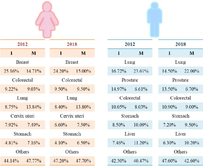
Figure 1.1. Distribution data, presented in percentage values, for the 5 most widespread cancer types in women (left) and men (right). The data represents the proportion of the total number of cancer cases, in terms of I (I=Incidence cases) and M (M=Mortality), age-standardized in 2012 to 2018 for its comparative analysis. The values were collected worldwide from 185 countries and estimated by IARC ... 6
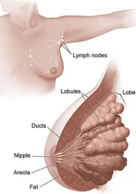
Figure 1.2. The female breast and adjacent lymph nodes and vessels. ... 7
Figure 1.3. Bar of 5-Year Relative Survival values in percentages for the period of 2009 – 2015 by the diagnosed localization of breast cancer in all races and categorized by ages. Stage at diagnosis is classified using SEER Summary Stage 2000 ... 9
Figure 1.4. Numbers of articles, presented in PubMed search on the request „metal compounds cancer‟ from 1967 to 2018 ... 12 Figure 2.1. The scheme of synthesis and structure of the copper-tropolone complex ... 21
Figure 2.2. The scheme of synthesis and structure of copper-hinokitiol complex ... 22
Figure 2.3. Microscope photo of MCF7 cells in a presence of DMEM (left) and in low and high density of cells (right) ... 24
Figure 2.4. Microscope photos of MDA-MB-231 cells in a presence of DMEM (left) and in low and high density of cells (right) ... 25
Figure 2.5. The irreversible reduction of non or weakly fluorescent resazurin (alamar blue) into pink highly fluorescent resorufin, the last one indicates by fluorescent spectrophotometry with the emission at 590 nm and excitation at 570 nm ... 32
Figure 3.1. The influence of Tropolone, Vanadium-tropolone and Copper-tropolone complexes, Copper chloride on the MCF7 (above) and MDA-MB-231(below) cell lines after 48h treatment. The results are expressed as the percentage of the basal and represent the values of mean ± SD *(p = 0.0003); **(p <0.0001) significant with respect to Basal ... 34
Figure 3.2. The effect of Hinokitiol and Copper-hinokitiol complexes on the MCF7 (left) and MDA-MB-231(right) cell lines after 48h treatment. The results are expressed as the percentage of
viii the basal and represent the values of mean ± SD. *(p = 0.0003); **(p <0.0001) significant with respect to Basal ... 35
Figure 3.3. The effect of Cu(hin)2 on migrating properties of MCF7 and MDA-MB-231 cells.
Results were obtained are measured in pixels an of healing before (0h) and after 48 h treatment and expressed in percentage of area, in a respect to control (0h) ± SD. *(p = 0.0003); **(p <0.0001) significant with respect to Basal ... 37
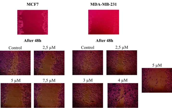
Figure 3.4. Microscope photos of the cell monolayers and scratches, done before introducing the drug (above) and the same areas after 48h of treatment (below) with different concentrations of Cu(hin)2 and a control ... 38
Figure 3.5. ROS production in the presence of increasing concentrations of Cu(hin)2 and
Cu(trp)2 of the MCF7 and MDA-MB-231 cell lines. The generation of ROS of the cells was measured by the absorption of oxidized DHR123 (dihydrorhodamine123) into rhodamine123. The conversion of DHR123 and the changes of the absorption indicate the presence of ROS in the cells. The values represent the mean ± SD and expressed as percentages with respect to the Basal level ... 39
Figure 3.6. The fluorescence, measured in a presence ctDNA, sodium chloride in water as a solvent and ethidium bromide (EB) with the various concentrations of the complexes. The data is presented corresponding with intensity to nm of the measured wavelength in the presence of Cu(hin)2 (left) and Cu(trp)2 (right). The control sample is measured as a blank sample without the presence of drugs. The shift of wavelength down and closer to the baseline characterizes the presence of the interaction into ctDNA in the presence of both of the complexes ... 41
Figure 3.7. The UV-vis absorption graphs of ctDNA interaction with drugs of interest. As a control baseline, marked as the green line, was a sodium chloride aqueous solution (0,9% w/w). The absorbance spectra is shown in a and b for Cu(trp)2 and Cu(hin)2 respectively. The
measurement was taken in a constant concentration of ctDNA ... 43
Figure 3.8. The UV-vis absorption graphs of ctDNA interaction with drugs of interest. As a control baseline, marked as the green line, was sodium chloride aqueous solution (0,9% w/w) together with ctDNA solution. The absorbance spectra is shown in c and d for Cu(trp)2 and
ix Figure 3.9. The UV-vis absorption graphs of ctDNA interaction with drugs of interest. As a control baseline, marked as the red line, was a sodium chloride aqueous solution (0,9% w/w). The absorbance spectra is shown in e and f for Cu(trp)2 and Cu(hin)2 respectively. The
measurement was taken in a constant concentration of complexes ... 45
Figure 3.10. The UV-vis absorption graphs of ctDNA interaction with drugs of interest. As a control baseline, marked as the red line, was sodium chloride aqueous solution (0,9% w/w) together with each of the complexes. The absorbance spectra is shown in g and h for Cu(trp)2 and
Cu(hin)2 respectively. The measurement was taken in a constant concentration of complexes ... 46
Figure 3.11. Effect of Cu(hin)2 on cell apoptosis in MCF7 cell line, determined by flow
cytometer with Annexin V-FITC/Propidium Iodide (PI) staining. The 48 h incubation after treatment with different concentrations of Cu(hin)2 was performed and analyzed by flow
cytometer afterward ... 48
Figure 3.12. The representation of the amount of early, late apoptotic and necrotic MCF7 cells, respectively in the presence of Cu(hin)2 compared to control. The measurement was performed
after 48h treatment with various concentrations of 2.5 µM, 5 µM and 7.5 µM of complex. The values represent the mean ± SD and are expressed as a number with respect to the Basal level .. 49 Figure 3.13. Microscope photos of MCF7 cellular spheroids growth in DMEM medium with FBS over 12 days ... 51
Figure 3.14. The microscope photos of MCF7 cellular spheroids in DMEM medium after 48h treatment of Cu(trp)2 (higher) and Cu(hin)2 (lower) ... 52
Figure 3.15. The effect of Cu(trp)2 and Cu(hin)2 complexes on the MCF7 cell spheroids after
48h treatment. The results are expressed as the percentage of the basal and represent the values of mean ± SD. *(p = 0.0003); **(p <0.0001) significant with respect to Basal ... 52
x List of Tables
Table 1.1. Overview on some physical, chemical, environmental properties and hazards, related to copper. *Data calculated by EPISuite; **Data from European Chemicals Agency (ECHA) ... 13
Table 1.2. Overview on some physical, chemical, environmental properties and hazards, related to tropolone and hinokitiol. *Data, calculated by EPISuite; **Data from European Chemicals Agency (ECHA). ... 17
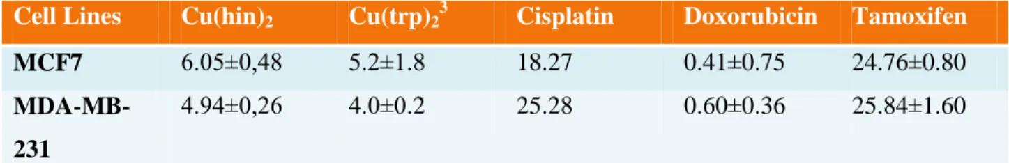
Table 3.1. The values of IC50, measured in µM and obtained after 48h of treatment in a presence of the Copper-hinokitiol complex. The results for complexes of the observation are the mean ± SD of independently performed experiments ... 36 Table 3.2. The values of IC50, measured in µM and obtained after 48h of treatment of MCF7 and MDA-MB-231 cell lines. The values represents effectiveness in a dose-response assessment for the Copper-hinokitiol and Copper-tropolone complexes versus commercial Cisplatin, Doxorubicin and Tamoxifen drugs. The results for complexes of the observation are the mean ± SD of independently performed experiments ... 36
1 Table of Contents
Acknowledgment ... i
Abstract (ENG) ... ii
Resumen (ES) ... iii
Resumen (PT) ... v
List of abbreviations and acronyms ... vi
List of Figures ... vii
List of Tables ... x
1. Introduction ... 4
1.1. Cancer ... 4
1.1.1. Breast Cancer ... 7
1.1.2. Treatment ... 10
1.2. Metal Complexes in Cancer Treatment ... 11
1.3. General Concerns on Copper Chemistry ... 13
1.3.1. Chemical and Physical Properties ... 15
1.3.2. Copper in Biological and Environmental Systems ... 15
1.3.3. Toxicity and Carcinogenicity Studies ... 16
1.4. General Concerns on Chemistry of Tropolone and Hinokitiol ... 17
1.4.1. Chemical and Physical Properties ... 19
1.4.2. Occurrence in the Environment ... 20
1.4.3. Cytotoxic Properties ... 20
1.5. Objectives ... 20
2. Experimental part ... 21
2 2.1.1. Synthesis ... 21 2.1.1.1. Complex Cu(trp)2 ... 21 2.1.1.2. Complex Cu(hin)2 ... 22 2.1.2. Dilutions ... 22 2.2. Biological part ... 23 2.2.1. Cell Lines ... 23 2.2.1.1. MCF7 ... 23 2.2.1.2. MDA-MB-231 ... 24 2.2.2. 2D Cell System ... 25
2.2.2.1. Cell Viability. MTT assay ... 25
2.2.2.2. Cell Migration ... 26
2.2.2.3. Mechanism of Action ... 27
2.2.2.3.1. Oxidative Stress. Determination of Reactive Oxygen Species (ROS Production) .. 27
2.2.2.3.2. Calf-Thymus Interaction (ctDNA) ... 28
2.2.2.3.2.1.1. Fluorescence studies ... 28
2.2.2.3.2.1.2. UV-vis studies ... 28
2.2.2.3.3. Apoptosis Study ... 29
2.2.3. 3D Cell Systems ... 30
2.2.3.1. Formation of spheroids ... 30
2.2.3.2. Cell viability of spheroids ... 31
2.3. Statistical Analysis ... 32
3. Results and Discussion ... 32
3.1. Results ... 32
3.1.1. Chemical part ... 32
3
3.1.2.1. 2D Observation System ... 33
3.1.2.1.1. Cell Viability ... 33
3.1.2.1.2. Cell Migration ... 37
3.1.2.1.3. Mechanism of Action ... 38
3.1.2.1.3.1. Oxidative Stress. Determination of Reactive Oxygen Species (ROS Production) .. 39
3.1.2.1.3.2. Calf-Thymus DNA Interaction (ctDNA) ... 40
3.1.2.1.3.2.1. Fluorescence studies ... 40
3.1.2.1.3.2.2. UV-vis studies ... 41
3.1.2.1.3.3. Apoptosis Study ... 47
3.1.2.2. 3D Cell Systems ... 50
3.1.2.2.1. Cell viability of spheroids ... 51
4. Conclusions and Discussions ... 54
Bibliography ... 56
Annexes ... 62
Annex 1. The measurement data of UV-vis absorption in the presence of Cu(trp)2 and Cu(hin)2 on the changes in the interaction to ctDNA ... 62
4 1. Introduction
1.1. Cancer
Among different sources, related to the research of cancer, such as the American Cancer Society (ACS), Cancer Research UK, National Cancer Institute and International Agency for Research for Cancer (IARC), most of them define the term „cancer‟ as a combination of diseases, related to abnormal cell change, which leads to uncontrolled cell growth, multiplicity and spreading in tissues, bones, blood, lymph system. Cell proliferation, controlled by different signaling pathways, is a mechanism that is responsible for the normal cell life cycle: from cell growth to cell division and death. While this mechanism undergoes changes, due to cell damage and/or genetic mutations, cells not anymore divide in the normal way, at the same time the cell elimination pathway is disturbed. Therefore, cancer is based on the dysfunction of the cell proliferation mechanism and unmanageable multiplicity of cells, abnormal cell mutations and migration throughout the organism. Cell mutations cause the appearance of tumors, the aggregated form of numerous cells, which could behave independently and differently. Depending on the cell´s origin, distinction is made between noncancerous, precancerous and cancerous tumors.1 In general terms, tumors are divided into two main groups2:
Benign tumors. Tumors that develop slowly and don‟t spread to other parts of the body. In general, they are not harmful, but due to their location in the body they could be life-threatening;
Malignant tumors. These are made up of cancer cells, growing faster, spreading into other tissues, causing damage and metastasis.
Currently, there are more than 100 different types of cancer with specific characteristics. They are differentiated mainly by location in the body, but also there are some types, which are distinguished by age, such as childhood and youth cancers. Some cancers are located in a specific part of the body and affect the organs only in those regions while some varieties are highly metastatic, meaning that cells move from their origin and tumors may appear out of the place of initial localization.
5
Carcinoma. Cancer starts in the skin surface or in tissues that cover internal organs. It has several subtypes, such as adenocarcinoma, basal cell carcinoma, squamous cell carcinoma, and transitional cell carcinoma;
Sarcoma. Cancer that begins in the connective and supportive tissues, such as muscle, fat, blood vessels, lymph vessels, and fibrous tissue (such as tendons and ligaments);
Leukemia. Cancer that forms in blood tissue, such as bone marrow with a further transition into the blood and does not cause solid tumors, but is associated with abnormal growth of white blood cells.
Lymphoma and myeloma. Cancer affects immune system cells (T-, B- or plasma cells) and does not form solid tumors;
Melanoma. Cancer beginning in melanocytes, where melanin is produced. Mainly, it appears in the skin surface, but melanomas could also form in other pigmented tissues, such as the eye.
Brain and spinal cord cancers. Cancer that affects the central nervous system. The reasons for its occurrence, as a disease itself, are a combination of factors, which are related to genetics, environmental factors, and lifestyle factors, such as smoking or physical activity.
Among genetic changes, which contribute to the appearance and development of cancer, there are three main genetic “drivers”: proto-oncogenes, tumor suppressor genes, and DNA repair genes. Proto-oncogenes are genes taking part in the normal cell growth cycle. Nonetheless, if these genes are modified in certain ways, mutated or demonstrate unusual activity, they may become oncogenes, which directly cause cancer. In this case, they enable cells to grow, divide, and proliferate. Tumor suppressor genes are the type of genes that make a protein that is responsible for the negative control of cell growth and division. Different types of changes in their tumor suppressor genes lead cells to divide in an uncontrolled way. In the event of damaged DNA, DNA repair genes are responsible for reversing genetic changes. In the case of failing, cells with such damages get mutated genes, which during division and formation of new cells are conveyed to newly formed cells. Those tend to develop additional mutations and, as a result, may cause the new cells to be potentially cancerous.3
6 From 1990 to 2015, environmental risks shifted from the second largest risk factor of mortality to the 8th largest risk factor, giving way to mostly metabolic and behavioral factors for mortality. Tobacco and alcohol use, unhealthy diet, and lack of physical activity are the highest risk factors of cancer morbidity and are responsible for more than 22% of deaths.4 And only near 15% of cancers diagnosed in 2012 were due to infectious agents.5
Figure 1.1. Distribution data, presented in percentage values, for the 5 most widespread cancer types in women (left) and men (right). The data represents the proportion of the total number of cancer cases, in terms of I (I=Incidence cases) and M (M=Mortality), age-standardized in 2012 to 2018 for its comparative analysis. The values were collected worldwide from 185 countries and estimated by IARC. 6,7
7 According to the World Health Organization (WHO), cancer is the second leading cause of mortality worldwide and 1 of 6 deaths is caused by cancer, accounting for approximately 9.6 million deaths in 2018. The most common are the following five types: lung (2.09 million cases, 1.76 million deaths), breast (2.09 million cases, 627 000 million deaths), colorectal (1.8 million cases, 862 000 million deaths), stomach (1.03 million cases, 783 000 million cases) and liver (782 000 million deaths). The ratio in different types of cancer in men and women is shown in Figure 1.1.
1.1.1. Breast Cancer
In 2018 breast cancer took second place among all other types of cancer for both sexes in age-standardized statistics, presented by IARC, and first place in the mortality rate for women. Breast cancer is the most common and leading cause of morbidity and mortality of women in Africa, North and South America, Asia, Europe, and Oceania. The highest rate of female patients with breast cancer is found in Belgium, Luxembourg and the Netherlands.8
8 In most of the cases, the formation of breast cancer tumors starts in the ducts, which carry milk during lactation from the lobules to the nipple. But in some cases, tumors may occur in other tissues of the breast, and these tumors are typically sarcomas or lymphomas. The majority of tumors cause lumps inside the breast, but not all do, and in this case, early detection plays the largest role in treating breast cancer and increasing the patient´s chances of survival. The anatomy of breast cancer schematically represented in Figure 1.2.
The stages of breast cancer development refer to the place, severity of mutation and size of the tumor. The ability of cancer cells to spread is another critical point in determining the stage of cancer. Lately, in 2018, the American Joint Committee on Cancer (AJCC) has proposed guidance on a categorization of breast cancer stages. If a malignant tumor was found in its place of origin, it‟s named as localized cancer and attributed to the stage I. This means that the area of abnormal cell production is situated only in one part of the breast. It is also considered non-invasive and the common treatment against the tumor is surgery, radiation or a combination of both. It is highly responsive to the treatments and depending on the type of cancer, hormone therapy may be considered effective. The spread to the lymph nodes varies this stage from IA to IB. Regional or distant are called the stages of cancer, when metastases are outside the place of origin, either in breasts or in other parts of the body. Stage II also divides in stage IIA and stage IIB, but like stage I, it has different variations. Stage IIA is diagnosed when tumor reached significant size (usually it‟s between 2 and 5 cm) and has not spread to lymph nodes, or tumor has the size of less than 2 cm and has spread to lymph nodes, or the tumor has moved from the ducts or lobules and appear only in the lymph nodes. Stage IIB means either the patient has been diagnosed with a tumor larger than 5 cm with no spreading to the lymph nodes, or the tumor is about 2-5 cm and cancer appears in the lymph nodes. Stage III is an invasive type and classifies more widely since it is correlated with migrational tumors spreading beyond the direct region of appearance and shifting to the lymph nodes, skin or bones, but not to nearby organs. Differentiation is determined by the size of the tumor and the location of the spreading of cancer, either to lymph nodes or surrounding tissues. Stage IIIA is diagnosed when the size of the tumor is less than 2 cm and cancer affects 4-9 lymph nodes, or the tumor is more than 5 cm and it is shown to have cancer clusters in a lymph node or the tumor is bigger than 5 cm and cancer has spread to lymph nodes, nearby bones or underarms. Stage IIIB is very likely to behave as an inflammatory type of cancer, which is determined by biopsy and distinguished by the following:
9 that tumor could be any size and cancer spread to 9 lymph nodes, and chest wall or breast skin. The stage IIIC characterizes itself by leaving the breast and migrating to the lymph nodes. It means that either cancer spread to more than 10 lymph nodes, or to lymph nodes near the collar bone, or to lymph nodes near underarms or the breastbone are affected. Stage IV is the most advanced stage of cancer, invasive and highly metastatic. It means there are metastases in other organs of the body, more likely in the brain, bones, lungs and/or liver. This stage is considered much more difficult to cure and in some cases incurable. However, depending on cancer type, stage IV could be treated as a chronic condition by numerous combined treatments.10
The 5-year survival rate of breast cancer between 2009 and 2015 was estimated at 89.9%. This value varies by individual case and health of patients, depending on several groups of factors, including treatment response and diagnosed cancer stage, which strongly correlates with the length of survival and the therapy method. Early detection has a crucial influence on survival and convalescence after the patient is diagnosed with cancer. Figure 1.3 represents the 5-year survival rate by the localization diagnosed, for all ages and between all races (both black and white) of breast cancer patients.11
Localized Regional Distant Unstaged/Unknown All Stages 0 20 40 60 80 100
Localized Regional Distant Unstaged/Un
known All Stages
50+ years 99,3 84,9 24,8 49,7 90
<50 years 97 86,9 37,1 75,1 89,6
All 98,8 85,5 27,4 54,5 89,9
10 Figure 1.3. Bar of 5-Year Relative Survival values in percentages for the period of 2009 – 2015 by the diagnosed localization of breast cancer in all races and categorized by ages. The stage at diagnosis is classified using SEER Summary Stage 2000
A distinction of breast cancer types is made by subtype, depending on characteristic receptors for each subtype. Across years of research, it's been determined that there are 4 to 6 various subtypes of breast cancer. From the receptors, different subtypes have different responses, defined as luminal A, luminal B, HER2 enriched, basal-like and claudin-low, and normal breast-like and molecular apocrine subtypes. The adapted and interpreted table in Annex 2 shows the classification of these subtypes. 12–14
Each breast cancer subtype varies either by invasive, migrational characteristics or on prognosis and treatment response. Moreover, several breast cancer subtypes have shown migratory behavior, its cells cause metastases in other parts of the body outside the original location. The most metastatic site among all subtypes was the bones, except for the basal-like subtype. For luminal A and HER2-enriched tumor subtypes, there are greater frequencies of metastasis in the brain, lung, and liver. The basal-like subtype has higher rates of lung, brain and distant nodal metastases.15
1.1.2. Treatment
Each diagnosed tumor type requires a specific treatment type. The most common treatments for cancer are surgery, chemotherapy, and radiation. Surgery is used by doctors to manually remove tumors and sometimes the nearby tissue as a whole, including organs or even an entire body part; and is a preferred treatment to prevent metastasis in other parts of the body. Radiation is the treatment type for cancer targets suppressing and preventing tumor or cell growth. Chemotherapy (or shortly “chemo”) is a common type of therapy used for killing cancer cells, inhibiting their growth, and preventing migration of cancer cells. Additionally, in most cases of chemotherapy prescription, a more effective treatment is a combination of two and more drugs and, in this way, the drugs should not influence the mechanism of action and/or interact between each other. As an additional or supportive therapy, or as a substitute for standard chemo, hormonal and targeted therapy, immunotherapy, stem cell transplantation or a combination of these treatment types could be used. The selection of a specific drug for treating a specific cancer
11 subtype is usually a complex issue and it should be based on several factors, which include: type and stage of cancer, patient‟s age and overall health, patient‟s and tumor‟s genetic characteristics, other serious health problems and past medical history.
For the last decades, the field of research and development of chemotherapy, which uses drugs to target and kill cancer cells and is focused on stopping or slowing the growth of cancer cells, brought a significant positive effect of the survival percentage of patients, as an example for breast cancer patients.16 However, to obtain this outcome a considerable price has been paid regarding different side effects, which influence not only the general health condition of the patient, but also the mental and psychological health of the patient, or could have a significant negative effect on other organs and systems. Specific nature-based compounds, which can be found in plants or animals, are gaining attention not only for their anti-inflammatory and antifungal properties, but their antitumor effects have been proved.17 These compounds and their complexes with other organic and inorganic substituents have much potential as anticancer drugs. In this regard, copper compounds, compared to other metal-based compounds, might take a priority over other metal-based drugs in minimizing the influence of its side effects on the body during cancer treatments.18
Among different types of chemotherapeutic drugs, alone or in combination, the main goal is either to kill existing cells, control and suppress multiplication, or not to allow cells to migrate to any other tissues in the body. Together with this, developing a drug that is able to distinguish the target between abnormal cancer cells and normal cells is still one of the most important issues in cancer treatment research today.
1.2. Metal Complexes in Cancer Treatment
Since discovery anticancer properties of cisplatin in 1967, broad attention was brought to platinum and metal compounds, in general. Nowadays, cisplatin is one of the most potent drug and still using for prescription in 50-70% cases alone or in combination with other drugs for treatment such types of cancer, as bladder, ovarian, cervical, testicular, ovarian, oropharyngeal, bronchogenic carcinomas, lymphoma, osteosarcoma, melanoma and neuroblastoma.19 Although, despite the successful positive anticancer activity, cisplatin raised the issue of high general toxicity in the body during treatment and also has less effective activity against many migrational
12 cell lines and metastatic cancers. Severe side effects are another concern in using cisplatin as an anticancer drug. During treatment, it was observed nephrotoxicity, cardiotoxicity, hepatotoxicity and hear loss, related to using cisplatin.20 In addition, some types of cell lines are resistant to cisplatin and some cancer types can acquire resistance.21 New generations of platinum-based compounds aim to increase efficacy on the treatment of many types of cancer, decrease toxicity and side effects influence, and become more effective anticancer drugs. All these factors lay the foundation of using metal ions in a complex with different other ligands, develop next generations of metal-based drugs, but also refocus from platinum to other metals and metal compounds as, potent chemicals for drug design. Due to exhibited characteristics of transition metals, like redox activity, different coordination modes and metal-ligand interaction, the interest in the potential activity of metal compounds against cancer increased.22
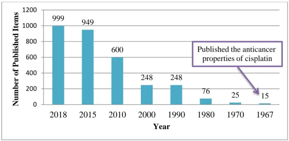
Figure 1.4. Numbers of articles, presented in PubMed search on the request ‘metal compounds cancer’ from 1967 to 2018.
Apart from platinum, the highest interest on research anticancer activity expresses interest to ruthenium, gold, titanium, vanadium, molybdenum and copper compounds. Complexes with these metals were synthesized and showed a certain success in cytotoxic activity and, even more, some of them have been already approved or currently undergoing clinical trials for drugs against many types of cancer. Among others, copper has been investigated mainly in a prospective of being less toxic metal for normal cells, but also effective in the treatment of cancer cells. For more than a decade, mixed chelate copper complexes were synthesized and reported. The group
999 949 600 248 248 76 25 15 0 200 400 600 800 1000 1200 2018 2015 2010 2000 1990 1980 1970 1967 N umber of P ub li shed It em s Year
Published the anticancer properties of cisplatin
13 of over 100 substances, patented under the name Casiopeinas®, substituted with bidentate anionic ligands, like diimine (N=C-C=N), amino acids (N-O) or O-O donors, has exhibited antineoplastic properties and evaluated as a possible anticancer agent. The design of these molecules used a structure of cisplatin as a model and was focused on containing essential metal, preferably cis-configuration of chelates around metal ion and mixed chelated may have different hydrophobicity. Experiments have shown that these compounds are related to the generation of ROS, inducing apoptosis, produce DNA fragmentation and, in terms of cytotoxicity, HeLa cells were more sensitive to the presence of copper compounds, than normal cells.23,24 DNA intercalation and ROS production have been proposed as two main mechanisms of action. Nevertheless, this group of compounds has shown respiratory and cardiovascular toxicity and reducing O2 consumption in in vivo studies. The second and third generations of this group of
drugs induced a significant higher effect on human lung cancer cells, higher selectivity among cancer and normal cells and caused overproduction of ROS in the mitochondria. Selection of more suitable ligands, control the thermodynamics of the reaction or kinetics of ligand substitution may lead to a more accurate design of new successful anticancer drug.25
The complex of Cu(II) with mixed cyano- and cyano-dithiolate ligands has been studied for its antiproliferative activity. Copper complexes with Schiff base ligand exhibit tumor-induced changes in mice and showed higher antitumor activity, compared to cisplatin. Few other copper complexes of ligands together with N-, O- donors and phosphine donors have proved their antitumor activity, being highly selective at the same time.26 Thereby the further research and studies on new copper-based compounds are highly potential.
1.3. General Concerns on Copper Chemistry
The main characteristics of data on copper are presented in Table 1.1.
Chemical Name Copper
Molecular formula Cu
Molecular weight, g/mol 63.55
CAS number 7440-50-8
14
Color Reddish
Physical state Metal
Boiling/melting point, ⁰C* 1083/2595
Density, g/cm3 8.96
Oxidation states I, II
Environmental Properties
Vapor pressure at 25⁰C -
Water Solubility at 25⁰C Insoluble
Log octanol-water k0w* -0.57
Half-Life* -
Ozone Reaction* No Reaction
Soil Adsorption Coefficient* -0.495 Total Removal In Wastewater
Treatment*
90.51%
Total Biodegradation in Wastewater*
0.02%
Safety, Hazards, and Toxicity Pictograms
GHS Hazard Statements** Among reported 56 notifications:
H302: Harmful if swallowed; H319: Causes serious eye irritation; H331: Toxic if inhaled;
H400: Very toxic to aquatic life;
H410: Very toxic to aquatic life with long-lasting effects H411: Toxic to aquatic life with long-lasting effects; H412: Harmful to aquatic life with long-lasting effects;
15
Codes** P304+P340, P305+P351+P338, P311, P321, P330,
P337+P313, P391, P403+P233, P405, and P501
Flammability, Fire Hazard In solid state non-combustible, in powdered form may ignite
Acute Toxicity LD50 (mice) = 110 mg/kg27
Table 1.1. Overview on some physical, chemical, environmental properties, and hazards, related to copper. *Data calculated by EPISuite; **Data from European Chemicals Agency (ECHA)
1.3.1. Chemical and Physical Properties
Copper is reddish color, soft, ductile and malleable metal with high electrical and heat conductivity. Also, it is a transitional metal, with an atomic number of 29 and an atomic weight of 63.546, takes place the period IV and group 11 in the periodic table. Copper is insoluble in the water but dissolves in most acids and alkalines. It reacts with the oxygen in the air, darkens and converts metal into its oxidative form. Together with oxygen from the air, water and carbon dioxide forms copper (II) carbonate hydroxide with its blue-green color, the product of exposure the metal to the air.
First discoveries of copper date in 9000 BC in the Middle East and taking its name from Latin (Cuprum) after the island of Cyprus, which was the mining area for Romans.
1.3.2. Copper in Biological and Environmental Systems
Copper is a trace element, prevalent in nature in elemental forms, forms of salts, complexes in the soil, water, plant and animals and essential nutritional element, for living organisms. It is one of the most common elements, which naturally occurs, spreads throughout the environment by natural processes.
Copper is an elemental part of enzymes and proteins with various biological functions. The metal ion is highly redox-active, being donor and acceptor of electrons while changing from one oxidative state to another (Cu+ ↔ Cu2+). Copper is an important element in reduction-oxidative processes in organisms as a part of several critical enzymes. One of the main roles of copper is its essential function in being a cofactor in various proteins. It has been reported about at least 10 proteins in prokaryotes that need copper for their function; among them are
16 cytochrome c oxidase (COX), NADH dehydrogenase-2 (ND-2) and others. Therefore, it takes irreplaceable place in enzymatic productivity.28 Copper is also involved in such biological processes in the body as pigment formation, free radical scavenging, iron metabolism, connective tissue synthesis, cell signaling, etc. The absorption of copper starts in the digestive system and then transfers to the liver, where enterocytes control the level of copper in the body by distributing copper to blood serum and other tissues. The suggested daily intake for adults stands for 19.10 mg, for adolescents, pregnant and lactating women this amount is a bit less, 8 mg. The excessive amount of copper is eliminated in the urine. 29
The deficiency of copper in the organism is associated with lots of diseases and critical states. Instability of copper level, its excess, and deficiency presented in metabolic disturbances. Copper is required for the body to metabolize iron and form red blood cells. Anemia is a common symptom of copper and iron deficiencies. In contrast, a high level of zinc leads to a decreased amount of copper. Several studies showed correlation between copper concentration in the body and amount of vitamin C and fat-soluble vitamins, like vitamin A and E. It is known about two genetic conditions, Menkes syndrome and Wilson‟s disease, where the mechanism of transportation of copper to the organs is disturbed and lead to excessive amount in the first case and toxic accumulation the metal in liver and brain in the second.29
1.3.3. Toxicity and Carcinogenicity Studies
As a trace nutrient, copper is vital to human health. It plays a role in iron metabolism, regulation of heart rate and blood pressure, activation of the immune system, and development and maintenance of bones, connective tissue, and organs. When the body is exposed to excessive amounts though, copper can produce reactive oxygen species (ROS), causing copper toxicity.
In general, the reported incidences of acute and chronic copper toxicity are quite rare, mainly related to accidents and/or contamination of drinking water. Chronic toxicity is a result of copper accumulation in the liver and brain, contributing to liver and brain damage, and develops into cirrhosis.30 In 1979-1980, a study was performed on the pro-angiogenic properties of copper salts, collected from tumors, cause the early step of angiogenesis by motility of endothelial cells.31 High level of copper in blood serum was studied as a possible cause of a variety of cancers; the increased copper level is incidental to progress some types of colorectal and breast
17 cancer. During a remission of patients with chronic lymphoid leukemia, non-Hodgkin‟s lymphoma, multiple myeloma and Hodgkin‟s lymphoma, the level of copper in serum has shown as below or normal levels. Excessive in 130-160% copper level in patients with advanced stages of cancer was related to drug resistance during chemotherapy.32
Despite its toxicity, copper was also investigated as a potential anticancer agent in the form of metal-coordinated compounds. Copper-coordinated compounds, both alone and in combination with other chemicals, have been observed in preclinical and clinical studies as effective agents against various cancer types in several different stages. Years of research have shown copper's angiogenetic properties, including binding to angiogenic growth, controlling the secretion of cytokines and inducing vascular endothelial growth factor (VEGF) expression. The mechanism of copper coordinated compounds targeting cancer cells was clearly described. Ionophores of copper force it to enter the cell, while chelators don‟t allow the metal to contact with the cell.30
1.4. General Concerns on Chemistry of Tropolone and Hinokitiol
The main characteristics data on tropolone and hinokitiol briefly present in Table 1.2.
Tropolone Hinokitiol
Chemical Name
2-Hydroxy-2,4,6-cycloheptatrien-1-one; 2-Hydroxytropone; Purpurocatechol β-Thujaplicin; 4-Isopropyltropolone; 2- Hydroxy-6-propan-2- ylcyclohepta-2,4,6-trien-1-one Structure Molecular formula C7H6O2 C10H12O2
Molecular weight, g/mol 122.12 164.204
CAS number 533-75-5 499-44-5
18
Color From white-beige to yellow Colorless to pale yellow
Physical state Solid Solid, crystals
Boiling/melting point, ⁰C* 80-84/50.8 140/50-52
Water solubility at 25⁰C 1.2 mg/ml
Environmental Properties
Log octanol-water k0w* 0.53 1.82
Ozone Reaction* 0.985×10⁻¹⁷cm3/molecule-sec
(Half-life: 1.881 h)
Not specified
Soil Adsorption Coefficient* 10.06 Not specified
Total Removal In Wastewater Treatment*
2.45% Not specified
Total Biodegradation in
Wastewater*
0.09% Not specified
Safety, Hazards, and Toxicity Pictograms
GHS Hazard Statements** H314: Causes severe skin burns
and eye damage
H315: Causes skin irritation H317: May cause an allergic skin reaction
H318: Causes serious eye damage H319: Causes serious eye irritation
H335: May cause respiratory irritation
H400: Very toxic to aquatic life H410: Very toxic to aquatic life with long-lasting effects
H302: Harmful if swallowed
19 Precautionary Statement Codes** P260, P261, P264, P271, P272, P273, P280, P301+P330+P331, P302+P352, P303+P361+P353, P304+P340, P305+P351+P338, P310, P312, P321, P332+P313, P333+P313, P337+P313, P362, P363, P391, P403+P233, P405, P501 P264, P270, P301+P312, P330, P501 Flash Point, ⁰C 112 140
Acute Toxicity LD50 (mouse, intraperitoneal) =
212 mg/kg LD50 (mouse, intravenous) = 106 mg/kg LD50 (mouse, intraperitoneal) = 85 mg/kg LD50 (mouse, intravenous) = 128 mg/kg
Table 1.2. Overview on some physical, chemical, environmental properties, and hazards, related to tropolone and hinokitiol. *Data, calculated by EPISuite; **Data from European Chemicals Agency (ECHA)
1.4.1. Chemical and Physical Properties
Tropolone is a 7-carbon aromatic organic compound of the terpenoid group. It has properties of both phenols and acids and weak ketone. Tropolone is weakly acidic, soluble in organic solvents and water. Easily react with metal ions, widely used as ligand precursors in chemical synthesis.
Hinokitiol is a tropolone derivative compound with pale yellow color crystals. It is moderately soluble in water, well soluble in alcohol and organic solvents; dissolves in concentrated sulfuric acid with further recover by neutralization. The sodium salts of hinokitiol are very stable in alkaline; acetate and methyl ether are easily hydrolyzed. Easily form complexes while contact with a metal ion.
20 1.4.2. Occurrence in the Environment
After it was discovered stipitatic acid as a metabolite Penicillium stipitatum, tropolone, which is produced by Pseudomonas bacteria with strong antimicrobial activity, was firstly isolated in 1980. At the same time, it was proven its antibacterial activity.33
In 1935 Tetsuo Nozoe extracted from the essential oil of Caldimonas taiwanensis the compound and gave its name „hinokitiol‟, which showed itself as a quite stable compound. 34 Since then it was found in various is trees and its essential oils of Thuja plicata (western red cedar), Cupressaceous family, such as Chamaecyparis obtuse (Japanese cypress).
1.4.3. Cytotoxic Properties
Tropolone due to its interesting chemical properties found significant application in coordination reaction with metals. Most of the naturally-occurred tropolones were isolated from plants and fungi. Many of tropolone derivatives and complexes with tropolone as a ligand, thanks to its interesting chemical structure and ability to bind with metals, found significant biological activities of tropolone such as antibacterial35, antifungal36, and antiviral activities. Some of the published recent data showed that tropolones could act as potent and selective inhibitors for enzymes.37 Already in the 1980s was studied the antitumor activity of both tropolone, hinokitiol, and their derivatives as well in vitro and in vivo.38
Hinokitiol accounts for various properties, similar to tropolone, including inhibition of apoptosis, antifungal36, anti-tumor, antibacterial39, anti-inflammatory, and cytotoxic activities. β-Thujaplicin promotes inhibition of migration of lung cancer cell lines through raising cytochrome c, activation of protease enzymes, caspase-3 and caspase-9, and antioxidants CAT and SOD.40 The complex of hinokitiol with copper inhibits apoptosis and replication of human influenza virus and studied as potential anti-influenza viral drug for treatment and prevention of spreading of influenza infection.41
1.5. Objectives
The aim of the work is to evaluate the cytotoxicity and the mechanism of action of two Copper (II) complexes with tropolone and hinokitiol ligands, Cu(trp)2 and Cu(hin)2, on breast
21 Cu(hin)2 complexes on MCF7 and MDA-MB-231 cell lines. Together with that, one of the
concerns is to estimate its mechanism of action on the cells together with an evaluation of its potential antitumor properties. The intention is to contribute to the design of new promising metal coordinated compounds for succeeding clinical investigations of new drugs for treating different cancer types.
2. Experimental part
2.1. Chemical part
2.1.1. Synthesis
The complexes of testing were synthesized and characterized by Prof. Dr. Enrique J. Baran and kindly provided by Prof. Ignacio E. León.
2.1.1.1. Complex Cu(trp)2
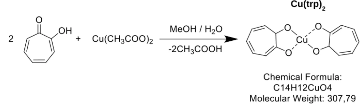
The Cu(trp)2 complex was synthesized and characterized by as it was reported and shown
in Figure 2.1.42
Tropolone was dissolved in methanol and after introduced in the flask by slowly mixing with the solution of copper acetate in methanol : water solution (1 : 6). Tropolone and copper acetate calculated in a 2:1 molar ratio. The obtained mixture refluxed while stirring for 2 hours. The occurred crystals were filtered off, washed several times with cold methanol and dried in vacuum over H2SO4.
The results of elemental analysis are following (%): Cu: 20.9, C: 55.2, H: 3.49; (calculated for Cu(trp)2 (%): Cu: 20.8, C: 55.0, H: 3.28)).42
Figure 2.1. The scheme of synthesis and structure of the copper-tropolone complex.
22 2.1.1.2. Complex Cu(hin)2
The complex of testing was synthesized and characterized as previously reported.43 The scheme of synthesis is shown in Figure 2.2.
After dissolving and during the continuous stirring of hinokitiol in ethanol : water solution (50 : 50), a solution of copper acetate was introduced. The molar ratio of concentrations of hinokitiol and copper acetate is 2:1 respectively. The blue-green mixture was stirring and gently heated for 2 hours. After that, the solution was standing for 1 week in order to crystals allow forming and growing from the product. The occurred crystals were filtered off, washed several times with cold methanol and dried in vacuum.
The results of elemental analysis are the following (%): (Calc. for Cu(hin)2): C: 61.6, H:
5.69. Selected IR data (cm-1): ν (C=O): 1600, 1574; ν (C=C): 1512; ν (C–O): 1286-1228.
Figure 2.2. The scheme of synthesis and structure of copper-hinokitiol complex.
2.1.2. Dilutions
The dilution of Cu(trp)2 and Cu(hin)2 complexes, hinokitiol, and tropolone typically were
done from 2 stock solutions. The first stock solution was prepared from the dissolving weighted powders using analytical weights (up to 2.00 mg), in dimethyl sulfoxide (DMSO) to reach the concentration of chemicals to 0.02M (20 000 µM). The first stock solution was used for preparing the second stock of 100 µM concentration in the DMEM medium by using aliquot from the first stock solution. The solutions of complexes of concentrations of testing were prepared by using the second stock solution and diluted to the desired concentration. The studies never exceeded 0.5% of DMSO in the solutions and control samples were treated with the lowest concentration of DMSO in order to standardize conditions of experiments.
23 2.2. Biological part
2.2.1. Cell Lines
Through years of research, the implication of cell lines tended to become the key element as in vitro models and in diagnostics and widely use in laboratory research. Moreover, it can imagine the drug development process without testing a potential drug on the targeted object, as for cancer research is mammalian cancer cells.
2.2.1.1. MCF7
First established by Dr. Soule in 1973 at the Michigan Cancer Foundation, where the name of the cell line comes, MCF7 becomes the most recognizable cell line among others in many aspects of laboratory research, and in vitro particularly. The MCF7 cells were derived from the pleural effusion, taken from 69-years old Caucasian female patient, who undergo right mastectomy from a benign tumor and radical left mastectomy for a malignant adenocarcinoma. Also, according to Dr. Soule reports, the patient was treated with a high dosage of synthetic estrogen diethylstilbestrol and after took a tamoxifen medication for cancer treatment. Later was reported that the influence of antiestrogen tamoxifen by inhibiting the growth of MCF7 cells could be reversed by estrogen activity. This fact and further investigations made the MCF7 the hormone-responsive breast cancer cell line44
This cell line becomes commonly used in breast cancer research, mainly because of being a proven suitable and sustainable model for testing. With the passage of time, it has been produced more data in molecular profiles through using MCF7 cells in experiments. It has been proven MCF7 cell line as an ER(estrogen positive and PR(progesterone receptor)-positive cancer line and relates to the luminal A molecular subtype. The growth of these cells is controlled either by ER and PR and by plasma-associated growth factor receptors, such as epidermal growth factor receptor (EGFR) and human epidermal growth factor receptor-2 (HER2). It is also low aggressive and showed low tendency to migration and invasion, due to low level of vascular endothelial growth factor (VEGF), a key mediator of angiogenesis in mechanisms of formation of new blood vessels and involves in growth and development of cancer. Without estrogen supplementation, this cell line doesn‟t tend to become tumorigenic and in mice do not induce metastasis.45 Among observed features of MCF7 cells, it was estimated its
24 ability for the formation of multicellular aggregated forms, which further growing into lumen-containing spheroids. 46 MCF7 reported to express the WNT7B oncogene. This oncogene was expressed at the same level in normal and benign tumors, but in the case of the second, it was found in approximately 30 times higher in 10% of tumors.47 MCF7 cell line is suitable to be investigated for anti-hormone therapy resistance, inasmuch as during cultivation, they keep the ER expression. Once the estrogen is removed, the rate of proliferation stays near a month after ER removal.48
Figure 2.3. Microscope photo of MCF7 cells in a presence of DMEM (left) and in a low and high density of cells (right).49
2.2.1.2. MDA-MB-231
Breast cancer adenocarcinoma cell line MDA-MB-231 was firstly established in 1973 and derived from single pleural effusion of the metastatic site was introduced from 51-years old Caucasian female patient with stage III adenocarcinoma, who went through a radical mastectomy and earlier received treatment of prednisone and fluorouracil, which apparently was ineffective.50 MDA-MB-231 cells belong to the basal subtype of breast cancer and have no expression of the ER and PR receptors. However, cells express the epidermal growth factor (EGF) and transforming growth factor-α (TGF-α).
In contrary to the MCF7 cell line, MDA-MB-231 is an ER-negative breast cancer cell line and does not respond to the anti-hormone treatment.48 This cell line has proved to be highly invasive in vitro. At the same time, MDA-MB-231 cells showed relatively low metastatic activity in vivo, but despite this, introduction cells into cultivation has been shown sufficient, which makes this cell line the useful model in experimental models.12 It has been expressed the
25 correlation between VEGF and aggressiveness in the behavior of the cells.45 MDA-MB-231 reported to express the WNT7B oncogene.47
Figure 2.4. Microscope photos of MDA-MB-231 cells in the presence of DMEM (left) and in a low and high density of cells (right).51
2.2.2. 2D Cell System
2.2.2.1. Cell Viability. MTT assay
The main purpose of this method is the evaluation of the effect of compounds with variable concentrations on cell viability and possible therapeutic effects. The assay is performing on the basis of reported by Mosmann‟s methodology with slight changes.52
The assay is based on the conversion of the water-soluble yellow dye of 3-(4,5-dimethylthiazol-2-yl)-2,5-diphenyltetrazolium bromide (MTT) to insoluble purple formazan, which is the product of the action of mitochondrial reductase. Due to the interaction of MTT with the capacity dehydrogenase and its following reduction, it causes the change of the solution color from yellow with MTT to the appearing of purple crystals of formazan. After formazan solubilizes, it is determined by optical density to be at 570 nm.
In order to evaluate the effect of two compounds on cell viability, the cells were grown and cultured for 24 hours in a CO2 incubator with the level of CO2 of 5% and 37⁰C in a plate of
96 wells per 250 000 cells in each. This creates a confluent monolayer. After it was placed in the culture medium (DMEM or DMEM/F12) with dilutions of various concentrations of the drugs and well plate was kept in a humidified atmosphere with 5% CO2 during 48 hours and 37⁰C. In
26 cells from the used medium. After PBS was eliminated from the plate, it was incubated with 100 µl per well of 0.5 mg/ml (10% v/v) 3-(4,5-dimethylthiazol-2-yl)-2,5-diphenyltetrazolium bromide (MTT) in DMEM solution in a humidified atmosphere with 5% CO2 during 3 hours and 37⁰C.
The change of color of the solution in the wells indicates the formazan creation. Subsequently, the formazan generated by mitochondrial enzymes was dissolved and extracted with DMSO. The plate was filled with 100 µl of DMSO into each well and put into the plate reader for the absorbance determination in the wavelength of 570 nm. The percentages of viable cells were calculated, according to the formula:
̅̅̅̅̅̅̅ ,
– absorption value of each of probation well value
̅̅̅̅̅̅ – average value of absorption for control samples. 2.2.2.2. Cell Migration
The wound-healing assay provides a reliable, valuable and accessible cell migration in vitro study of combined cell traveling-like behavior or migration and proliferation in a monolayer. Also, among those processes, cell interaction in the presence of a chemical has taken place. The approach of the assay is the observation of the behavior of cells in a confluent monolayer after making a gap or scratch (wound) and incubate it free (control) and in the presence of the drugs. The cell-free gap is created in the monolayer of cells by either direct manipulation or physical exclusion. Each manipulation could be performed and destroy the area of the monolayer by using mechanical, electrical, chemical or thermal tools.53
Cells were grown and cultured in a 24 wells cell culture plates of (300 000 – 350 000 cells/ml) for 24 hours in CO2 incubator with the level of CO2 of 5%, humidified atmosphere and
temperature of 37⁰C with complete medium DMEM for MCF7 cells or DMEM/F12 for MDA-MB-231 cells, including 10% FBS, until 90-100% of confluence. After removing the medium, the monolayer was scratched with a sterile 200 µl pipette tip and washed after with PBS in order to remove non-adherent cells. At this point, it was taken digital images by an inverted microscope for the wound by using AmScope software. Then, the cells were treated with copper complexes for 48 hours in a CO2 incubator with the level of CO2 of 5%, a humidified atmosphere and
27 temperature of 37⁰C, dissolved in DMEM for MCF7 cells or DMEM/F12 for MDA-MB-231 cells, including 5% FBS. After treatment time and discarding the medium, the monolayer was washed with PBS and stained with Giemsa solution (5%) for 10 min. Digital images were taken using an Olympus BX51 inverted microscope with a digital camera. The inhibition of cell migration was analyzed with ImageJ software. The percentage (%) of migration was calculated using the following formula:
2.2.2.3. Mechanism of Action
The exact mechanism of action is unknown for any of Cu(trp)2 and Cu(hin)2 complexes. Moreover, copper complexes with similar chemical structure and its influence on cancer cells have not been reported in the literature, thus it was proposed to observe the occurrence of oxidative stress, calf thymus interaction and apoptosis studies for evaluation of possible way of mechanism action. In this study, the ROS production and calf thymus interaction were taken into the scope of the drug-cell and drug-DNA assays to determine the effect and influence of these drugs on mammalian cells.
2.2.2.3.1. Oxidative Stress. Determination of Reactive Oxygen Species (ROS Production)
The appearance and formation of ROS is an aerobic process, which could be caused either by genetic factors, nutritional intake, hormonal or environmental influence. Even if it is an invalid process, the overproduction of ROS leads to oxidative and nitrosative stress, damage of DNA and attack cellular components by production of reactive species after the interaction with proteins and lipids in the living organisms. Therefore, the generation of ROS is a mutagenic process and is used as a potential carcinogenic marker.54
Cells were grown and cultured in a 24 wells cell culture plates of (150 000 cells/ml) for 24 hours in a CO2 incubator with the level of CO2 of 5%, humidified atmosphere and temperature
of 37⁰C with complete medium DMEM, including 10% FBS, until 90-100% of confluence. After the medium was discarded, complexes were introduced into wells with an increasing amount of the concentration of the drug and incubated for 48 hours in the incubator with the level of CO2 of