Recebido a 28/05/2014 Aceite a 05/09/2014 Correspondência Ângela Figueiredo Serviço de Radiologia
Centro Hospitalar Tondela-Viseu Av. Rei D. Duarte
3504-509 Viseu
e-mail: ang_figueiredo@hotmail.com
1 - Serviçode Radiologia do Centro Hospitalar Tondela - Viseu
Diretor: Dr. Duarte Silva
2 - Ser viço de Imagem Médica do Centro Hospitalar e Universitário de Coimbra Diretor: Prof. Doutor Filipe Caseiro Alves
DOENÇAS INFECIOSAS DO RIM – REVISÃO PICTÓRICA
INFECTIOUS DISEASES OF THE KIDNEY – A PICTORIAL REVIEW
Ângela Figueiredo1, Luísa Andrade2, Hugo Correia1, Nuno Ribeiro1, Rui Branco1, Duarte Silva1Artigo de Revisão / Review Article ACTA RADIOLÓGICA PORTUGUESA
Maio-Agosto 2014 nº 102 Volume XXVI 37-43
Introduction
Urinary tract infections are the most common urologic disease. Acute bacterial pyelonephritis is the most frequent renal infection and typically occurs as a result of bacterial invasion of the renal parenchyma from ascending infection originating in the lower urinary tract. Escherichia coli is the most common organism involved. Haematogenous spread to the kidneys is less common [1].
Abstract
Acute pyelonephritis is the most common renal infection but a variety of other infectious processes can be seen in the kidney. Although radiologic evaluation is not necessary in cases of uncomplicated pyelonephritis, it plays an important role in high-risk patients and in those who do not respond to therapy or whose clinical presentation is atypical.
Although ultrasonography (US) is relatively insensitive in early stages of acute pyelonephritis, it is considered the first level investigation technique for its availability and lack of radiation use.
Computed tomography (CT) has higher sensitivity when compared to US and is considered the gold standard in identifying focal parenchymal abnormalities, extension of the disease and its complications. Magnetic resonance imaging (MRI) with diffusion-weighted sequences is a good alternative in patients in which iodinate contrast is contraindicated and in children and pregnant women.
Using iconographic material from the authors departments, a pictorial review is performed to review the US and CT imaging findings of some common and not so common infectious diseases of the kidney, including acute pyelonephritis, pyonephrosis, emphysematous pyelonephritis, xanthogranulomatous pyelonephritis and tuberculosis.
Keywords
Urinary tract infections; Pyelonephritis; Multidetector Computed Tomography; Ultrasonography
Resumo
A pielonefrite aguda é o tipo de infeção renal mais frequente, no entanto, o rim pode ser afetado por vários outros processos infeciosos. Embora a avaliação imagiológica não seja necessária nos casos de pielonefrite não complicada, pode desempenhar um papel importante nos doentes de risco, nos que não respondem de modo adequado à terapêutica e naqueles com uma apresentação clínica atípica.
A ecografia, embora pouco sensível nas fases iniciais da pielonefrite, é o exame de primeira linha por ser uma técnica acessível e não utilizar radiação ionizante. A tomografia computorizada (TC) é mais sensível que a ecografia, sendo considerado o método de referência na deteção de anomalias focais do parênquima renal, na avaliação da extensão da doença e suas complicações. A ressonância magnética (RM), incluindo estudo por difusão, é uma alternativa válida nos doentes em que a utilização de contraste iodado esteja contra-indicada bem como nas crianças e grávidas.
Recorrendo a material iconográfico proveniente dos departamentos dos autores, é efetuada uma revisão pictórica das características imagiológicas (em ecografia e TC) de alguns processos infeciosos do rim, incluindo a pielonefrite aguda, a pionefrose, a pielonefrite enfisematosa, a pielonefrite xantogranulomatosa e a tuberculose renal.
Palavras-chave
Infeções do trato urinário; Pielonefrite; Tomografia Computorizada; Ecografia
The diagnosis is usually based on a combination of typical clinical features (urinary frequency, dysuria, flank pain and a high grade fever accompanied by rigors) and laboratory findings (pyuria, white cell casts, bacteriuria, positive urine culture and elevation of acute-phase reactants including erythrocyte sedimentation rate (ESR), C-reactive protein (CRP) and blood white cell count) [1] .
Routine radiologic investigation is usually not required for diagnosis and treatment of uncomplicated cases in adults [2] - those occurring in a nonpregnant woman aged between 15 and 65, with no functional or anatomical abnormalities of the
urinary system, no sign of obstruction, no recent intervention to the urinary system, no recent or recurring episodes of pyelonephritis, and no current illness affecting the patient’s immune status [3].
The role of imaging is to assist in the diagnosis of acute pyelonephritis when the patient doesn’t respond to appropriate therapy within the first 72 hours; to look for any complication that would need specific therapeutic management (such as obstruction of the collecting system; renal or perirenal abscess); to assess those patients at significant risk for more severe life-threatening complications (eg diabetic, elderly or immunocompromised patients); to look for previously occult structural or functional abnormalities; and to identify rare forms of pyelonephritis in atypical clinical presentations or with atypical laboratory results [2, 3].
Pyonephrosis is a suppurative infection that occurs in the setting of a hydronephrotic obstructed kidney and is considered a urologic emergency requiring urgent drainage [4]. If it is left untreated, a rapid decline in renal function may result and patients may develop septic shock [2, 5, 6]. Pyonephrosis should be suspected in any patient with a known urinary tract obstruction accompanied by fever and flank pain. The obstruction may arise from calculi, tumor, complications from pyelonephritis (sloughed papilla) or strictures [2]. Emphysematous pyelonephritis is a life-threatening gas-for ming infection resulting in necrosis of the renal parenchyma. Escherichia coli, Klebsiella pneumonia and Proteus
mirabilis are the most commonly responsible organisms [2, 4]. Patients present with symptoms of severe acute pyelonephritis, urosepsis, or shock [4] and the majority of them (approximately 90%) have poorly controlled diabetes. No diabetic patients are typically immunocompromised or have associated urinary tract obstruction by stones, neoplasm or sloughed papilla [2]. Xanthogranulomatous pyelonephritis is an unusual form of chronic pyelonephritis in which the renal parenchyma is destroyed and replaced by lipid-laden foamy macrophages. It usually affects middle-aged women with a history of recurrent urinary tract infections, diabetes or kidney stones [7]. Symptoms are often nonspecific (low-grade fever, malaise). Flank pain and haematuria may also be present [2].
The urinary tract is the most common extrapulmonary site of tuberculosis, being affected in 4 to 8% of patients with evidence of pulmonary tuberculosis [4, 8]. Almost all cases result from haematogenous dissemination of Mycobacterium
tuberculosis to the kidney after initial pulmonary inoculation. Despite this presumed route of spread from the lungs to the kidney, less than 50% of patients with urinary tract tuberculosis have abnormal chest radiography [2]. Clinical diagnosis is usually delayed because of the insidious onset and nonspecific symptoms. Haematuria and culture-negative pyuria may be seen at urinalysis.
Imaging findings
Acute bacterial pyelonephritis
In early stages of infection there may be isolated inflammation of the renal pelvis mucosa (pyelitis) which appears on sonography as thickening and hyperechogenicity of the collecting system walls. This thickening is also seen on CT and MRI scans, but sonography is usually sufficient to point to this diagnosis. When the urinary epithelium is involved, the
infection may then spread to the renal parenchyma (pyelonephritis) [3]. In most patients with clinically suspected pyelonephritis ultrasound imaging shows a normal appearing kidney. When positive findings are found at US they can include renal enlargement (kidney length at least 1,5 cm longer than the unaffected side); loss of renal sinus fat due to edema; loss of corticomedullary differentiation; changes in renal echogenicity with either hypoechoic or hyperechoic areas; hydronephrosis and areas of hypoperfusion visible with Doppler evaluation (figs.1, 2) [2, 9].
Fig. 1 - Acute pyelonephritis. Ultrasound scan (sagittal view) shows an enlarged right kidney with diffuse hyperechogenicity of the parenchyma and loss of differentiation.
Fig. 2 - Acute pyelonephritis. (a) US scan (sagittal view) shows a wedge-shaped hyperechoic focus (arrows) in the upper pole of the right kidney related to acute bacterial pyelonephritis. (b) US image (sagittal view) demonstrates diminished flow through the involved area.
CT is the modality of choice for evaluating acute bacterial pyelonephritis. The imaging protocol should include an unenhanced scan because it can detect calculi, gas formation, haemorrhage, parenchymal calcifications, obstruction, renal enlargement and inflammatory masses. However, the kidney will appear normal on unenhanced CT in many cases, and contrast-enhanced scans should be performed. Parenchymal changes are better depicted during the nephrographic phase (preferably 90 to 120 seconds after the injection of contrast) [3]. An excretory phase should also be performed if urinary obstruction is suspected [1].
The classic CT finding on contrast-enhanced studies is the so-called striated nephrogram which appears as one or multiple wedge-shaped areas of hypoattenuation demonstrating a striated appearance, extending from the papilla to the renal cortex. However the presence of these hypoattenuating areas is not pathognomonic of pyelonephritis and they may also appear in renal infarction, renal lymphoma / metastases and vasculitis [10]. Additional CT findings include focal or global renal enlargement, thickening of Gerota fascia, perinephric stranding and urothelial thickening (figs.3, 4) [3].
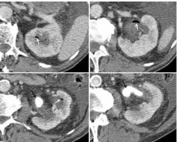
Acute pyelonephritis can also present as a focal alteration with a masslike appearance mimicking a neoplasm. Clinical history is fundamental in suggesting the diagnosis of focal pyelonephritis and follow-up imaging after appropriate therapy may be necessary to exclude a renal neoplasm (figs.5, 6, 7) [4].
MRI is a good alternative to CT imaging because its high spatial and contrast resolution allows optimal evaluation of the renal parenchyma and adjacent structures. This imaging technique has some important advantages when compared to CT including the absence of ionizing radiation (which is particularly useful in children and pregnant women) and the use of paramagnetic contrast medium (which is better tolerated by patients and has a lower incidence of allergic reactions when compared to iodinate contrast material) [11]. Typically an affected area will have low signal intensity on T1-weighted images and increased signal intensity on T2-weighted images and after gadolinium administration MRI features mimic those of CT [1]. However MRI has also some limitations – it is relatively time consuming; it presents high costs and, even if to a lesser extent than iodinated contrast medium, the administration of gadolinium is not recommended in patients with a history of allergic reaction. Besides that, studies in recent years about systemic fibrosis due to gadolinium contraindicate its use in patients with renal impairment [12].
Some recent studies have focused on the value of diffusion-weighted MRI. They concluded that it could be an additional tool for routine renal MR imaging protocol showing that this technique is effective in diagnosing acute pyelonephritis and can be a reasonable alternative to contrast-enhanced MRI especially when contrast media administration is contraindicated (such as in patients with renal insufficiency and pregnant or lactating women). The affected areas appear hyperintense on diffusion-weighted sequences with high b value and hypointense on ADC maps [11, 12, 13].
Renal abscess
In cases of untreated or inadequately treated pyelonephritis, tissue necrosis and liquefaction can occur resulting in abscess formation [14]. Diabetic patients are predisposed to abscess formation, with 75% of all renal abscesses occurring in this population [2].
Ultrasonography is less sensitive than CT in evaluating for the presence of an abscess and small microabscesses, which are common in early acute infection, are frequently missed by this technique (fig. 8) [2]. When positive imaging findings are found US demonstrates a fluid-filled mass with distinct walls, sometimes with internal echoes (fig. 9). However, in the acute phase it may show indistinct margins with edema in the surrounding renal parenchyma [8].
Fig. 3 - Acute pyelonephritis. Contrast-enhanced axial CT images (nephrographic phase) show several wedged-shape areas of decreased enhancement consistent with a striated nephrogram (white arrows). They also demonstrate a large stone in the renal pelvis (*), perinephric stranding (arrowheads) and thickening of Gerota fascia (grey arrow).
Fig. 4 - Acute pyelonephritis. Contrast-enhanced axial CT (excretory phase) shows several wedged-shape areas of decreased enhancement consistent with a striated nephrogram (arrows).
Fig. 6 - Masslike appearance of acute bacterial pyelonephritis. US scan (sagittal view) demonstrates a geographic, slightly lobulated, hyperechoic “mass” (arrows) in the mid pole of the right kidney, a finding that is worrisome for a solid tumor.
Fig. 5 - Focal acute pyelonephritis. (a) US scan (sagittal view) demonstrates a hypoechoic “mass” (arrows) in the upper pole of the right kidney, a finding that is worrisome for a solid tumor. (b) Power Doppler image (sagittal view) demonstrates diminished flow through the involved area.
Fig. 7 - Focal acute pyelonephritis. Axial (a) and coronal (b) contrast-enhanced CT images (excretory phase) show a nodular hypodense area in the upper pole of the right kidney, representing a focus of pyelonephritis.
Contrast-enhanced CT shows round or geographic low-attenuation collections with enhancing walls. Abscess cavities may be single or multiple and either intra or extraparenchymal. Gas within the collections may or may not be present (fig.10, 11, 12) [4].
Pyonephrosis
Ultrasonography may show dilatation of pelvicalyceal system, thickening of the walls of the renal pelvis and the presence of echogenic debris in dependent portions of the collecting system [9, 3] (fig.13).
CT may demonstrate dilatation of the collecting system, higher than usual attenuation values of the fluid within the renal collecting system, thickening of renal pelvis wall (> 2 mm) and parenchymal or perinephric inflammatory changes (fig.14) [2].
Both the techniques (US and CT) may identify the cause of obstruction of the collecting system.
A caveat to CT evaluation is that it is often difficult to distinguish simple hydronephrosis from pyonephrosis on the basis of fluid attenuation measurements [2]. Some studies have
focused on the value of diffusion-weighted MRI and apparent diffusion coefficient (ADC) maps in distinguish pyonephrosis from hydronephrosis with promising results (in pyonephrosis ADC values of the renal pelvis were found to be lower than those of renal pelvis of hydronephrotic kidneys) [15]. Emphysematous pyelonephritis
Ultrasonography demonstrates an enlarged kidney with parenchymal high-amplitude echoes with posterior acoustic Fig. 8 - Acute pyelonephritis with microabscesses. Axial (a and b) and coronal
(c) contrast-enhanced CT images (nephrographic phase) show several wedged-shape areas of decreased enhancement (*) and small collections representing microabscesses (arrows) which were not seen on ultrasound scan.
Fig. 10 - Renal abscess. Coronal (a) and axial (b) contrast-enhanced CT images (corticomedullary phase) show a round low-attenuation collection (*) with enhancing walls (arrows), in the mid pole of the left kidney.
Fig. 9 - Renal abscess. US scan of the left kidney (sagittal view) shows a well-defined fluid-filled lesion with a distinct wall in the lower renal pole (arrows).
Fig. 11 - Multiple renal abscesses. Axial (a, b and c) and coronal (d) contrast-enhanced CT images (nephrographic phase) show an enlarged and heterogeneous left kidney, with multiple hypodense collections representing abscesses (white arrows). There is also dilatation of several calyces in the lower pole, which are filled with dense material (pyonephrosis) (black arrows).
Fig. 16 - Emphysematous pyelonephritis. Axial (a and b) and coronal (c and d) contrast-enhanced CT images (nephrographic phase) show multiple air bubbles within both the renal parenchyma and the collecting system, with associated fluid collections (*).
dirty shadowing, but it may underestimate the extent of parenchymal involvement (fig.15) [2, 16, 17].
CT is the modality of choice for evaluating patients with emphysematous pyelonephritis. Findings include renal enlargement and destruction, small bubbly or linear streaks of gas, fluid collections, gas-fluid levels and tissue necrosis, with or without abscess (fig.16) [2, 17, 18].
It is important to distinguish emphysematous pyelonephritis from emphysematous pyelitis because the latter has a better prognosis [19]. In emphysematous pyelitis gas is limited to the renal collecting system. US findings are typically nondependent high-amplitude echoes within the renal sinus or calices, representing foci of air. CT findings include a dilated collecting system, gas bubbles or gas-fluid levels within the collecting system, and the lack of parenchymal gas (fig.17) [2, 18, 19]. Xanthogranulomatous pyelonephritis
Ultrasonography features of xanthogranulomatous pyelonephritis include multiple hypoechoic round masses in the affected kidney, which can demonstrate internal echoes. Global enlargement with relative preservation of the renal contour is usually seen with diffuse disease. In focal segmental xanthogranulomatous pyelonephritis a mass-like lesion may be demonstrated. In addition, there is usually evidence of obstruction and renal calculus (85% of cases) (fig.18) [9, 20]. CT is the mainstay of diagnostic imaging for xanthogranulomatous pyelonephritis as it usually shows a high Fig. 12 - Extraparenchymal abscess. Axial contrast-enhanced CT images
(corticomedullary phase) show a well-defined low-attenuation collection (*), with enhancing walls (arrows) and gas (curved arrow), in the right retroperitoneal region, near psoas muscle.
Fig. 13 - Pyonephrosis. US scan images (sagittal view) show a dilated collecting system that is partially filled with echogenic debris (*). There is also thickening of the collecting system walls (arrow).
Fig. 14 - Pyonephrosis. Axial (a and b) and coronal (c) contrast-enhanced CT images (nephrographic phase) show a dilated collecting system (*) with thickening of their walls (white arrows). There is also a perinephric abscess with extension to psoas muscle (arrowhead), and a large stone in the proximal ureter (black arrow).
Fig. 15 - Emphysematous pyelonephritis. US images (sagittal view) show multiple hyperechoic foci with dirty shadowing consistent with gas, in the left kidney.
Fig. 17 - Emphysematous pyelitis. Axial contrast-enhanced CT images (nephrographic phase) show a dilated collecting system with multiple air bubbles (white arrows). Dilatation is caused by a large staghorn-shaped stone. There is no evidence of gas collections in the renal parenchyma.
Fig. 18 - Xanthogranulomatous pyelonephritis. (a) US scan (sagittal view) shows an enlarged right kidney with distention of the collecting system secondary to inflammatory debris (*). (b) A central shadowing calculus is also seen (arrow).
specific set of findings that allow a confidence diagnosis. It also accurately assesses the extent of extrarenal disease, if present, and aids in surgical planning.
In the diffuse form of disease CT usually demonstrates renal enlargement, a central calculus (often staghorn-shaped) within a contracted renal pelvis and replacement of the renal parenchyma by multiple oval hypodense areas representing dilated calices and abscess cavities filled with pus and debris. Areas of fat attenuation can be present because of lipid rich xanthogranulomatous tissue. Renal function (excretion) is rarely seen at the time of diagnosis (fig. 19) [4, 7].
A less common manifestation of xanthogranulomatous pyelonephritis is a focal form which is seen in approximately 10% of patients [2].
Renal tuberculosis
Although both the kidneys are usually seeded with the
Mycobacterium tuberculosis organisms, clinically relevant disease is usually limited to one side. Initial small granulomas form in the renal cortex, adjacent to the glomeruli [21]. The upper and lower poles of the kidney are more commonly affected [22].
Ultrasonographic evaluation may demonstrate granulomatous lesions as masses of mixed echogenicity, with or without necrotic areas of caseation and calcifications. Mucosal
Fig. 19 - Xanthogranulomatous pyelonephritis. Axial (a and b) and coronal (c and d) contrast-enhanced CT images (excretory phase) demonstrate an enlarged right kidney with multiple oval hypodense collections replacing renal parenchyma. Renal function (excretion) is not seen in the affected side.
thickening and stenosis of the calyces may also be detected by US as well as mucosal thickening of the renal pelvis and ureter, ureteral strictures and hydronephrosis. Additionally bladder changes as mucosal thickening and reduced capacity are commonly seen [14].
CT imaging findings in renal tuberculosis depend on the stage of disease. The earliest findings are focal hypoperfusion areas on contrast-enhanced CT, with a striated nephrogram. As the parenchymal granulomas coalesce, CT can demonstrate a masslike lesion (tuberculoma) with central low attenuation representing caseous necrosis [4, 23]. A moth-eaten calyx secondary to papillary necrosis is another finding that may be seen.
As the disease progresses the host launches a fibrotic reaction in response to infection, causing stricture formation of the calyceal infundibula, which leads to uneven caliectasis and eventually incomplete opacification of the calyx (phantom calyx) [4, 24].
Calcification is present in a large number of patients (40-70%) [25]. The extension of calcifications is variable ranging from thin rims surrounding low attenuation areas of focal cortical inflammation to diffuse, uniformly radiodense areas that replace portions or all of the renal parenchyma in late stage disease (autonephrectomy) (fig. 20) [2].
Sometimes fistulization may occur in renal tuberculosis. Fistulas involving the kidney may communicate with the bowel, skin, blood vessels; lymphatics or thoracic cavity (pleura or bronchus). Renal fistulas may be classified into those communicating with the calyces via the parenchyma and those that communicate with the renal pelvis. CT is the most useful diagnostic modality in these cases [22].
CT may also demonstrate the involvement of the ureter and urinary bladder. Granuloma formation within transitional epithelium of the ureter causes fibrosis with consequent ureteral shortening and strictures, and wall calcifications. Strictures are more common in the distal one-third of the ureter and at sites of normal anatomic narrowing (ureteropelvic junction, pelvic brim and ureterovesical junction). The initial
Fig. 20 - Renal tuberculosis (late stage disease). Axial (a, b, c and d) and coronal (e and f) contrast-enhanced CT images (nephrographic phase) demonstrate bilateral dilatation of the collecting system, with marked reduction of renal parenchyma thickness. The left kidney shows multiple thin calcifications (white arrows). The bladder is contracted (black arrow). A stenosis in the distal right ureter is also seen (white curved arrow).
involvement of urinary bladder usually manifests as interstitial cystitis, with mucosal ulceration and mural thickening. In later stages, wall fibrosis and scarring lead to decreased of bladder capacity. Wall calcification and fistula formation are rare [21].
Conclusion
Imaging evaluation is not necessary in most patients with suspected pyelonephritis but it plays a role in high-risk patients or in those who do not respond to conventional therapy. Usually the first imaging modality in these cases is ultrasonography. However contrast-enhanced CT is the preferred technique as it performs better than sonography at detecting parenchymal changes and identifying most complications of pyelonephritis. MRI is a good alternative when iodinate contrast is contraindicated and in children and pregnant women.
Acute pyelonephritis is the most common renal infection but a variety of other infectious conditions can be seen.
Many of these processes require urgent management so it is imperative for the radiologist to be familiar with their imaging findings.
References
1 - Stunnell, H.; Buckely, O.; Feeney, J.; Geoghegan, T.; Browne, R.F.; Torreggiani, W.C. - Imaging Of Acute Pyelonephritis In The Adult. Eur Radiol, 2007, 17:1820-1828.
2 - Craig, H. D.; Wagner, B. J.; Travis M. D. - From The Archives Of The AFIP:
Pyelonephritis: Radiologic - Pathologic Review. Radiographics, 2008, 28:255-276. 3 - Ifergan, J.; Pommier, R.; Brion, M. C.; Glas, L.; Rocher, L.; Bellin, M. F. –
Imaging In Upper Urinary Tract Infections. Diagnostic and Interventional Imaging, 2012, 93:509-519.
4 - Hammond, N. A.; Nikolaidis, P.; Miller, F. H. – Infectious And Inflammatory
Diseases Of The Kidney. Radiol Clin N Am, 2012, 50:259-270.
5 - Subramanyam, B. R.; Raghavendra, B. N., Bosniak, M. A.; Lefleur, R. S.; Rosen, R. J.; Horii, S. C. - Sonography Of Pyonephrosis: A Prospective Study. AJR Am J Roentgenol, 1983, 140:991-993.
6 - Yoder, I. C.; Pfister, R. C.; Lindfors, K. K.; Newhouse, J. H. - Pyonephrosis:
Imaging And Intervention. AJR Am J Roentgenol, 1983, 141:735-740.
7 Loffroy, R.; Guiu, B; Watfa, J.; Michel, F.; Cercueil, J. P.; Krause, D.
-Xanthogranulomatous Pyelonephritis In Adults: Clinical And Radiological Findings In Diffuse And Focal Forms; Clinical Radiology, 2007, 62:884-890.
8 - Kawashima A.; Sandler C. M.; Goldman S. M.; Raval, B. K.; Fishman E. K. - CT Of Renal Inflammatory Disease. Radiographics, 1997, 17:851-866. 9 - Vourgant, S.; Agarwal, P. K.; Bodner, D. R.; Dogra, V. S. – Ultrasonographic
Evaluation Of Renal Infections; Ultrasound Clin, 2010, 5:355-366.
10 - Federle, M. P.; Jeffrey, R. B.; Desser, T. S.; Anne, V. S.; Eraso, E.; Chen, J. J. S. - Diagnostic Imaging Abdomen. Amirsys Ink, Salt Lake City, 2004. 11 - Faletti, R.; Cassinis, M. C.; Fonio, P.; Grasso, A.; Battisti, G.; Bergamasco, L. et al. - Diffusion–Weighted Imaging And Apparent Diffusion Coefficient Values
Versus Contrast–Enhanced MR Imaging In The Identification And Characterisation of Acute Pyelonephritis. Eur Radiol, 2013, 23:3501-3508.
12 - Pascale, A.; Piccoli, G. B.; Priola, S. M.; Rognone, D.; Consiglio, V.; Garetto, I. et al. - Diffusion-Weighted Magnetic Resonance Imaging: New Perspectives
In The diagnostic Pathway Of Non-Complicated Acute Pyelonephritis. Eur Radiol, 2013, 23:3077-3086.
13 - Vivier, P. H.; Sallem, A.; Beurdeley, M.; Lim, R. P.; Leroux, J.; Caudron, J. et al. - MRI And Suspected Acute Pyelonephritis In Children: Comparison Of
Diffusion-Weighted Imaging With Gadolinium-Enhanced T1-Weighted Imaging. Eur Radiol, 2014, 24:19-25.
14 - Papanicolaou, N.; Pfister R. C. - Acute Renal Infections. Radiol Clin North Am, 1996, 24:545-569.
15 - Cova, M.; Squillaci, E.; Stacul, F.; et al. - Diffusion-Weighted MRI In The Evaluation Of Renal Lesions: Preliminary Results. BrJ Radiol, 2004, 77:851-857.
16 Allen, H. A.; Walsh J. W.; Brewer, W. H.; Vick, C. W.; Haynes, J. W.
-Sonography Of Emphysematous Pyelonephritis. J Ultrasound Med, 1984,
3:533-537.
17 - Wan, Y. L.; Lee, T. Y.; Bullard, M. J.; Tsai, C. C. - Acute Gas-Producing
Bacterial Renal Infection: Correlation Between Imaging Findings And Clinical Outcome. Radiology, 1996, 198:433-438.
18 - Grayson, D. E.; Abbott, R. M.; Levy, A. D.; Sherman, P. M. - Emphysematous
Infections Of The Abdomen And Pelvis: A Pictorial Review. RadioGraphics, 2002, 22:543-561.
19 - Roy, C.; Pfleger, D.; Tuchmann, C. M.; Lang, H. H.; Saussine, C. C.; Jacqmin, D. - Emphysematous Pyelitis: Findings In Five Patients. Radiology, 2001, 218:647-650.
20 - Tiu, C. M.; Chou, Y. H.; Chiou, H. J.; Lo, C. B.; Yang, J. I.; Chen, K. K. et al. Sonographic Features Of Xanthogranulomatous Pyelonephritis. J Clin Ultrasound, 2001, 29(5):279-285.
21 - Wong, A.; Dhingra, S.; Surabhi, V. R. - AIRP Best Cases in Radiologic –
Pathologic Correlation - Genitourinary Tuberculosis. RadioGraphics, 2012, 32:839-844.
22 - Merchant, S.; Bharati, A.; Merchant, N. - Tuberculosis Of the genitourinary
system-Urinary tract tuberculosis: Renal tuberculosis - Part II. Indian J Radiol Imaging, 2013, 23:64-77.
23 - Matos, M. J.; Bacelar, M. T.; Pinto, P.; Ramos, I. - Genitourinary Tuberculosis. Eur J Radiol, 2005, 55:181-187.
24 - Engin, G.; Acunas, B.; Acunas, G; Tunaci, M. - Imaging Of Extrapulmonary
Tuberculosis. Radiographics, 2000, 20:471-488.
25 - Muttarak, M.; ChiangMai, W. N.; Lojanapiwat, B. - Tuberculosis Of The
Genitourinary Tract: Imaging Features With Pathological Correlation. Singapore Med J, 2005, 46:568-574.