110 Radiol Bras. 2013 Mar/Abr;46(2):110–116
Central nervous system lymphoma: iconographic essay
*
Linfoma do sistema nervoso central: ensaio iconográfico
Fabiano Reis1, Ricardo Schwingel2, Felipe Barjud Pereira do Nascimento3
The authors illustrate the present pictorial essay about central nervous system lymphoma with magnetic resonance images obtained in their institution over the past 13 years. Some of the main radiological findings in primary and secondary presentations of this type of lymphoma are discussed. Central nervous system lymphoma is a relatively uncommon tumor, but magnetic resonance imaging findings may suggest the diagnosis.
Keywords: Lymphoma; Central nervous system; Magnetic resonance imaging.
Ilustramos este ensaio iconográfico de linfoma do sistema nervoso central com imagens de ressonância magnética obtidas em nosso serviço nos últimos 13 anos e discutimos algumas das principais características radiológicas deste tipo de linfoma, primário e secundário. O linfoma sistema nervoso central é um tumor relativamente infrequente, mas alguns achados na ressonância magnética podem sugerir este diagnóstico.
Unitermos: Linfoma; Sistema nervoso central; Ressonância magnética.
Abstract
Resumo
* Study developed at Hospital das Clínicas da Faculdade de Ciências Médicas da Universidade Estadual de Campinas (Uni-camp), Campinas, SP, Brazil. Financial support: Fundação de Amparo à Pesquisa do Estado de São Paulo (Fapesp) – Process No. 2010/01939-0.
1. PhD, Teacher responsible for the Sector of Neuroradiology, Professor at Faculdade de Ciências Médicas da Universidade Estadual de Campinas (Unicamp), Campinas, SP, Brazil.
2. Graduate Student of Medicine (6th year) at Faculdade de Ciências Médicas da Universidade Estadual de Campinas (Uni-camp), Campinas, SP, Brazil.
3. MD, Radiologist, Fellow of Neuroradiology, Hospital Israelita Albert Einstein, São Paulo, SP, Brazil.
Mailing Address: Dr. Fabiano Reis. Faculdade de Ciências Médicas, Universidade Estadual de Campinas, Departamento de Radiologia. Rua Tessália Vieira de Camargo, 126, Cidade Uni-versitária Zeferino Vaz. Caixa Postal: 6111. Campinas, SP, Bra-zil, 13083-887. E-mail: [email protected].
Received July 16, 2012. Accepted after revision November 5, 2012.
Reis F, Schwingel R, Nascimento FBP. Central nervous system lymphoma: iconographic essay. Radiol Bras. 2013 Mar/Abr;46(2):110– 116.
In immunocompetent patients, CNS lymphoma tends to present as a single and solid large mass, with tumor cells which tend to be monoclonal. Often, the patient’s age at the disease onset is higher and the survival is longer. Such single lesions, with high cellularity and generally without ne-crosis, present preponderantly hyposignal (or isosignal) on T2-weighted and isosignal on T1-weighted images, and demonstrate intense enhancement after intravenous in-jection of paramagnetic contrast agent.
The presence of this kind of lesion in-volving the corpus callosum is highly sug-gestive of lymphoma (Figure 1)(5).
Addi-tionally, not only the image but also the frequent subependimal dissemination can demonstrate the common perilesional edema which occurs in varied degrees and the mass effect (Figure 2A)(4–7).
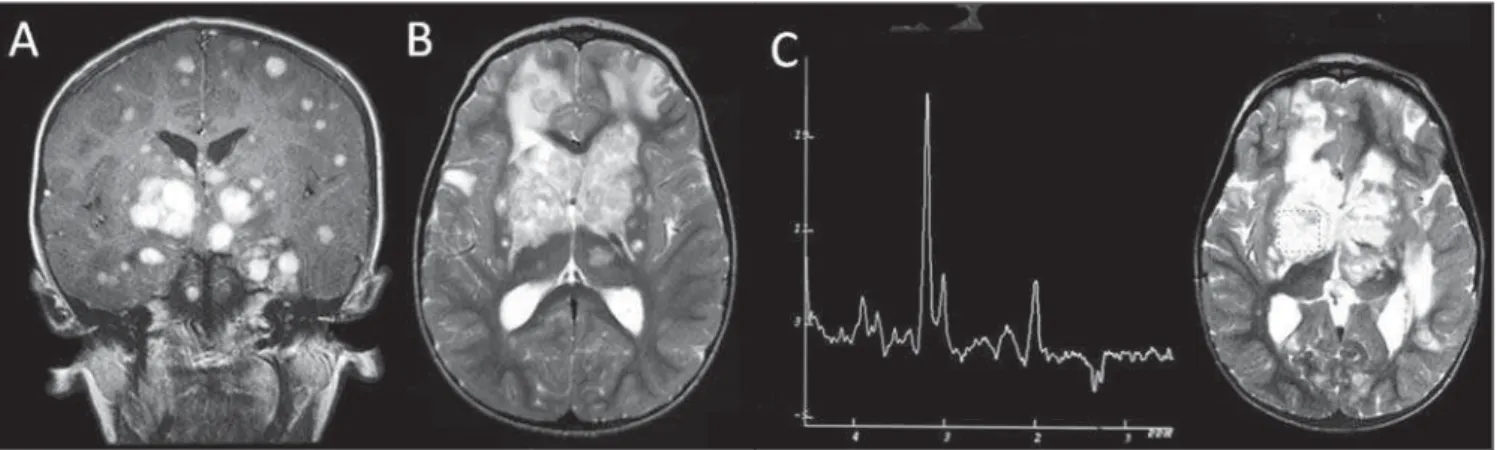
Several significant similarities may be found between patients with acquired im-munodeficiency syndrome and patients with other forms of immunosuppression. In such cases, there is a higher probability of PCNSLs presenting as multifocal lesions (Figures 3A and 3B) with necrosis (Figures 4A and 4B) and differentiation between them and toxoplasmosis may be difficult. In regard to contrast-enhancement, sev-eral series have demonstrated that its ab-sence can be considered as being rare in CNS lymphomas (Figure 5)(5,6).
lymphomas. Primary or acquired immuno-deficiency is also associated with higher risks for development of primary or sec-ondary CNS lymphoma(2).
The early diagnosis of CNS lymphomas depends upon the understanding by radi-ologists of their varied presentations at magnetic resonance imaging (MRI). The present study reviews images obtained over the past 13 years in the Radiology Service at Hospital das Clínicas – Universidade Es-tadual de Campinas (Unicamp). The diag-noses were duly confirmed by histopathol-ogy, and the project was approved by Com-mittee for Ethics in Research of the School of Medical Sciences of Unicamp.
RADIOLOGICAL CHARACTERISTICS
As regards the location of lesions, su-pratentorial region is generally more fquently affected than the infratentorial re-gion. In cases of deep intra-axial lesions, striatocapsular regions and the corpus cal-losum are affected with a certain frequency. However, lesions may be observed in other sites such as the hypophysis, cavernous sinus, hypothalamus, pineal gland, and pos-terior fossa. Periventricular or superficial lesions, in contact with the meningeal or ventricular surfaces (intraventricular lesions for example), are commonly found(2–7). INTRODUCTION
Central nervous system (CNS) lym-phoma may present as a primary disease, however, it is more frequently found as a secondary disease. In a survey carried out in the USA between 2004 and 2008, lym-phomas represented 2.3% of CNS primary tumors, most commonly found in men (1.38:1) and after the sixth decade of life(1).
Typically, primary CNS lymphoma (PCNSL) is a B-cell non-Hodgkin’s lym-phoma but occasionally it may manifest as T-cell lymphoma, intravascular lymphoma-tosis or Hodgkin’s lymphoma(2,3). On the
Reis F et al. Central nervous system lymphoma: iconographic essay
Figure 3. Male, 3-year-old patient. Post-contrast coronal MRI T1-weighted image (A) shows the presence of centroencephalic, cortical and subcortical lesions with nodular homogeneous contrast enhancement. Subependymal dissemination is observed. On the axial T2 weighted image (B) the lesions present isointense signal, some of them with a hyperintense center. Also, moderate perilesional edema is observed. At spectroscopy with ET of 135 ms (C), increased choline, decreased NAA levels and inverted lactate peak are observed.
Figure 2. Female, 19-year-old patient. Post-contrast coronal MRI T1-weighted image (A) reveals presence of a left periventricular lesion, with homogeneous contrast uptake. Subependymal dissemination is observed. Spectroscopy with ET of 135 ms (B) demonstrates increased choline levels, decreased NAA levels and inverted lactate peak (double peak of 1.3 ppm). In this case, CNS was the primary site of involvement by lymphoma.
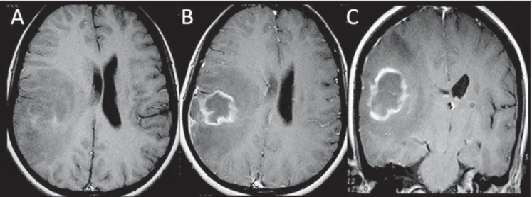
Figure 4. Female, 32-year-old, HIV positive patient. Axial T1-weighted image (A) demonstrates right parietotemporal lesion with high signal components (hem-orrhage). B, C: Post-contrast axial and coronal T1weighted images demonstrate peripheral contrast uptake and moderate perilesional vasogenic edema. Cen-tral non-enhancing component corresponds to necrosis.
Reis F et al. Central nervous system lymphoma: iconographic essay
Pachy- or leptomeningeal involvement is more commonly found in secondary lym-phomas or in immunocompromised pa-tients. Additionally, appearance of dural/ pachymeningeal lesions may be similar to that of meningiomas (Figures 6 and 7)(4).
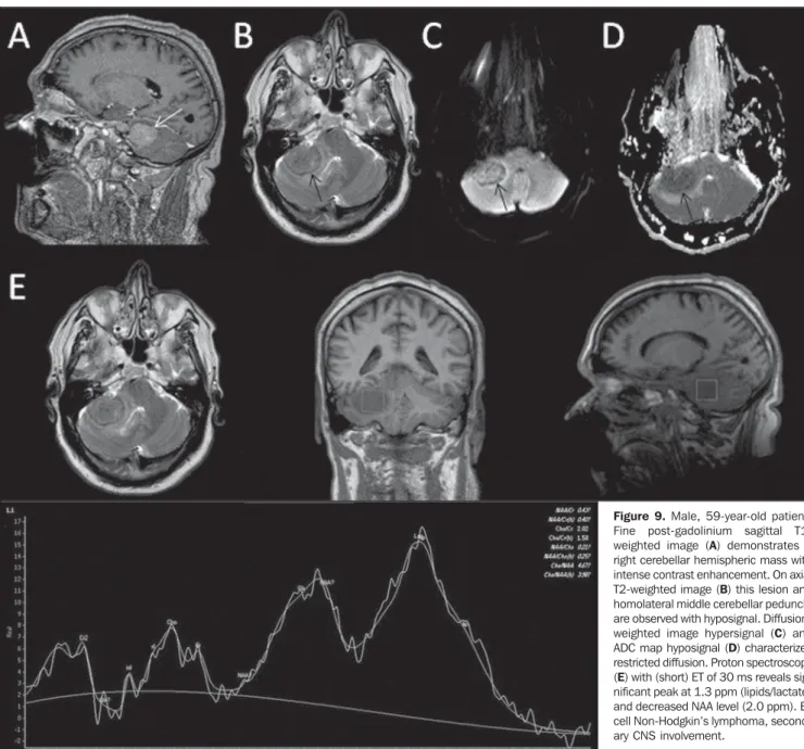
Secondary CNS involvement by non Hodgkin’s lymphoma generally occurs by leptomeningeal infiltration (found in up to two thirds of cases) and infiltration of peri-vascular spaces resulting in parenchymal lesions (observed in one third of cases – Figures 8 and 9)(2,3). Lesions affecting
cra-nial nerves are relatively rare and may be solitary or multiple (Figure 10)(3,4,8). On the
other hand, in Hodgkin’s lymphoma, pri-mary CNS involvement is rare and second-ary involvement represents a late manifes-tation of disseminated disease (Figure 11).
SPECTROSCOPY
Proton magnetic resonance spectros-copy provides graphs which allow the analysis of specific tumor tissue metabo-lites, particularly in cases of parenchymal lesions. With this complementary method, it is possible to evaluate the levels of cho-line (peak ant 3.2 ppm), which marks cell membranes biosynthesis; N-acetylaspartate (NAA) (2.0 ppm), related to neuronal and axonal number and viability; creatine (3.02 and 4.0 ppm), utilized as a reference for its stability and a marker of oxidative phos-phorylation of normal brain metabolism; lactate (1.3 ppm), related to anaerobic me-tabolism observed in anaerobiosis; lipids (usually between 0.9 and 1.3 ppm), related to necrosis(9). In CNS lymphomas, it is
common to observe a pattern of increased choline levels, decreased NAA, increased lipids (moreover in the solid component without necrosis, an information that helps the differential diagnosis with glial lesions, in which such peak is usually observed in
Figure 7. Female, 42-year-old patient. Contrast-enhanced, coronal MRI T1-weighted sequence pre-sents a homogeneously enhanced solitary, extra-axial lesion in the right temporal fossa. Previously to the intracranial involvement, chronic sinusitis was present, whose cause was later identified by means of biopsy of the right maxillary sinus, as diffuse large B-cell non-Hodgkin’s lymphoma.
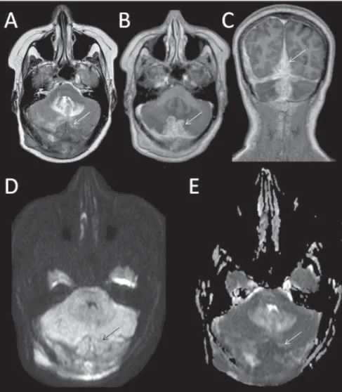
Figure 6. Female, 52-year-old patient. Images show diffuse supra and infratentorial dural (pachymeningeal) thickening at median right region, with hyposignal on T2-weighted image, homogeneous contrast-en-hancement (C), hypersignal on diffusion-weighted image (D) and ADC map hyposignal – restricted diffu-sion (E). High-histologic-grade B immunophenotype non-Hodgkin’s lymphoma.
the necrotic component) (Figures 2B, 3C, 4C and 9E). It has been suggested that spectroscopy might be useful in differen-tiation between lymphoma, glioblastoma multiforme and metastases(10). It is
impor-tant to observe that with echo time of 135-144 ms, lactate is characterized by a double peak inverted below baseline. However this peak can be demonstrated at approximately 1.3 ppm above baseline with short echo time (30 or 35 ms) and with very high echo time (270 or 288 ms) lipids are suppressed and lactate is seen as a double peak above the baseline(4,5,9).
DIFFUSION
Diffusion-weighted MR images mea-sure the apparent diffusion coefficient (ADC) which is inversely proportional to cell density, presumably resulting from the tortuosity of the interstitial space and con-sequential limitation of water movement. Although not always observable, CNS lym-phoma tends to present a low ADC value at diffusion-weighted MRI because of its high cellularity, characteristically present-ing restriction in those lesions without ne-crosis (Figures 6D, 6E, 9C and 9D)(2,5,10).
PERFUSION
Perfusion-weighted MRI techniques evaluate the microcirculation allowing the calculation of physical parameters such as cerebral blood volume (CBV), cerebral blood flow, and mean transit time. Typi-cally, lymphomatous lesions present low CBV (Figure 12). The maximum relative CBV measured in the tumor and its rela-tionship with the contralateral parenchyma is low as compared with that of other brain tumors(2).
Reis F et al. Central nervous system lymphoma: iconographic essay
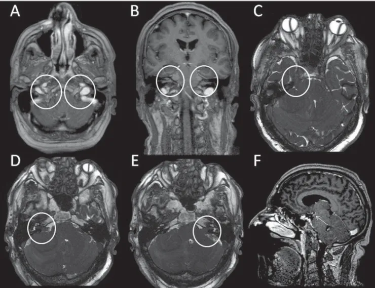
Figure 10. Same case as on Figure 9. Post-contrast axial T1-weighted image (A) demonstrates thickening and enhancement of the cranial nerve XI, bilaterally. Post-contrast coronal T1-weighted image (B) shows bilateral thickening and homogeneous enhancement of the cranial nerves V and VII-VIII are observed. Axial 3D CIS image demonstrates bilateral thickening of the cranial nerves V (C) and VII/VIII (D,E). Post-contrast sagittal T1-weighted image (F) demonstrates en-hancement and thickening of the III cranial nerv.
Figure 11. Male, 22-year-old patient. Coronal T1-weighted image (A) demonstrates frontal left extra-axial lesion with diffuse contrast-enhancement. This lesion is hypointense on extra-axial T2-weighted image (B). Marked edema is observed. The extension to CNS was secondary to Hodgkin’s disease/ type II nodu-lar sclerosis diagnosed by means of cervical lymph node biopsy.
CONCLUSION
CNS lymphomas may present with var-ied forms, however some findings can con-tribute to facilitate the differential diagno-sis which includes gliomas, metastases, and inflammatory diseases. The finding of an expansile supratentorial solid lesion hypointense on T2-weighted images, with no signs of necrosis, lipids peak in the solid component, low perfusion and diffusion restriction, favors the possibility of such diagnosis.
REFERENCES
2. Haldorsen IS, Espeland A, Larsson EM. Central nervous system lymphoma: characteristic find-ings on traditional and advanced imaging. AJNR Am J Neuroradiol. 2011;32:984–92.
3. Slone HW, Blake JJ, Shah R, et al. CT and MRI findings of intracranial lymphoma. AJR Am J Roentgenol. 2005;184:1679–85.
4. Besada C, Schvartzman P, Paganini L, et al. Neuroimágenes estructurales y funcionales en la caracterización del linfoma del SNC. Rev Argent Radiol. 2010;74:147–53.
5. Küker W, Nägele T, Korfel A, et al. Primary
cen-tral nervous system lymphomas (PCNSL): MRI features at presentation in 100 patients. J Neurooncol. 2005;72:169–77.
6. Schwingel R, Reis F, Zanardi VA, et al. Central nervous system lymphoma: magnetic resonance imaging features at presentation. Arq Neuro-psiquiatr. 2012;70:97–101.
7. Nacif MS, Jauregui GF, Mello RAF, et al. Lin-foma adrenal primário bilateral com envolvi-mento do sistema nervoso central: relato de caso. Radiol Bras. 2005;38:235–8.
8. Garcia MM, Martins JCT. Avaliação por imagem
Figure 12. Male, 63-year-old patient. Nodular lesion located at left mesial temporal region, with isosignal on T2-weighted image (A), marked contrast-en-hancement (B), hypersignal on DWI (C) and hyposignal on ADC map (D), characterizing diffusion restriction. The CBV map (D) does not demonstrate increased perfusion characterized by the quantitative analysis in form of curves (E,F). B image also demonstrates contrast-enhancement foci on the surface of the ependymal lining of right temporal horn, characterizing cerebrospinal fluid dissemination. (Case kindly assigned by Dr. Cassio Iwakura, from Hospital Vera Cruz, Campinas, SP, Brazil).
das lesões isoladas do III par craniano. Radiol Bras. 2005;38:219–23.
9. Horská A, Barker PB. Imaging of brain tumors: MR spectroscopy and metabolic imaging. Neuroimaging Clin N Am. 2010;20:293–310. 10. Chawla S, Zhang Y, Wang S, et al. Proton