1
UNIVERSIDADE DE LISBOA
FACULDADE DE CIÊNCIAS
DEPARTAMENTO DE BIOLOGIA ANIMAL
Dynamics of embryo axis elongation in
amniotes vs. anamniotes: The role of the
notochord
Tomás Augusto Barreiros Pais de Azevedo
MESTRADO EM BIOLOGIA EVOLUTIVA E DO
DESENVOLVIMENTO
2
UNIVERSIDADE DE LISBOAFACULDADE DE CIÊNCIAS
DEPARTAMENTO DE BIOLOGIA ANIMAL
Dynamics of embryo axis elongation in
amniotes vs. anamniotes: The role of the
notochord
Tomás Augusto Barreiros Pais de Azevedo
MESTRADO EM BIOLOGIA EVOLUTIVA E DO
DESENVOLVIMENTO
Dissertação orientada pelos
Doutor Gabriel G. Martins (FCUL)
Prof.ª Doutora Sólveig Thorsteinsdóttir (FCUL)
3
INDEX
ACKNOWLEDGEMENTS ... 4 SUMMARY ... 6 RESUMO ... 7 INTRODUCTION ... 10MATERIALS AND METHODS ... 16
General considerations... 16
Zebrafish embryo culture and live-imaging ... 16
Chick embryo culture and live-imaging ... 18
Image analysis and quantification ... 21
Testing the autonomous elongation of chick embryos ... 24
Chick embryo notochord excision experiments. ... 24
Immunohistochemistry and confocal imaging of chick embryos. ... 27
Results ... 28
Dynamics of zebrafish embryo axis elongation... 28
Dynamics of chick embryo axis elongation... 29
Axis elongation in chick embryo is autonomous ... 33
No portion of the notochord is essential for embryo extension ... 35
DISCUSSION ... 39
4
ACKNOWLEDGEMENTS
Foram muitos os que me ajudaram neste longo caminho que levou à conclusão da minha tese de mestrado. Aqui quero realçar alguns deles.
Em primeiro lugar e provavelmente mais importante, ao Gabriel. Com ele construí o meu primeiro grande projecto profissional. Agradeço-lhe todos os conhecimentos que me transmitiu, fossem eles de microscopia, embriologia ou simplesmente a arte de sobreviver ao trabalho num laboratório. Agradeço-lhe também o ter-me introduzido no mundo da ciência, com tudo o que isso traz. Desde as sessões de “brainstorming” no seu gabinete de onde nasceram muitas das ideias aqui discutidas, até às alegrias das experiências bem sucedidas e fracassos das experiências falhadas.
Um agradecimento geral ao grupo de Biologia do Desenvolvimento do CBA da FCUL, por tão bem me acolherem dentro do grupo e por toda a ajuda que me deram com diferentes pontos de vista sobre o meu trabalho
À professora Sólveig, pela sua calma e serenidade que sempre me transmitiu e pelas valiosas ideias e maneira lúcida de ver as coisas que ajudaram este projecto a tornar-se uma realidade.
À professora Gabriela pela sua simpatia e sorriso constantes e pela doçura com que sempre me tratou.
À Rita por toda a amizade com que me recebeu no grupo e por me ajudar com conselhos e incentivos desde o primeiro dia até às vésperas de entrega da tese.
Às minhas colegas de mestrado dentro do grupo, Ana Lina e Raquel, pelo apoio que sempre me deram, sobretudo quando tudo me parecia mais sombrio do que realmente era e pelas experiências que fomos trocando no decorrer das nossas teses.
Ao António por todos os ensinamentos que me passou sobre o trabalho com peixe-zebra, desde o descorionamento à manutenção dos aquários.
Ao Rifes, pelos seus conhecimentos de embriões de galinha e me ter conseguido convencer finalmente a ler o “Senhor dos anéis” em inglês.
Ao Luís pelo companheirismo nomeadamente em várias noites longas de trabalho e pelas conversas “geeks” sobre ficção científica que finalmente consegui ter.
Às novas membros do grupo Ana Bernardo e Ana Martins pela frescura que trouxeram ao laboratório.
À Ana Gaspar pelos momentos de boa disposição que sempre trouxe ao laboratório, com as suas sempre originais obras de arte.
5 À Carla, pela sua amizade e pelos seus conselhos sobre os cuidados a ter com uma “fish facility”.
À Marta Santos e à professora Margarida Matos pelas preciosas ajudas na estatística realizada e ensinamentos sobre como usar o Statistica 8
À Dona Branca, por toda a ajuda que me deu no laboratório e por ser alguém com quem conversar sobre o “nosso” Benfica.
Ao Joaquin Rodriguez-Léon e Catarina Certal do grupo de organogénse do IGC e à Raquel Mendes do IMM pelos seus valiosíssimos conselhos sobre a técnica de New, sem os quais nunca conseguiria ter feito muitas das experiências apresentadas.
Aos meus grandes amigos Bárbara, Miguel Cunha e Miguel Santos por todos os bons momentos partilhados durante a tese que ajudaram a descomprimir do stress diário.
Às minhas grandes amigas Célia, Isabel e Rita pelos “grupos de apoio” que criámos para facilitar a escrita da tese.
À Catarina, ao Menezes e ao Miguel por me lembrarem constantemente que há um mundo lá fora para além da tese.
Finalmente, aos meus pais e minha avó de quem sempre tive apoio incondicional e uma paciência infinita para me restaurarem o ânimo perdido e para partilharem comigo todas as alegrias. Um agradecimento especial à minha mãe pela história da Rã que me manteve à tona neste final de ano…
6
SUMMARY
In this thesis, we sought to analyze the dynamics of amniote and anamniote embryo axis elongation and the possible role of the notochord. For that, we chose the chick and zebrafish as representative model organisms of each group. We found that in the zebrafish embryo, elongation has two different phases during the stages analysed, from “shield” to 15 somite stage. The first phase is characterized by a constant (i.e., linear) growth rate which is faster than that observed during the second phase. The first phase occurs simultaneously with the epiboly movements of the basltoderm and yolk-syncytial layer (YSL). In chick embryos, we also find a constant (linear) growth phase which spans from stage HH4-11 (Hamburger & Hamilton 1951), a period during which the chick embryo forms structures homolog to those formed by zebrafish embryos during the first (linear) growth phase. Furthermore, in chick embryos, we found that the cranial portion elongates at a slower rate than that of two other portions of the embryo: the “segmented” and “pre-somitic” portions of the embryo, which contain all tissues adjacent to the segmented and pre-somitic mesoderm, respectively. Using micromanipulation techniques we show that elongation in the chick is independent of the primitive streak and a direct connection of Hensen’s node to the extra-embryonic tissues. This contrasts with zebrafish embryo development where it has been well established that axis extension only occurs if there is an intact connection between the embryo and the YSL. We propose that the structures that could contribute to axis extension are: the notochord, the neural tube and the paraxial mesoderm. Again, using micro-manipulation techniques, we show that none of three different portions of the notochord (cranial, “segmented” and “pre-somitic”) are essential for embryo axis elongation. However, preliminary experiments suggest that the presence of the whole notochord might contribute to the process of embryo axis extension. This work shows that the notochord is not the main driver of early embryo axis extension in amniotes, a hypothesis that had been advanced in the literature often but had not yet been formally tested. Clearly, the forces that shape the early vertebrate embryo into its typical elongated shape come from tissues inside the embryo itself, other than the notochord. In amniotes, these forces have evolved in order to allow the embryo to extend autonomously, without the “additional” force produced by expanding extra-embryonic tissues, as still seen in most anamniotes (e.g., fish and amphibians).
7 RESUMO
A gastrulação é o processo pelo qual o embrião forma as três camadas germinativas, endoderme, mesoderme e ectoderme. É durante este processo que muitos dos organismos formam os seus três eixos embrionários: Dorso-ventral, antero-posterior e esquerda-direita. Após o estabelecimento do eixo anterio-posterior, este sofre um alongamento que depende da arquitectura e modo como o embrião internaliza os tecidos, da quantidade de vitelo, entre outros. Muito pouco se sabe ainda sobre os mecanismos que levam à extensão dos embriões. No caso do peixe-zebra (um anamniota), o embrião é composto por uma grande célula não dividida com uma zona cortical que consiste num sincício denominada “Yolk Syncytial Layer” (YSL). Após as primeiras divisões do embrião, formam-se duas camadas celulares, a Enveloping Layer (EL) composta por células epiteliais que estão ligadas pela sua margem à YSL e, dentro destas, a Deep Layer (DL) formada por células mesenquimatosas. No princípio da gastrulação, as células do embrião migram para o “equador” e a blastoderme cobre aprroximandamente 50 a 75% do embrião. É na zona da margem das células da blastoderme que se localiza o “organizador” de peixe-zebra, um espessamento denominado “embryonic shield”
Apesar de se saber pouco sobre as forças que contribuem para o alongamento do embrião, existe a ideia que elas podem ser geradas por diferentes estruturas do embrião (Keller e tal. 2003). Uma das possibilidades é que esta força seja gerada dentro da própria blastoderme (a “Deep layer”) através dos rearranjos celulares que poderiam produzir extensão. No entanto, trabalhos realizados com mutantes no tail (Glickman, 2003) provaram que os movimentos de intercalação médio-lateral não produzem extensão, pelo que outros tecidos poderão ser responsáveis pela extensão do embrião, nomeadamente os mecanismos de epibolia (Keller et al. 2003).
Em relação ao embrião de amniotas, como o de galinha, o conhecimento sobre os mecanismos que levam ao seu alongmento estão ainda mal estudados. A galinha produz um tipo de gastrulação drasticamente diferente, sendo que neste caso, os movimentos de epibolia dos tecidos extra-embrionários (nomeadamente a área opaca) se tornam independentes da gastrulação. Outra diferença verificada que acompanha a primeira é o facto de, no embrião de galinha, haver uma externalização da fonte de nutrientes (o vitelo) e uma desacoplação dos movimentos de epibolia do crescimento do embrião. Assim, sem a contribuição dos movimentos de epibolia para o seu alongamento, os embriões de amnionas (como a galinha) teriam então de possuir outra fonte de produção de força que os levasse a alongar. Tendo estudado na literatura que a notocorda possui
8 uma função estrutural em muitos organismos, contribuindo para conferir rigidez aos embriões em alongamento, e baseando-nos em experiências que provam que a notocorda possui um alongmento autónomo, a notocorda poderia ser uma das fontes geradoras de força para o alongamento.
Para esta tese propusemo-nos então analisar as dinâmicas de alongamento dos embriões de peixe-zebra e galinha (representativos de anamniotas e amniotas).
Começámos por analisar a dinâmica de alongamento no embrião de peixe-zebra, num período de tempo desde o “shield stage” até ao estádio de 15 sómitos. Durante este período, está estabelecido na literatura que não existe alongamento do embrião (Kimmel et al. 1995). Ao medirmos o comprimento desde a região mais rostral dos tecidos que sofreram involução rostral até à fronteira caudal do “shield”, apercebemo-nos que existe efectivamente alongamento durante esta fase e que este possui duas dinâmicas durante este período, uma com um alongamento linear (desde “shield” até ao estádio de 4 sómitos) e outra com uma dinâmica logarítmica desde 4 até 15 sómitos.
Em seguida analisamos a dinâmica de alongamento do embrião de galinha. Ao analisar o comprimento dos embriões ao longo do tempo, descobrimos que a dinâmica no período de tempo analisado (estádio HH4 até aproximadamente estádio HH 11) é linear. É importante referir que a dinâmica de alongamento do embrião de galinha apenas pode ser directamente comparada com a primeira dinâmica (linear) do embrião de peixe-zebra, uma vez que correspondem aos momentos de formação de estruturas homólogas. Outro ponto interessante que resultou dos nossos dados foi a conclusão de que, contrariamente ao que se pensava (Keller et al. 2003), a maior parte da extensão do embrião de galinha não ocorre aquando da extensão da linha primitiva. O embrião alonga mais do que o triplo do comprimento da linha primitiva, sem a substituir completamente, pelo menos até estádio HH11. Já que no embrião de galinha, as várias porções do embrião são facilmente identificáveis, sendo elas a craniana (desde a cabeça até ao primeiro sómito), a segmentada (desde o primeiro ao último sómito) e a pré-somítica (do último sómito formado até ao nó de Hensen) decidimos então analisar as dinâmicas destas diferentes porções. Descobrimos que todas elas têm taxas de alongamento significativamente diferentes, sendo que a porção craniana alonga muito pouco comparada com as restantes porções. Descobrimos também que a porção segmentada é a que alonga com maior valor e que, contrariamente ao que seria de esperar, a porção pré-somítica alonga também. Em seguida, decidimos verificar uma ideia corrente da literatura, segundo a qual o alongamento do de embrião de galinha não
9 necessitaria de ligação aos tecidos pré-somíticos (como a área opaca) ou da linha primitiva. Para tal, removemos uma grande porção de linha primitiva, fazendo também ao mesmo tempo um corte na conexão com a área opaca. Os nossos resultados provaram que o embrião é capaz de produzir alongamento independente da linha primitiva e dos tecidos extra-embrionários.
Tendo descoberto este facto, tornou-se óbvio que as forças que podem produzir alongamento no embrião de galinha teriam de provir da região rostral em relação ao nó. As três estruturas candidatas são a notocorda, o tubo neural e a mesoderme pré-somítica. Pelas razões já mencionadas, resolvemos testar o papel da notocorda no alongamento do embrião através de experiências em que esta era removida. Como tínhamos já descoberto que as diferentes porções do embrião alongam a taxas diferentes, decidimos começar por retirar porções da notocorda correspondentes às do embrião. O efeito no alongamento não é estatisticamente significativo, pelo que concluímos que as diferentes porções da notocorda não contribuem para o alongamento. No entanto, experiências preliminares em que retirámos uma porção maior da notocorda (correspondente ao conjunto de duas porções anteriormente retiradas), mostraram um efeito significativo no alongamento do embrião, sugerindo que a notocorda como um todo, pode ter uma pequena contribuição. Gostaríamos de realizar mais experiências para testar esse facto, uma vez que apenas obtivemos um n=2 para este tipo de manipulação. Estes resultados mostram claramente que há diferenças na forma como os embriões de anamniotas e amniotas alongam durante as fases iniciais.
10
INTRODUCTION
"It is not birth, marriage, or death, but gastrulation, which is truly the most important time in your life."
Lewis Wolpert (1986)
As this famous sentence from Lewis Wolpert states, gastrulation is a fundamental process for all embryo development. It is during gastrulation that the blastula undergoes a reorganization of its cells in order to form and position the three germ layers, which will later form the organs of the adult. It is also during this process that the embryo transforms from a simple mass of undifferentiated cells and begins acquiring the shape of an organism with three main embryonic axes: the antero-posterior (AP), the dorso-ventral (DV) and the left-right (LR). Upon establishing these axes, the AP axis suffers a dramatic elongation which sets the characteristic body pattern of the vertebrate species with long trunks and tails. Although this elongation occurs in both amniotes and anamniotes, the two groups use different extension mechanisms, which depend on several factors, including tissue internalization mechanisms during gastrulation, the size of yolk and the architecture of the tissues.
In zebrafish (Danio rerio), the early embryo consists of a large undivided yolk cell with a cortical layer consisting of a syncytium, the Yolk Syncytial Layer (YSL); (D'Amico & Cooper, 2001) The first cleavages are meroblastic and create a blastodisc which later organizes into two layers, the Enveloping Layer (EL) composed of epithelial cells and connected to the YSL at the margin, and the Deep Layer (DL) composed of mesenchymal cells, which lay inside the EL. After the first cleavages, and at the onset of gastrulation, the blastoderm cells initiate a movement of epiboly which will eventually encircle the whole yolk
Gastrulation begins only when the movements of epiboly reach the “equator” and the blastoderm covers approximately 50-70% of the yolk. At this time, the cells at the margin of the epibolizing blastoderm begin involuting, causing a circumferential thickening called the germ ring, formed by the hypoblast and epiblast. At a specific sector of this ring, which will later become the dorsal side of the embryo, further intercalation of hypoblast and epiblast cells forms an enlarged structure known as the “embryonic shield” which acts as the zebrafish embryo organizer (Gilbert 2006). After the formation of the shield, cells of the lateral portions of the germ ring converge to the shield and ingress towards the centre of the blastoderm beginning the extension
11 movements of the embryo. At the same time, the cells of the EL continue epiboly until they completely cover the yolk.
Although the cellular movements that elongate the embryo following the formation of the embryonic shield have not been well studied, it has been suggested that these “extensive” forces can be generated either by rearrangement of cells within the axial and paraxial mesoderm or as the result of “pulling” by the blastoderm during its movement of epiboly over the yolk, which occurs at the same time as the embryo is elongating (Keller et al., 2003). Through the study of the notail (ntl) mutants, Glickman and colleagues (Glickman, 2003) showed that extension of the embryo could occur in the absence of medio-lateral intercalation of dorsal mesoderm cells, thus suggesting that mechanism(s), other than medio-lateral intercalation could also drive embryo extension. Experiments done by Trinkaus (1951) with Fundulus (another teleost like zebrafish) showed that by breaking the connection between the YSL and blastoderm, the latter stops its movement towards the vegetal pole while the YSL continues epiboly. These results support the hypothesis that epiboly normally drives early embryo extension.
In chick (Gallus gallus), gastrulation movements are different from those observed in zebrafish and most anamniotes. The egg includes an enormous mass of yolk (much larger than that of fish or amphibian embryos), and the meroblastic cleavage originates a blastodisc, which is “small” when compared to the size of the yolk cell. This blastodisc is composed of an area pellucida (Gilbert 2006) which covers an acellular subgerminal cavity, and by the area opaca located at the periphery of the area pellucida and attached to the yolk cell. The most peripheral zone of the area pellucida is the Marginal Zone (MZ) and in a particular location of the edge of the area pellucida (which will later become the caudal side of the embryo) a structure characteristic of amniotes emerges - the primitive streak. The streak is gradually formed from a thickening of epiblast cells on top of the Koller’s Sickle; these cells begin digesting the extracellular matrix located underneath and produce convergent and intercalating movements towards the midline (Lawson & Schoenwolf, 2001a; Lawson & Schoenwolf, 2001b; Voiculescu et al., 2007) which, together with oriented cell division parallel to the anterior-posterior axis (Wei & Mikawa, 2000) produce an extension of the streak. After the primitive streak has fully extended, a thickening of cells at its rostral end forms the chick organizer - the Hensen’s node. From this moment on, the internalization movements of the cells that will give rise to the three embryonic germ layers begin, while the Hensen’s node regresses caudally. First, the epiblast cells
12 themselves undergo an epithelial-to-mesenchymal transition and ingress through the streak to form the endoderm of the embryo. Simultaneously, the area opaca and margin of the area pellucida continue to spread and form extra-embryonic tissues which will gradually cover the yolk (eventually surrounding it, but only much later in development; (New 1959; Stern, 2006),a movement that is considered reminiscent of the blastoderm epiboly of zebrafish and amphibian embryos (Gilbert 2006). As the node regresses caudally, several cells ingress into the blastocoel and originate the mesoderm. It is in chick embryos that these movements are best understood: The cells that enter through Hensen’s node and migrate rostrally are “layed-down” in a cord-like structure in the midline, the notochord, while the cells that enter through the posterior part of the node migrate laterally, converging towards on the centre and contributing either to the notochord or somites. The cells that enter through the rostral-most primitive streak originate the paraxial, intermediate and lateral plate mesoderm, and the cells that enter through the caudal-most primitive streak originate extra-embryonic mesoderm (Psychoyos & Stern, 1996). Keller et al. (2003) consider gastrulation as divided in two phases in amniotes, each characterized by its own major morphogenetic movement and type of extension of the AP axis in the chick embryo; the first corresponding to the elongation of the primitive streak and the second corresponding to the regression of the node and elongation of the notochord and neural tube. While results of several studies have already shed light on the mechanisms by which the embryo elongates during the first phase of gastrulation (Lawson & Schoenwolf, 2001a; Lawson & Schoenwolf, 2001b; Wei & Mikawa, 2000; Voiculescu et al., 2007), so far no studies have formally addressed the mechanisms of extension during the second phase. Located at the centre of the elongating AP axis and intimately related with this second phase of elongation is the notochord, a structure characteristic of all species of the phylum Chordata, ranging from ascidians to humans. Although this structure has a transient existence in higher vertebrates, it has both structural and functional relevance for the embryo (Stemple, 2005):Structurally, it has been suggested that, because of its flexibility, it is an essential part of the mechanical system required for swimming in amphibian tadpoles and zebrafish larvae (Nishikawa & Wassersug, 1989; Stemple, 2005). Other studies point to an important role in the straightening of amphibian embryos due to its osmotic swelling capabilities (Adams et al., 1990; Koehl, 2000). As for the final destiny of the notochord, it will later give rise to the nucleus pulposus of the intervertebral discs, thus participating in the formation of the vertebral column. Functionally, it produces several signalling cues that provide both positional and cell-fate determination information. For
13 example, the notochord produces the protein Sonic hedgehog (Shh) which induces different neural progenitor cell populations in the neural tube in chick embryos (Briscoe & Ericson, 1999) and the formation of slow muscle from medial cells of the somite in zebrafish (Blagden et al., 1997). In the somite of chick embryos, Shh secreted from the notochord also induces sclerotome formation in the somite (Cossu et al., 1996; Christ et al., 2004).
Several different mechanisms seem to contribute to the extension of the notochord, although the relative contribution of each one varies in different species (Keller et al., 2003). These mechanisms include i) accretion (the addition of cells from Hensen’s node (Sausedo & Schoenwolf, 1993), ii) antero-posterior oriented cell division (Keller et al., 2003) and iii) cell intercalation movements like medio-lateral intercalation (Delarue et al., 1998; Keller et al., 2003). In anamniotes such as zebrafish (Glickman, 2003) and Xenopus (Wilson et al., 1989), it seems that medio-lateral intercalation generates the main force driving axial extension of the notochord. In amniotes, like chick and mice, notochord extension occurs mainly through accretion and cell division within the notochord (Sausedo & Schoenwolf, 1994; Sausedo & Schoenwolf, 1993). In chick, although there are cellular rearrangements within the notochord itself, the authors conclude that these do not account for the full extension of the notochord, mainly because its average diameter decreases only slightly from stage HH7 to stage HH12 (stages as in Hamburger & Hamilton, 1951). Instead, they propose the “toothpaste model” whereby the ingression of a large number of cells from the Hensen’s node to the notochord (structures with a large and smaller diameter, respectively) forces the cells to rearrange themselves just as they pass the border between the Hensen’s node and the notochord. They propose that these rearrangements which occur at the interface between the notochord and Hensen’s node are the cause of lengthening of the notochord. With similar experiments, (Sausedo & Schoenwolf, 1994) showed that the mechanism of notochord extension in the mouse embryo is similar.
Altogether, these facts point to an increase of the role of cellular division and accretion, and a less important role of cellular rearrangements within the notochord, as the drivers of notochord extension in higher vertebrates. That the notochord can extend by different mechanisms is not surprising given that several lines of evidence show the notochord has an autonomous capacity to extend. In the half baked zebrafish mutants, the epiboly of the deep cells of the blastoderm arrests between 60-80%, while epiboly of the YSL and EL continues. This mutation affects the zebrafish homolog of the adhesion protein E-cadherin (Kane et al., 2005), which is necessary for the cellular
14 rearrangements that spread the blastoderm over the yolk. Although these embryos eventually die, some of them survive and Kane et al (1996) showed that they develop until 20 to 30 hours, even showing somite-like structures and, curiously, a bent notochord, i.e., the notochord still extends, even though the embryo cannot elongate. Another experiment (Baumann & Sander, 1984) in which embryos were treated with a teratogen (α-amanitin) that blocks epiboly of the YSL and EL also showed formation of bent notochords which extend beyond the point where the epiboly stopped. When reviewing these two results, Keller et al (2003) concludes that the notochord has an autonomous capacity to extend, although lacking the stiffness to become fully straightened.
This capacity was also discovered in chick embryosbyWaddington (1932). When blastoderms of embryos were ablated shortly before the appearance of the head process and just posterior to the primitive pit, he obtained a “tail” with somites, notochord and neural tube. A similar result was obtained later (Spratt, 1947); when a small portion of the anterior side of the embryo, containing the Hensen’s Node and 0,4mm of the anterior primitive streak was explanted in plasma clots, the embryo still elongated and produced a “tail” posterior to the cut site. This tail had a neural tube, notochord and somites which formed at the correct time of development (as in Hamburger & Hamilton, 1951).
Spratt also showed that the growth rate of the notochord was very similar to the growth rate of what he called the anterior pellucid area (spanning form the anterior margin of the area pellucida to the anterior tip of the node; (Spratt, 1947). He also showed that the notochord growth rate paralleled the rate of regression of the node and primitive streak, which meant that the faster the rate of notochord growth, the faster the node and primitive streak regressed. This led Spratt to suggest that the different regions of the embryo are interdependent and that extension of the notochord could be an active process, unlike the regression of the primitive streak and node which could, in turn, be dependent on the extension of the notochord. This idea was later proposed also by Keller (2003), who suggested that the notochord, besides being an active extender, could be pushing the node caudally in avian embryos, a proposal that, to my knowledge, has not yet been formally tested. Could the extension of the amniote embryo be related to the extension of the notochord as Spratt and Keller suggest for the regression of the node and primitive streak? And if so, why would this be prevalent in amniotes, but not in anamniotes?
15 During the course of evolution, two major changes in the process of gastrulation occurred from anamniotes to amniotes. First, there was a considerable increase in the amount of yolk (particularly evident in reptiles and birds), which was accompanied by an externalization of the yolk, now included in an extra-embryonic compartment (Shook & Keller, 2008). Secondly, there was a dramatic change in the major movements during gastrulation (which could also be a consequence of the first): In anamniotes, these occur by involution and internalization of cells following the movements of epiboly around the yolk at the marginal zone of zebrafish and at the blastopore lip in Xenopus (anamniotes), while in amniotes (like chick and mouse) the gastrulation movements became independent of the epiboly and occur at a more central location mainly through ingression at the primitive streak.
Because embryonic and extra-embryonic tissues are tightly connected in zebrafish embryos, the expansion of the YSL may provide a driving force for the initial elongation of the embryo; the observation that without epiboly the embryo does not elongate (Trinkaus, 1951) supports this assertion. Because the chick embryo cannot rely on those movements (epiboly of the extra-embryonic tissues around the massive yolk is uncoupled from embryo extension (Arendt & Nübler-Jung, 1999; Stern, 2006), other forces are necessary to drive axis elongation. Based on the evidence that the notochord has an autonomous capacity to extend, on the possibility that it could be pushing Hensen’s node caudally because it has been shown that it has a structural role in other embryos, we propose that this property might have been co-opted to aid in the elongation of the embryo AP axis in amniote embryos.
Thus the aims of this thesis were: i) to characterize and make a comparative analysis of the dynamics of zebrafish and chick embryo elongation; ii) to test the role of the notochord in driving elongation in chick embryos.
16
MATERIALS AND METHODS
General considerations
In this project we studied the dynamics of embryo axis elongation in two different species of vertebrates using embryo culture and live-imaging techniques. In order to compare the dynamics of embryo axis elongation in anamniotes vs. amniotes and to address the role of the notochord in embryo early elongation we chose one animal model for each group. As an amniote model we used chick embryos for their accessibility and possibility of in vitro culture (New, 1945), which facilitates the observation of development over the course of several hours. We chose zebrafish as the model for anamniotes because they are easily maintained and have a highly reproductive rate, which facilitates collection of embryos. Also, because of their transparency and external development, it was easy to observe inner structures during development.
Zebrafish embryo culture and live-imaging
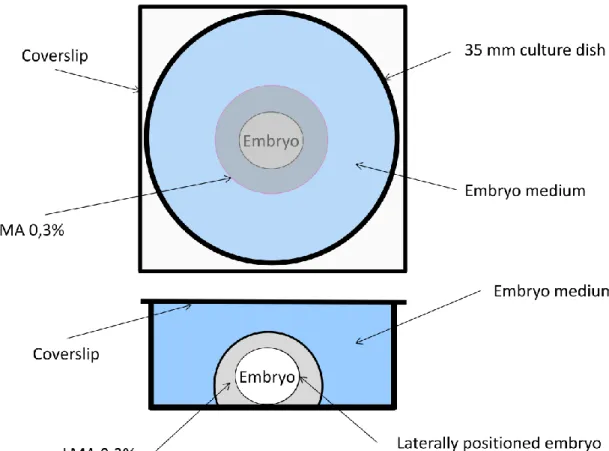
Adult zebrafish were acquired from ZFIN and maintained in an aquarium with artificial “fish water” (Westerfield, 1995). The animals were regularly fed either live, or frozen or lyophilized brine shrimp (Artemia salina). The fish were subjected to a circadian cycle of 14 h light /10 h darkness at a constant temperature of 28ºC to ensure maximum fertility, and were mated once a week at most. Embryos from different broods were obtained in different days from natural spawning in mating boxes with perforated bottoms to collect the eggs and prevent predation from the adults. Collected embryos were placed in “embryo medium” (Westerfield, 1995) and then maintained in an incubator at 28ºC, to allow normal development. Using a stereoscope, we selected embryos for imaging and identified their stages of development according to Kimmel et al. (1995) by the degree of epiboly completed and time post-fertilization at 28,5ºC. Selected embryos were manually dechorionated and mounted in 0,3% low-melting agarose (LMA) in embryo-medium, for immobilization (see Figure 1). This concentration was chosen because it provided the best compromise between the time it took the LMA to polymerize and the freedom of movements needed for normal embryo development. After dechorionation, the embryos were embedded in LMA by transferring them to a Falcon 15ml tube containing melted LMA. A drop of solution
17 containing each embryo was then transferred to 35mm Petri dishes and allowed to polymerize at room temperature. Freshly made embryo medium was then added to the Petri dish up to the top and a coverslip was added (see figure 1 for a schematic view of the mounted embryo). This prevented both dehydration of the embryo and condensation on the coverslip. The room temperature was kept at 28,5ºC.
Fig 1. Schematic diagram of the culture camber used to image zebrafish embryos (top and side views)
Time-lapse images were acquired with a Zeiss Stereo LUMAR stereoscope starting at shield stage (6hpf) (n=2) or at 75% epiboly stage (8hpf) (n=2) (Kimmel et al., 1995) and ending at approximately 15 somite stage (~ 16,5hpf). At 15 somite stage the zebrafish embryo has formed the tissues which in the adult will give rise to the whole head and the pre-caudal vertebrae. A total of nine embryos were filmed, however only three were selected for analysis since they developed without abnormalities and maintained the lateral orientation (Fig 2) necessary for measurements of axis elongation. The starting stage was selected because it is the first stage when the dorso-ventral polarity can be identified, making it possible to orient the embryo in a sagittal plane, thus facilitating measurement of the AP axis (see movie 1 in annex). We chose to stop the time-lapses at fifteen somite stage because at that stage, the embryo’s tail reached
18 the head and there were physical constraints inside the LMA to allow proper embryo development. The interval between time-points was set at 5 minutes.
Fig 2. Example of positioning and “straightening” of digital images of zebrafish embryos.
A) Original image, before “straightening” with area selected in yellow. B) Resulting image, used for linear measurements of embryo axis extension.
Chick embryo culture and live-imaging
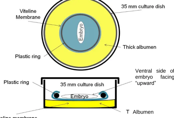
Fertilized chicken eggs (Gallus gallus) were placed in an incubator at 38°C. Eggs were windowed and embryos staged according to Hamburger and Hamilton (Hamburger and Hamilton, 1951). Embryos at stage 3-6 were collected in different days and transplanted into the “New” culture system (New, 1955) for further culture and observation, as well as time-lapse image acquisition (Figure 4). However, instead of glass rings, we used plastic rings made from cutting 50ml Falcon tubes with 2,5 cm of inner diameter and 2,8 cm of outer diameter. These rings provided an ideal diameter that allowed for enough growth of the area opaca and fitting of the vitelline layer (Stern & Bachvarova, 1997).
Some embryos were cultured in an cell-culture incubator kept at 38ºC with saturated humidity and normal atmosphere. Images of these embryos were obtained at
19 the beginning and end of the culture period using an Olympus C-4040ZOOM camera coupled to the eyepiece of at Nikon SMZ645 stereoscopic microscope.
Fig 4. Schematic diagram of the culture camber used to image chick embryos. Top and side views.
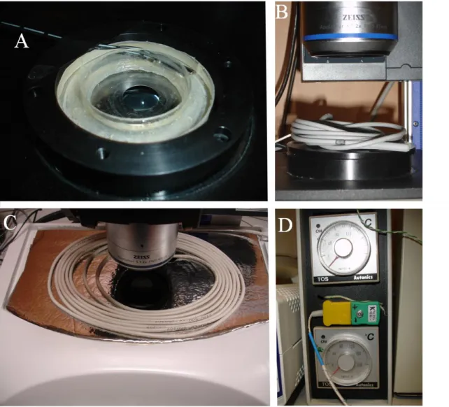
Other embryos were time-lapse imaged using a fully automated Zeiss Stereo LUMAR stereoscope + Cooled CCD Axiocam, set to record an image each 6min over the course of 14-24h (see movie 2 in annex). This interval between time-points was chosen because chick embryos grow slower than zebrafish embryos. To maintain proper environmental conditions we built a custom made incubation chamber (Figure 4) which consisted of a small round box (Figure 4A) whose interior contained a “donut-shaped” reservoir filled with water (to maintain a humid environment) and an inner chamber where the Petri dish with the embryo in “New” culture was placed. The box was then covered with a lid (Figure 4B) containing a large nº0 coverslip to allow episcopic visualization of the embryo. The temperature inside the chamber was maintained by heating the microscope stage with a terrarium heater (Figure 4C) placed under it and by placing another heater on top of the incubation chamber (Figure 4B). This ensured a uniform temperature inside the chamber and also avoided condensation on the lid, which would have made viewing the embryo impossible. The heating wires were connected to two temperature controllers (Figure 4D) with two probes. The stage heater was set for 38ºC and the probe was placed in the water reservoir while the temperature
Ventral side of embryo facing “upward”
20 of the top heater was set slightly higher to prevent condensation. A digital thermometer recorded the temperature throughout the time-lapse image acquisition period and showed that the temperature variations did not exceed +-1ºC. A total of 12 embryos were filmed, of which we selected the best 7 for analysis of the dynamics of embryo axis elongation.
Fig 4. Custom made incubation chamber used to culture chick embryos at 38ºC during
time-lapse imaging. A) Circular-shaped metal box with the inner chamber where the embryo is filmed and the “donut-shaped” water reservoir for maintaining a saturated environment inside the chamber. Inside the water reservoir is the probe for the temperature recording system. B) Box lid covering the chamber with one of the terrarium heaters used for avoiding condensation on the upper window glass. C) Terrarium heater placed under the microscope stage to maintain the temperature inside the culture box. D) Temperature controllers with which the temperature was set. The top window glass was kept at a slightly higher temperature to avoid condensation.
21 Image analysis and quantification
Because not all movies started with the embryos at the same developmental stages, direct comparisons of embryo lengths were not possible. To solve this, we determined the most unambiguous developmental stages, which in zebrafish embryos were the 13 somite stage and in chick embryos the 4 somite stage (HH8) in chick embryos, and considered that these stages had been reached as soon as the characteristic features of each developmental stage were observed (e.g., in chick embryos we looked for the features described in Hamburger and Hamilton, 1951). All movies and measurements were analyzed by synchronizing all embryos to these developmental stages.
Measurements of zebrafish embryo length were made using ImageJ (http://rsb.info.nih.gov/ij/). To facilitate the measurements and comparisons with chick embryos (which grow in a straight line) we used the straighten plugin for ImageJ to obtain a “non-curved” image of the embryo (Figure 3). The measurements were made every two time-points (i.e., every 10 min) from 50% epiboly (Kimmel et al., 1995) up to 16,5 hours post fertilization (hpf) and plotted in a graph. The embryo length was measured from the rostral-most end of involuted tissues to the caudal-most edge of the shield, as represented in figure 5.
22 Fig 5. Examples of measurements (yellow lines) made of the embryo axis length in “straightened” images time-lapse imaged embryos. The length measured (yellow line) was from the rostral-most end of involuted tissues (left side) to the caudal-most edge of the shield (right side). A-F) Are representative stages of development as in Kimmel et al (1995): A) Shield B) 75% epiboly C) bud D) 2 somites E) 8 somites F) 15 somites. Scale bars are 200μm.
Chick embryos’ lengths were measured from the tip of the head process to the middle of Hensen’s node as shown in Figure 6. Although the primitive streak is part of the embryo, it is a structure from which the cells will migrate to form the embryo proper and thus we did not consider it to be part of the elongating embryo axis. Because in chick embryos it is easier to recognize inner structures during early development, we also measured three portions of the embryo separately: The anterior portion spanning from the anterior-most extremity of the head to the first somite and thus corresponding to the cranial mesoderm, the mid portion spanning from the first to the last somite (corresponding to the segmented mesoderm) and the posterior portion starting at the last somite formed and ending at the Hensen’s node, thus corresponding to the pre-somitic mesoderm. This allowed us to analyse separately the dynamics of extension of these portions. These measurements were also made using ImageJ and started with the first time-point of each movie. Statistica 8 was used to analyze and compare the dynamics of the different portions of the embryo. All embryos (n=7) were measured every five time-points which means that we had a value of the length of the embryo every 30 min.
23 Fig 6. Examples of the measurements made of the embryo axis length in six representative HH stages. The measurements were made between stages HH4 and HH13 and the length measured was from the tip of the head process to the middle of Hensen’s node. A) HH5 B) HH6 C) HH7 D) HH8 E) HH9 F) HH10. White scale bars represent 500μm.
Despite the synchronization of all movies, in both models we noticed that not all embryos reached the same developmental stages at the same times of incubation. This heterochrony has been described in the literature. (Kimmel et al., 1995) reports an asynchrony in the rate at which different zebrafish embryos reach the same developmental stages, even if fertilization occurs simultaneously, with this effect being
24 more pronounced in older embryos. Heterochrony is also described in chick embryos by Hamburger & Hamilton (1951): Usually this time varies between 2-5h (depending on stage considered) and this variation occurs even between embryos maintained in the same incubator. However, this does not affect our results of the rates of elongation because the interval between time-points was kept constant.
Testing the autonomous elongation of chick embryos
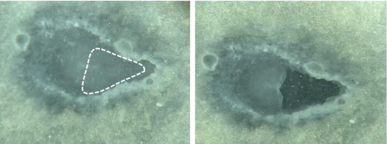
To assess the autonomous elongation of axial structures in chick embryos, we performed experiments to break the connection between the embryo and the area opaca (extra-embryonic tissue). Chicken embryos were collected at stages HH4-5 and prepared for the “New” system, and before re-incubation we excised at least 50% of the primitive streak and adjacent area pellucida, leaving only the Hensen’s node and a small portion of the primitive streak (Figure 7), as well as the whole area opaca. Two types of culture substratum were tested during these experiments: either albumen or a mixture 1:1 of agarose and albumen. In both cases, the results were comparable Out of the 16 embryos produced in both experiments, eight of them produced the “tails” described by (Spratt, 1947). Of these, we decided to analyse the two that produced the most extension.
Fig 7. Example of manipulated embryo in which the primitive-streak and area pelucida were excised (dashed area).
Chick embryo notochord excision experiments.
To test the role of the notochord in chick embryo axial extension we cultured embryos using the ”New” culture system which exposes the embryo’s ventral side, thus facilitating micro-manipulation of the notochord with minimal effects on adjacent tissues. Four different types of excisions were performed using sharpened tungsten
25 needles to “scrape out” the notochord. In three sets of experiments, portions of the notochord were removed as shown in Figure 8, and whichcorresponded to the portions in which we divided the embryo for the analysis of AP axis elongation dynamics. These were i) the cranial notochord (Figures 8A and 8A’) (n=4), ii) the “mid” notochord (adjacent to the segmented mesoderm) (Figures 8B and B’) (n=3), and iii) the posterior notochord (adjacent to the pre-somitic mesoderm) (Figures 8C and C’) (n=3). In other embryos (n=2), we removed the largest portion of notochord which we thought would not produce severe damage to the embryo. This included the mid and posterior notochord and about 1/3 of the cranial notochord (Figures 8D and 8D’). Thus we designated these experiments as “mid+posterior”, because only these portions were entirely removed. Embryos were manipulated between HH7-9 (Hamburger and Hamilton, 1951) and images were collected using the Wild stereoscope + Olympus camera with intervals of three or four hours starting from the moment when the embryo was micro-manipulated and ending as long as HH11 (13 somites). Images were used to measure the length of micro-manipulated embryos and the measurements from each set of excision experiments, as well as controls (n=5), were used to calculate net growth rates which were compared with an ANOVA using Statistica 8.
26
Fig 8. Examples of notochordectomies performed in chick embryos. The portion of the
notochord to be removed is indicated between arrowheads on the left panels (A, B, C and D) and the right side panels show the resulting embryo immediately after the procedure (between asterisks is the portion of the notochord removed, in A’, B’, C’ and D’). A) and A’) Removal of the cranial portion. B) and B’) Removal of the mid portion (adjacent to segmented mesoderm). C) and C’) Removal of the posterior portion (adjacent to pre-somitic mesoderm). D) and D’) Removal of the mid+caudal portion. Scale bars represent 1000 μm.
27 Immuno-histochemistry and confocal imaging of chick embryos.
Manipulated embryos were fixed and subjected to immuno-histochemistry as described in (Martins et al., 2009) to observe the results of notochord excision experiments using confocal microscopy. Briefly, embryos were fixed, permeabilised and incubated with ToPro3 and Phalloidin to stain for nuclei and F-actin cytoskeleton (respectively) and then cleared and mounted in methylsalicylate. Images of these embryos were acquired on a Leica SPE confocal system operating on a Leica DMI4000 microscope and 40x 1,3NA lens, and 3D reconstructions made using Amira v4.1.2 (Visage Imaging, Inc.). These images were used to show the results of notochord excision, and to show that the notochord does not regenerate.
28
RESULTS
Dynamics of zebrafish embryo axis elongation
Analysis of the dynamics of zebrafish axis extension shows that there are two distinct phases of embryonic growth between shield (6hpf) and 15 somites (~16.5 hpf) stages. The first phase spans from shield stage to approximately 4 somites stage, while the second phase starts at approximately 4 somites stage and continues to, at least, 15 somite stage (Figure 9A). For each “synchronized” time-point (see Materials and Methods) we plotted the average lengths and corresponding standard deviations (Figure 9B), which allowed us to establish that the turning point between the two phases is at the 4 somite stage (the time-point with the highest standard deviation during the period of 4-5h since the beginning of the culture). To better analyze the growth rates of each phase, we separated the measurements into two sets (one with measurements up to 4 somite stage, and another from 4 somite stage onwards) and plotted the values against a time series (Figure 9C). The first growth phase is clearly linear, i.e., the embryo elongates at a constant rate. Toshow this we computed the corresponding trendline and the linear model was the best adjusted with an R2 above 0,99. The average elongation rate (AER) was 310,78μm/h. However, during the second phase, the best adjusted trendline was logarithmic and the AER was considerably smaller with an average of 90,68μm/h.
(Kimmel et al., 1995) considers that there is no increase in zebrafish embryo length (which he considers to be the “embryo’s longest linear dimension”) before 14 somite stage. Our results show that there is an actual elongation before 14 somite stage and that this elongation is bi-phasic. When comparing the values of the elongation rates analyzed here and the ones proposed by (Kimmel et al., 1995), we estimate a growth rate for the first phase (310,78μm) which is actually higher than the one determined by Kimmel (1995) for the period between 16hpf (14 somites) and 32hpf (prim-stage) which was of 125μm/h.
29 Fig 9. Plots of embryo axis extension in zebrafish embryo. XX is time and or synchronized stages in hours and YY is length in μm. A) Represents the growth of the three embryos analysed. B) Represents the averages and standard deviations for each “synchronized” time point. C) Plots of the two phases of growth separately with the corresponding trendlines and R2.
Table I. Embryo lengths and standard deviations of different stages of zebrafish development
±0 means that only one embryo was measured for the corresponding stage.
Dynamics of chick embryo axis elongation
Having established the dynamics of zebrafish embryo axis elongation, we next analyzed the dynamics of chick embryo axis elongation between stages HH4-11. Initially we sought to make this analysis through a longer period of time [until at least stage HH13 (19 somites)], to allow the study of the formation of all pre-thoracic vertebrae and associated tissues. Due to constraints associated with the “New” culture
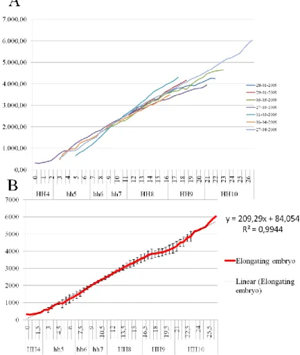
30 technique, we could not reach this stage, as all embryos ceased growing and begun presenting severe abnormalities after stage HH11. At this stage the chick embryos had just formed the first 13 somites, which in the adult chicken correspond to nearly half of the cervical vertebrae (Christ et al., 1998). Therefore, comparisons between the chick and zebrafish embryos were only possible for the first stages of growth. Plotting the chick embryo lengths against a time series reveals that embryo extension is linear during stages HH4-11 (computed trendlines using a linear model yielded R2 above 0,97 for all seven embryos analysed; see graph in Figure 10A). Plotting the averages of embryo lengths for each “synchronised” time-point (see Materials and Methods) and the corresponding standard deviations, produced a trendline R2 of 0,99 (Figure 10B) and revealed an AER of 213,74 μm/h. Table II shows the average embryo lengths at developmental stages between HH5-11 and corresponding standard deviations, as well as the AER for the whole embryo.
Fig 10. A) Representation of the measurements of the length of the seven time-lapse movies produced. XX is hours and HH stages. YY is embryo length in μm. B) Represents average embryo length and standard deviations for each “synchronised” time-point at which embryos were measured. AER estimated is 209,29 μm/h.
31 We next sought to analyze the dynamics of the elongation of the different portions of the embryo to find whether they grow linearly also, or whether some portions grow faster than others during the stages studied. The length of these portions (see Materials and Methods above) are plotted in a bar graph (Figure 11) representing the average embryo lengths over time. Table II shows the lengths of the three portions of the embryo and corresponding standard deviations in the period of time analyzed, as well as the AERs for each portion.
Table II. Embryo lengths of the entire embryo and of its portions in 5-11HH stages
(*) Although only none of the embryos reached the completed HH11 (13 somites) we present the value of the predicted time-point for stage HH11. The embryo measured at this stage had 12 somites and was almost forming the 13th somite.
±0 means that only one embryo was measured for the corresponding stage.
To analyze the different rates of elongation, we compared them using an ANOVA (Figure 11). For all portions, a linear model fit showed an R2 above 0,94. During this period, the cranial portion elongated from an average 1249,76 μm to 1848,73μm which represents an approximate 1,5 fold increase, at a rate of 26,88 μm/h. This elongation rate is significantly less than that of the segmented and pre-somitic portions (P<0,05), which elongate from average lengths of 349,81μm to 2195,42μm (segmented portion) and 248,85μm to 1850,30μm (pre-somitic portion). These correspond to an increase in length of approximately 6,3 and 7,5 fold, respectively, with AERs of 104,24 and 74,5 μm/h. Between the two portions, the elongation rate of the segmented portion was significantly higher than the pre-somitic. (P<0,05)
32 Fig. 11. Graphic produced by the ANOVA analysis of the AERs
of the different portions of the chick embryo. The three portions elongate at significantly different rates (P<0,05).
Because it has been suggested that in chick embryos most of the axis extension occurs in the first stage of gastrulation in the form of primitive streak elongation (Keller 2003), we measured the length of the primitive streak and its shortening rate to that of embryo axis extension. Figures 12A and 12B contain complementary information that represent this relationship and reveal that: i) the elongating embryo increases its length well beyond the length of the primitive streak, surpassing it between stages HH6 and HH7 and eventually becoming three fold longer than the primitive streak, and that ii) the primitive streak shortens at a slower rate (49,16 μm/h vs. 213, 74 μm/h). This shows that there is a considerable extension of the embryo during the regression of the node which is independent in the presence of the primitive streak. Interestingly, we noticed that the primitive streak is not all “replaced” by the growing embryo, at least, until HH11.
33 Fig. 12. Graphic representation of the comparison of the elongating embryo and of the shortening primitive streak. A) Bar graph representing the different dynamics throughout time. B) Plot of the two lengths, in which
Axis elongation in chick embryo is autonomous
Considering our observation that the embryo elongates more rapidly than the primitive streak regresses we wanted to test the capacity of chick embryos to extend independently of the primitive streak. To do this, we removed the streak as explained in “Materials and Methods”, thus severing of the connection between the node and the caudal part of the area opaca, i.e., portion of the embryo that elongates the embryo lost its connection to the extra-embryonic tissues. The result in all experiments was that embryos were still capable of produce elongation. One of the embryos analysed
34 survived 12h and reached a total length of 2557μm, with 2281μm beyond the cut edge (Figure 13A). This shows that, contrarily to what has been shown in zebrafish, where embryo axis extension only occurs if there is epiboly of the YSL and a continuous connection between the growing embryo and the YSL (Trinkaus 1951), the chick embryo does not require a connection to the extra-embryonic tissues to extend. In the portion of the embryo which grew beyond the cut edge, we could identify a head and trunk with a notochord, neural tube and somite-like structures. Having been cultured for 12 hours, it would be expected that this embryo had reached stage HH9, and had extended approximately 3640μm, considering our earlier observation of control embryos (Table I). One other embryo analyzed survived for 22h and extended well beyond the cut edge, with a trunk where somites were clearly identified (Figure 13B). It had 12 well formed somites, which is close to the expected 13 (this embryo was manipulated at stage HH5 and cultured for an additional 22h). In this embryo, the cut was made farther from the node, thus leaving approximately 50% of the primitive streak. The resulting embryo spanned 969μm beyond the cut edge to a total length of 3210μm. These results, once again, clearly demonstrate that elongation of the embryo axis is independent on the presence of, or physical connection with, the primitive streak. It further suggests that the forces that elongate the embryo are produced either within the Hensen’s node itself or in the tissues rostral to the node. The tissues that could produce forces rostral to the node are the notochord, the neural tube or the paraxial mesoderm (as these were the tissues formed in our “amputated” embryos).
Fig. 13. Pictures of the embryos whose primitive streak and area pellucida were removed, thus breaking the physical connection between the Hensen’s node and the extra-embryonic tissues (see also Fig 7). A) This embryo (head is towards left side) survived for 12h and reached a total length of 2557μm. The “outgrown part” contained a head and trunk with somite-like structures, neural tube and notochord. B) This embryo (head is towards the right side) survived for 22 hours and reached a final length of 3210μm. The number of somites formed(12) was close to the predicted number for this culture time (13).
35 No individual portion of the notochord is essential for embryo extension
After demonstrating that chick embryos have an autonomous capacity to elongate, we tested the hypothesis that the notochord could be contributing to embryo axis elongation. For that, we excised portions of the notochord in four different experiments, as described in the “Materials and Methods” section. Other embryos were cultured without any manipulation and served as control to these experiments (Figure 14). Different embryos were manipulated at different stages, however, since we had pre-established that embryo axis elongation is linear from HH4-11, we compared the results of these experiments by comparing the average AERs per type of experiment with an ANOVA (Figure 15A). The test did not reveal significant differences between the three types of experiments + control group (P=0,16). However, we compared the rate of elongation with each type of the excision using the “Planned comparisons” module of the ANOVA. This allowed us to compare each of the manipulations directly with the control measurements. Results of this analyzes are shown in figure 15B. When we removed the cranial portion of the notochord (Figure 14A) the embryo developed normally and there was no significant effect on the embryo axis elongation (Figure 14A’; P=0,75). The removed portion of the notochord did not regenerate and was absent in embryos fixed and immuno-stained at the end of culture and imaged using confocal microscopy (Figure 15). The removal of the mid section of the notochord (the one corresponding to the segmented mesoderm; Figure 14B) and of the caudal (corresponding to the pre-somitic mesoderm; Figure 14C) also did not produce any significant effects and the embryos developed normally with AERs of 145,04 μm/h and 172,06 μm/h, respectively (Figure 14B’ and 14C’, respectively). An ANOVA revealed no significant differences between these values and those of controls (P=0,16 and 0,20 respectively). In these cases, we also verified that the notochord did not regenerate after the treatment and culture.
Interestingly, the embryos in which we removed the mid+posterior portions of the notochord (Figure 14D) also developed normally (Figure 14D’) but with an AER of 123,47μm/h, which was significantly different (P=0,03) from the other experiments, suggesting that a cumulative effect of the removal of different portions of the notochord influences embryo axis extension.
36
Fig. 14 Examples of chick embryos in which the notochord was excised before culture. The
portion previously removed is indicated between arrowheads (left panels). The lacking portion is indicated between asterisks. A), B), C) and D represent embryos before culture and A’), B’), C’) and D’) are images of the same embryos after 4,5 to 6 hours. A) and A’) Removal of the cranial portion. B) and B’) Removal of the mid portion. C) and C’) Removal of the posterior portion. D) and D’) Removal of the mid+caudal portion. Scale bars represent 1000 μm.
37
Fig 15. Comparison of the different rates of elongation of manipulated and control
embryos.
A) Graphic produced by the ANOVA statistical test which compared all the effects simultaneously. None of the excision types produced a significant (P=0,16) effect. B) Table produced by the “Planned comparisons” module comparing the extension rate of each type of excision to the control. The only procedure which produced a significant effect was the excision of the Mid+posterior portions of the notochord (P=0,03).
Curiously, we also observed that notochord cells accumulated in a round mass at the caudal end of the remainder cranial portion of the notochord (Fig 16), suggesting that notochord cells are motile and move caudally during embryo axis extension. In the embryos where the caudal-most portion of the notochord (adjacent to the pre-somitic mesoderm) was removed, although the caudal border of the excision was made next to the node, new notochord continued to form and the node continued to regress, showing that a direct connection between the Hensen’s node and the previously formed notochord is not essential for axis extension.
38 Fig 16. 3D reconstruction produced from a series of confocal images of a chick embryo fixed and stained to show cell’s nuclei. This embryo was manipulated to remove the cranial portion of the notochord prior to culture. This is an anaglyph 3D image which can be viewed using anaglyph red-green glasses. The notochord does not regenerate and a mass of its cells can be seen at the caudal end of the remainder cranial portion of the notochord (yellow circle). Ventral side is oriented toward the viewer, and rostral side is to the left.
39
DISCUSSION
In this work we sought to analyze the dynamics of embryo axis elongation in amniotes and anamniotes and the possible role of the notochord in the extension of amniotes using zebrafish and chicken embryos as representative models. Our hypothesis was that the notochord plays an important role in amniote axis elongation as opposed to anamniotes embryos, taking advantage of its structure and autonomous capacity to extend.
Comparison of the different dynamics of zebrafish and chick embryo axis elongation
We first analyzed the dynamics of axis elongation in the zebrafish embryo. Our first conclusion was that, contrary to what is established in (Kimmel et al., 1995) there is an actual elongation in the period of time spanning from shield stage to 15 somite stage. Although it is not clear what he considered the “embryo’s longest linear dimension”, as he states in his paper, we used a new approach to analyze embryo length during this period of development. By digitally “straightening” the embryo we adapted the concept of linear dimension and were able to discern what was happening in this phase and determine a pattern that repeats itself. This pattern was composed of two different dynamics of elongation, with the first one having a higher rate of extension than the second. We found that the border between these two dynamics was located at around the 4 somite stage, which is only 1h after bud stage, when epiboly is considered to end (Kimmel et al., 1995). Several experiments (Trinkaus, 1951; Glickman, 2003) have shown that epiboly of the YSL could be driving extension of the embryo axis. Based on our results, we suggest that epiboly is contributing to the extension of the embryo in the first phase, which translates in a high extension rate. In the second phase, when epiboly has ceased, the embryo extension is driven only by other forces, e.g., convergent extension of mesodermal tissues. After 15 somites stage, the extension rate is estimated to be 125μm/h (Kimmel et al., 1995), which is considerably slower than the rate we measured during the first stage (310,78μm/h). If, in fact, the epiboly mechanism drives extension in the first phase of elongation, this force seems to be stronger than whatever forces are driving extension after 15 somite stage. Curiously, the border between the two phases of early extension corresponds to the border between the cranial somites and pre-caudal somites (Holley, 2007).
40 We next analyzed the dynamics of chick axis elongation, first looking at the whole embryo and then to its different portions. Our first conclusion was that between stages HH4-10, the extension is linear. This result was unexpected at first, according to our initial hypothesis that the notochord could be driving axis elongation. The notochord only changes from a bi-laminar organization (when it is composed of two layers of cells) to a rod-like organization which is achieved at approximately stage HH9 (Bancroft & Bellairs, 1976). Thus, we would expect to see a change in the dynamics of axis elongation at this stage, which was not the case. Keller (2003) considers that most of the chick embryo axis extension comes in the form of the primitive streak extension. He also suggests that the continuing extension after stage HH4 is absorbed as the primitive streak regresses. Our results show that not only is this extension not all absorbed in the regression of the node, but it has a higher rate than the shortening of the streak itself. This leads us to suggest that the elongating embryo in front of the node is actually pushing the streak and node backwards as the streak shortens. To analyze these dynamics, we would like to perform time-lapse movies using confocal microscopy, which would give us a better idea of the movements of the cells and tissues in play.
Because we could identify the different portions of the embryo in the chick time-lapse movies, we decided to analyze these and their relative extension. The first curious fact was that the extension in the cranial portion was much smaller than that of the segmented and pre-somitic portions. Thus, most of the extension of the embryo seems to occur in the region caudal to the first somite. As we analyzed the time-lapse movies the cranial portion was becoming progressively thinner with cell rearrangements in the form of convergence movements (data not shown). If convergent is occurring and not much lengthening is happening in the cranial region, than this could translate into dorso-ventral thickening. As before, confocal time-lapse movies showing DV movements of cells could confirm these observations. Another interesting observation is that the pre-somitic portion of the embryo elongates (although with a significantly smaller rate when compared to the segmented portion). While it is not surprising that the segmented portion of the embryo is elongating, the same cannot be said about the pre-somitic portion. If the segmented portion is constantly recruiting cells from the pre-somitic portion to form somites, it would be expected that this portion would be decreasing its length or at least maintaining its length, so there is always enough cells to form somites. It is known that 10 to 12 prospective somites are always contained inside the pre-somitic mesoderm of 48 hour embryos (Packard, 1976; Palmeirim et al., 1997). These prospective somites have been proposed to be the somitomeres, which are segmented
