325
ana teResa tiMóteo, J. PeReiRa albino, luísa MouRa bRanCo, nuno banazol, Paula ColaRinha, nuno tavaRes Jalles, Rui feRReiRa
Serviço de Cardiologia, Hospital de Santa Marta, Lisboa, Portugal Rev Port Cardiol 2009; 28 (3): 325-332
Recebido para publicação: Abril de 2008 • Aceite para publicação: Outubro de 2008 Received for publication: April 2008 • Accepted for publication: October 2008
ABSTRACT
Primary Idiopathic Chylopericardium
Chylopericardium is a rare entity which is usually associated with trauma (thoracic duct lesions), neoplasia or filaria infection. Primary forms are even rarer and are caused by lymphatic circulation malformations. We present the case of a 52-year-old male patient with primary chylopericardium. We discuss the diagnostic approach, as well as the favorable clinical evolution with conservative management.
Key words
Primary chylopericardium; Lymphatic metabolism; Mediastinal cyst
RESUMO
O quilopericárdio é uma entidade rara, que é encontrada mais frequentemente associada a traumatismos (com lesão do canal torácico), neoplasias e infiltrações por filária. As formas primárias são mais raras e associam-se a malformações da circulação linfática. Apresenta-se o caso clínico de um doente de 52 anos, sexo masculino, com o diagnóstico de quilopericárdio primário, sendo discutida a abordagem diagnóstica, assim como a evolução clínica favorável com tratamento conservador.
Palavras-Chave Quilopericárdio primário; Metabolismo linfático;
Quisto mediastino.
INTRODUÇÃO
O
quilopericárdio é uma entidade clínica rara, cujo diagnóstico é dado pelas carac-terísticas típicas do líquido pericárdico. A sua abordagem etiológica e terapêutica também têm muitas particularidades, pelo que entendemos apresentar o caso de um doente de 52 anos, com quilopericárdio primário e discutir a sua evolução com recurso a um tratamento conservador.INTRODUCTION
C
hylopericardium is a rare clinical entity which is diagnosed on the basis of typical characteristics of the pericardial fluid. Etiology and the therapeutic approach also have unusual features, and so we present the case of a 52-year-old male patient with primary chylopericardium and discuss the clinical evolution with con-servative management.326
CASE REPORT
A 52-year-old male, asympatomatic, with no risk factors for coronary artery disease and no history of heart disease or other systemic pathology (including trauma, surgery, filariasis, tuberculosis or radiotherapy) had a cardiological chek-up). He was not routinely taking any medication. The patient was normotensive, cardiac examination was normal with no signs of heart failure, and the ECG was normal. Chest X-ray showed a cardiothoracic ratio of around 70% (an exam performed three months previously was normal).
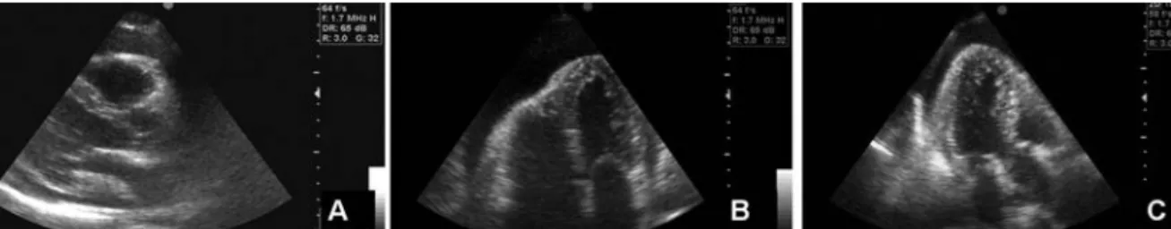
Echocardiography revealed a large pericardial effusion, with no evidence of hemodynamic compromise (Figure 1), and no other significant
abnormalities. Therapy was begun with diuretics and non-steroidal anti-inflammatory drugs for three months, after which there was no improvement in the effusion and the patient remained asymptomatic. Preliminary outpatient laboratory tests for renal and thyroid function and autoimmune study were normal. In view of the persistent effusion and for etiological study, the patient was admitted and therapy discontinued. All immunological studies, tumor markers, thyroid function, C-reactive protein and cardiac viral serology were negative. The Mantoux test was also negative. Pericardiocentesis was performed, with a total of 2500 ml of milky fluid being drained over a period of three days. At discharge, the patient had only a small pericardial effusion. Pathological study was negative for neoplastic cells. The pericardial biopsy showed moderate lymphocytic inflammatory infiltrate, with no granulomas. Biochemical analysis
CASO CLÍNICO
Doente de 52 anos, sexo masculino, em avaliação de check-up cardiológico, assin-tomático, sem factores de risco para doença coronária. Nega antecedentes de patologia cardíaca ou outra patologia sistémica, inclusive traumatismos, cirurgias prévias, filaríase, esta-dias em África, tuberculose ou radioterapia. Nega também utilização regular de fármacos. O doente apresentava-se normotenso, com observação cardíaca normal, sem evidência de sinais de insuficiência cardíaca. O ECG era normal. A radiografia do tórax apresentava um índice cardio-torácico de cerca de 70% (sendo o doente portador de um exame prévio efectuado cerca de 3 anos antes, normal).
Efectuou ecocardiograma, que revelou vo-lu mo so derrame pericárdico, sem qualquer evi-dência de compromisso hemodinâmico (Figura
1). Sem outras alterações a valorizar. Foi
iniciada terapêutica com diuréticos e anti-infla-matórios não esteróides durante 3 meses, sem evidência de melhoria do derrame, mantendo-se o doente assintomático. A avaliação laboratorial preliminar em ambulatório, com função renal, estudo de doença auto-imunes e estudo da função tiroideia foram normais. Por persistência do derrame e para estudo etiológico, foi internado e entretanto foi suspensa a terapêutica em curso. Todo o estudo imunológico, marcadores tumorais, estudo da função tiroideia, proteína C reactiva e serologias virais cardíacas foram negativas. O teste de Mantoux foi também negativo. Foi então efectuada pericardiocentese, que drenou um total de cerca de 2500 ml de líquido com características leitosas, tendo mantido drenagem
Figura 1. Grande derrame pericárdico na admissão do doente, antes da pericardiocentese (A – para-esternal esquerdo, eixo longo; B – apical 4 câmaras; C – apical 3 câmaras).
Figure 1. Large pericardial effusion at admission, before pericardiocentesis (A: left parasternal long-axis view; B: 4-chamber apical view; C: 3-chamber apical view).
327
was compatible with a chylous effusion (total cholesterol 42 mg/dl; triglycerides 1230 mg/dl; total protein 6.2 g/dl; albumin 3.4 g/dl; LDH 413 uI/l; ADA 6.2 mg/dl).
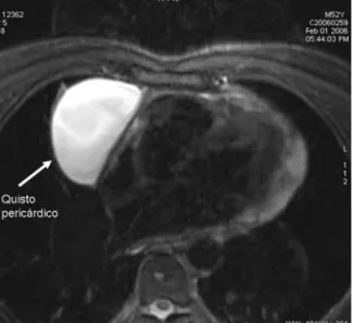
In order to fully characterize the pericardial effusion, cardiac magnetic resonance imaging (MRI) was performed, which detected a large pericardial cyst filled with a similar fluid and a millimetric image of a possible fistula, but which was not conclusive (Figure 2). Radionuclide lymphoscintigraphy was performed as a noninvasive method of obtaining functional information on the course of lymphatic drainage and the presence of lymphatic leak and its output. This showed the lower limb lymphatic system to be permeable and functional, and confirmed a communication with the venous system and systemic circulation through visualization of hepatic activity (Figure 3). An abnormal con-centration of the radioisotope was observed in the left supraclavicular and mediastinal region, with a slight deviation towards the left inferior mediastinum. Involvement of the mediastinum is unusual but the etiology of the effusion remained unclear. The slight increase in concentration would suggest a low-volume effusion.
Echocardiography was repeated a month after the patient’s discharge (Figure 4) and again showed worsening of the pericardial effusion, although the patient still had no signs of hemodynamic compromise or symptoms. He durante 3 dias. À data da alta, apresentava apenas
ligeiro derrame pericárdico. O estudo anatomo-patológico foi negativo para células neoplásicas. A biópsia pericárdica mostrou moderado infiltrado inflamatório linfo-plasmocitário, sem granulomas. O estudo bioquímico foi compatível com derrame quiloso (colesterol total: 42 mg/dl; triglicéridos: 1230 mg/dl; proteínas totais 6,2 g/ dl; albumina: 3,4 g/dl; LDH: 413 uI/L; ADA 6,2 mg/dl).
Foi realizada ressonância magnética car-díaca, para melhor caracterização do derrame pericárdico, tendo-se identificado volumoso quis-to pericárdico, preenchido pelo mesmo tipo de líquido e imagem muito duvidosa (milimétrica) de eventual fístula, contudo não conclusiva (Figura
2). Efectuou linfocintigrafia isotópica, que permite
obter de modo não invasivo informação funcional quanto ao percurso da drenagem linfática, á presença ou ausência de derrame linfático, assim como quanto ao seu débito. Este exame mostrou permeabilidade e perfil funcional do sistema linfático dos membros inferiores, confirmando-se comunicação com o sistema venoso/circulação sistémica através da visualização de actividade hepática (Figura 3). Foi observada acumulação anómala de radiofármaco a nível da região
Figura 2. Ressonância magnética cardíaca, mostrando derrame pericárdico, com volumoso quisto pericárdico preenchido.
Figure 2. Cardiac MRI, showing pericardial effusion and a large pericardial cyst filled with fluid.
Figura 3. Linfanciocintigrafia, com captação mediastínica aumentada.
Figure 3. Lymphoscintigraphy, showing increased medias-tinal uptake.
328
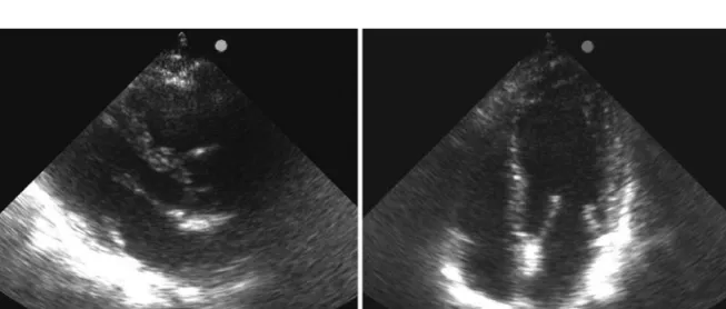
was then assessed for vascular surgery, but in the absence of an identifiable etiology, we opted for a conservative approach, with dietary fat restriction and preferential use of butter. Six months after drainage, the effusion had stabilized. Control echocardiograms at 9, 12 and 18 months (Figure
5) showed that the effusion had completely
disappeared. The patient remained asymptomatic throughout, with no change in weight and with a normal lipid profile, with no specific treatment. supraclavicular esquerda e mediastínica, com
ligeiro desvio para a vertente infero-esquerda do mediastino, sendo que a actividade mediastínica não é habitual, mas sendo inconclusiva quanto à etiologia do derrame. Esta acumulação de ténue intensidade sugeria uma situação de derrame de baixo débito.
O doente repetiu o ecocardiograma um mês após a alta (Figura 4), tendo mostrado novo agra vamento do derrame pericárdico, embora sem sinais de compromisso hemodinâmico, e continuando assintomático. Foi entretanto ava-liado por Cirurgia Vascular. Na ausência de etiologia identificável, optou-se por uma dieta com restrição lipídica e utilização preferencial de manteiga como lípidos na dieta. Seis meses
Figura 4. Derrame pericárdico 3 meses após drenagem pericárdica (A – paraesternal esquerdo, eixo longo; B - para- -esternal esquerdo, eixo curto; C – apical 4 câmaras).
Figure 4. Pericardial effusion three months after pericardial drainage (A: left parasternal long-axis view; B: left parasternal short-axis view; C: 4-chamber apical view).
Figura 5. Derrame pericárdico 18 meses após drenagem pericárdica (A – para-esternal esquerdo, eixo longo; B – apical 4 câmaras).
Figure 5. Pericardial effusion 18 months after pericardial drainage (A: left parasternal long-axis view; B: 4-chamber apical view).
329
DISCUSSION
This rare case of idiopathic chylopericardium is an opportunity to review this pathology. Chylous fistulas are unusual but easily diagnosed, being immediately identified by the milky nature of the exudate(1). A review of the
literature since 1977 in PubMed revealed only 24 cases of chylopericardium in adults, 15 of them primary. Etiology can be primary or the result of various clinical situations, particularly trauma (thoracic duct lesions), neoplasm (primary such as lymphangioma or through invasion of the lymphatic system by other neoplasms) and filaria infection(2). Primary forms are the result of
malformation of the intestinal lymph circulation and its relationship with the systemic circulation, resulting in megalymphatics that develop fistulas following even minimal trauma and that can be located at atypical sites in the body(2). In the
abdomen, lower limb lymphatics containing high-protein lymph come in contact with intestinal lymphatics that transport lipids(1).
Among lymphatic lipids, long-chain fatty acids (a constituent of chylomicrons) pre-dominate. Short- and medium-chain fatty acids (<12 carbon atoms) are more soluble in water and are absorbed passively by the gastric mucosa, forming complexes with albumin and passing directly into the portal circulation(3).
Gastric lipases also act more strongly on short- and medium-chain fatty acids(4). Long-chain
fatty acids are less soluble and are incorporated into triacylglycerols and phospholipids in chylomicron particles transported in the lymph(3).
Lymph also contains coagulation factors, which can under certain flow conditions and content cause it to coagulate in the same way as blood, as shown by studies that found an increase in activated factor VII linked to increased post-prandial lipid levels(5).
Of the dietary fats, butter has the highest content of short- and medium-chain fatty acids (20-25%), which reduces secretion of triacylglycerols into the lymph(6). Meals
con-taining butter lead to lower post-prandial lipid levels and a lower concentration of chylomicrons in the circulation than those containing olive oil or sunflower oil(6). Degrace et al.(7) also found that
butter, compared to other fats, leads to lower triacylglycerol output into the lymph due to greater portal absorption. In another study in humans(8),
após a drenagem, verificou-se estabilização do derrame. No ecocardiograma de controlo aos 9, 12 e 18 meses (Figura 5), verificou-se desaparecimento completo do derrame. O doente manteve-se sempre assintomático, sem variação ponderal e com perfil analítico lipídico normal, sem qualquer terapêutica específica.
DISCUSSÃO
Aproveitamos a identificação desta caso raro de quilopericárdio idiopático para fazermos uma revisão desta patologia. As fístulas quilosas são entidades clínicas pouco usuais, no entanto, de diagnóstico fácil, na medida em que o aspecto leitoso do exsudado permite de imediato a sua identificação(1). uma análise da literatura médica
publicada desde 1977 (PubMed), identificou ape nas 24 casos descritos de quilopericárdio em adultos, dos quais 15 eram primários. Podem ser de etiologia primária ou consequentes a numerosas situações clínicas, das quais se destacam os traumatismos (com lesão do canal torácico), as neoplasias (primárias como as linfangiomatoses ou por invasão do sistema linfático por outras neoplasias) e as infiltrações por filária(2). As
causas primárias resultam da existência de malformações da circulação entérica da linfa e a sua relação com a circulação sistémica, com megalinfáticos, que, ao mínimo traumatismo, de-senvolvem fístulas, que podem ter localizações muito atípicas no corpo(2). A nível abdominal, os
linfáticos dos membros inferiores, com linfa de alto conteúdo proteico, entra em contacto com os linfáticos provenientes do sistema intestinal e que transportam lípidos(1).
Relativamente aos lípidos linfáticos, os áci-dos goráci-dos de cadeia longa (constituinte áci-dos quilomicrons) são os que predominam. Os ácidos gordos de cadeia pequena e média (<12 átomos de carbono) são mais solúveis na água, sendo a bsorvidos passivamente na mucosa gástrica, com incorporação em complexos com a albumina e passam directamente para a circulação portal(3).
As lipases gástricas são também mais activas nos ácidos gordos de cadeia curta e média(4).
As cadeias longas são menos solúveis e são incorporadas em triacilgliceróis e fosfolipidos localizados nas partículas de quilomicrons transportadas na linfa(3). A linfa possui também
330
replacing the short- and medium-chains of butter by long-chain fatty acids increased the post-prandial chylomicron response. In the case described above, the patient was prescribed a diet with short- and medium-chain triglycerides (such as found in butter), which are absorbed less by the lymphatic system, leading to increased protein content in the lymphatic duct and thus encouraging healing and closure of the fistula.
If this approach fails, total parenteral nutrition can be prescribed in order to reduce flow in the lymph duct and increase its protein content(2), or alternatively the somatostatin analog
octreotide(9). Somatostatin acts by decreasing
intestinal absorption of lipids and reducing triglyceride concentrations in the thoracic duct and flow in the major lymphatic ducts. Surgery is also a treatment option(10), but in the case
presented, failure to clearly identify the site of the fistula meant this approach could not be used. Radionuclide lymphoscintigraphy can be useful in detecting fistulas or anomalous lymphatic drainage and for functional characterization, but since it is a functional rather than an anatomical exam it does not enable direct visualization of fistulas(11). In the case of our patient, a
conservative approach was very effective, and there was thus no need to consider higher-risk and more aggressive therapeutic options.
With regard to the paracardial cyst detected by MRI in our patient, differential diagnosis is required of mediastinal cysts containing chylous fluid between pericardial cysts, thoracic duct cysts and cystic lymphangioma.
Thoracic duct cysts are rare; they can occur anywhere along the course of the duct within the mediastinum, in either the costovertebral sulcus or the visceral compartment of the posterior and superior mediastinum(12); this was not the
case in our patient. Cystic lymphangioma (also known as hygroma or lymphatic cyst) arises from malformations in the development of the lymphatic system. Around 90% are diagnosed before the age of two, and most are found in the head or neck region (75% of cases) or in the axillary region (20%)(13,14). The rare cases
diagnosed in adulthood are usually detected on routine exams in asymptomatic patients and are located in the thoracic cavity(13,14).
Mediastinal lymphangiomas account for 0.7 to 4.5% of all mediastinal tumors(13,15). Symptoms
occur when the tumor grows and compresses or determinadas circunstâncias de fluxo e conteúdo,
levar à coagulação de modo idêntico ao sangue, em particular alguns estudos que verificaram o aumento do factor VII activado em relação com o aumento lipídico pós-prandial(5).
A manteiga é a gordura alimentar com maior composição de ácidos gordos de cadeia curta e média (cerca de 20 a 25%), pelo que se traduz numa menor secrecção de triacilglicerois na linfa(6). Com efeito, refeições com manteiga
resultam numa lipémia pós-prandial inferior e em menor acumulação de quilomicrons na cir-culação, comparativamente com o azeite e o óleo de girassol(6). Também Degrace e col.(7)
verificaram que a manteiga, comparativamente com outros óleos, condiciona um menor débito de triacilgliceróis na linfa, explicado pela maior absorção portal. Num outro estudo em humanos(8),
a substituição das cadeias curtas e médias da manteiga por outras de cadeia longa, aumentou a resposta pós-prandial de quilomicrons. Pro-porcionou-se por isso ao doente aqui descrito uma dieta com triglicéridos de pequena e média cadeia (como a existente na manteiga), sendo assim menos absorvidos pelo sistema linfático, permitindo um aumento do conteúdo proteico no canal linfático, facilitando assim a cicatrização e encerramento da fístula.
Quando esta abordagem falha, a utilização de dieta parentérica total (de modo a reduzir o fluxo do canal linfático e aumentar o seu conteúdo proteico) pode ser utilizada(2), ou
em alternativa, a utilização de somatostatina (octreótido)(9). A somatostatina actua por
diminuição da absorção intestinal dos lípidos, redução da concentração de triglicéridos no canal torácico e redução do fluxo linfático nos canais linfáticos maiores. A opção cirúrgica(10) é
também uma possibilidade terapêutica, mas no presente caso, a não identificação da localização da fístula não permitiria este tipo de abordagem. A linfocintigrafia isotópica poderá ser útil na identificação de fístulas ou drenagens linfáticas anómalas e na sua caracterização funcional, mas por ser um exame funcional e não anatómico, não permite a visualização directa de fístulas(11).
Relativamente ao nosso doente, a abordagem conservadora foi muito eficaz, não tendo havido por isso necessidade de recorrer às outras opções terapêuticas com maior risco, e também mais onerosas.
para-car-331
invades adjacent structures. Chylothorax and chylopericardium are other possible com pli-cations(13). Such cysts are well defined, either
unilocular or multilocular, and filled with fluid(16). They can occur anywhere but are most
commonly found in the superior mediastinum. On rare occasions, they are found between the anterior thoracic wall and the right heart(16), as
was the case in our patient, and this can make it difficult to differentiate from pericardial cyst.
Mesothelial pericardial cysts are uncommon and benign anomalies, accounting for 6% of mediastinal masses and 33% of mediastinal cysts, and can be of various etiologies including congenital, lymphomatous and infective(17).
Peri-cardial cysts are typically unilocular containing fluid, and are found in the mediastinal fat along the right or left cardiophrenic angle or in the anterior mediastinum, inside or outside the pericardial sac(16). The cyst wall is composed of
connective tissue with a single layer of mesothelial cells, and may communicate with the pericardial space(16). In our patient, the fact that the cyst was
not present on previous exams, thus excluding a congenital origin, its location and unilocular nature, its communication with the pericardial space and its appearance following resolution of the pericardial effusion indicate that it was a pericardial cyst; however, only histological study after excision of the cyst can confirm its type. díaca identificada na ressonância magnética,
impõe-se o seu diagnóstico diferencial. Dentro dos quistos mediastínicos com conteúdo quiloso, o diagnóstico diferencial coloca-se entre quisto pericárdico, quisto do canal torácico e linfangioma quístico.
Os quistos do canal torácico são raros. Podem ocorrer em qualquer localização ao longo do trajecto do canal pelo mediastino, podendo localizar-se a nível do sulco costo-vertebral ou no compartimento visceral do mediastino (posterior e superior)(12); deste modo, não se
enquadra no quisto descrito no nosso doente. Os linfangiomas quísticos (também designados por higromas ou quistos linfáticos) são malformações do desenvolvimento do sistema linfático. Cerca de 90% dos casos são diagnosticados até aos dois anos de idade, ocorrendo principalmente na região da cabeça e pescoço (75% dos casos) e na região axilar (20%)(13,14). Os casos raros
que são diagnosticados em idade adulta são habitualmente achados detectados em exames de rotina em doentes assintomáticos e com localização intra-torácica(13,14). Os linfangiomas
mediastinicos representam cerca de 0,7% a 4,5% de todos os tumores medias tínicos(13,15).
Os sintomas surgem quando o tumor cresce e comprime ou invade as estruturas adjacentes. O quilotoráx e o quilopericárdio são também complicações que podem ocorrer(13). Estes quistos
são bem delimitados, com cavidade única ou multiloculada, preenchida por liquido(16). Podem
ocorrer em qualquer localização, mas são mais frequentes no mediastino superior. Raramente podem ser detectados entre a parede torácica anterior e o coração direito(16). Nesta localização
(como é o caso do nosso doente), pode ser difícil distinguir de quisto pericárdico.
Os quistos pericárdicos mesoteliais são anomalia benignas raras (6% das massas me-diastinicas e 33% os quistos mediastínicos), podendo ter etiologias variadas desde congénita, linfomatosa ou infecciosa(17). Os quistos
pericár-dicos são caracteristicamente uniloculados e com fluido no seu interior, localizado na gordura mediastínica ao longo do ângulo cardio-frénico direito ou esquerdo ou no mediastino anterior, no interior ou exterior do saco pericárdico(16).
A parede do quisto é composta por tecido conjuntivo e uma camada única de células mesoteliais, podendo comunicar com o espaço pericárdico(16). No nosso doente, o facto de
332
Address for reprints: Pedido de separatas para: AnA TeresA TimóTeo
Av. Miguel Torga, nº 27, Edifício C, 9ºA 1070 – 183 Lisboa
e-mail: ana_timoteo@yahoo.com estar presente em exames prévios (excluindo
origem congénita), a sua localização, o facto de ser uniloculado, a comunicação com o espaço pericárdico e a resolução desta imagem com a resolução do derrame pericárdico apontam para que se trate de um quisto pericárdico, contudo, apenas uma avaliação histológica após excisão do quisto poderá confirmar o tipo de quisto.
BIBLIOGRAFIA / REFERENCES
1 – Guyton A and Hall J. Digestion and absorption in the gastrointestinal tract. In: Textbook of Medical Physiology. 1996, W. B. Saunders: Philadelphia. p. 833-844.
2 – Svedjeholm R, Jansson K, Olin C. Primary idiopathic chylopericardium – a case report and review of the literature. Eur J Cardio-Thorac Surg. 1997; 11: 387-90.
3 – Jensen M, Christensen M, Hoy C. Intestinal absorption of octanoic, decanoic, and linoleic acids: effect of triglyceride structure. Ann Nitr Metab. 1994; 38: 104-16.
4 – DeNigris S, Hamosh M, Kasbekar et al. Secretion of human gastric lipase from dispersed gastric glands. Biochim Biophys Acta.1985; 836: 67-72.
5 – Sanders T, Berry S, Miller G. Influence of triacylglycerol structure on the post-prandial response of factor VII to stearic acid-rich fats. Am J Clin Nutr. 2003; 77: 777-82.
6 – Mekki N, Charbonnier M, Borel P et al. Butter differs from olive oil and sunflower oil in its effects on postprandial lipemia and triacylglycerol-rich lipoproteins after single mixed meals in healthy young men. J Nut. 2002; 132: 3642-9.
7 – Degrace P, Caselli C, Rayo J, Bernard A. Intestinal lymph absorption of butter, corn oil, cod liver oil, menhaden oil and eicosapentaenoic and docosahexaenoic acid ethyl esters in rats. Lipids. 1996; 31: 405-14.
8 – Tholstrup T, Sandstrom B, Hermansen J, Holmer G. Effect of modified dairy fat on postprandial and fasting plasma lipids and lipoproteins in healthy young men. Lipids. 1998; 33: 11-21.
9 – Demos N, Kozel J, Scerbo J. Somatostatin in the treatment of chylothorax. Chest. 2001; 119: 964-6.
10 – Riquet M, Badia A. Surgery for chylothorax. Rev Pneumol Clin. 2004; 60: 104-8.
11 – Wang C, Yen T, Ng K, et al. Pedal 99mTc-sulfur colloid lymphoscintigraphy in primary isolated chylopericardium. Chest. 2000; 117:598-601.
12 – Turkyilmaz A, Eroglu A. A giant thoracic duct cyst: an unusual cause of dysphagia. J Thorac Cardiovasc Surg. 2007; 134 (4): 1082-3.
13 – Saleiro S, Magalhães A, Souto Moura C, Hespanhol V. Linfangioma cístico do mediastino. Rev Port Pneumol. 2006; 12 (6): 731-5.
14 – Kavunkal AM, Ramkumar J, Gangahammaiah S, Parimlazhagan KN, Cherian VK. Isolated mediastinal cystic lymphangioma in a child. J Thorac Cardiovasc Surg. 2007; 134 (6): 1596-7.
15 – Park JG, Aubey MC, Godfrey JA, Midthun DE. Mediastinal lymphangioma: Mayo clinic experience of 25 cases: Mayo Clin Proc. 2006; 81 (9): 1197-203.
16 – Bernasconi A, Yoo SJ, Golding F, Langer JC, Jaeggi ET. Etiology and outcome of prenatally detected paracardial cystic lesions: a case series and review of the literature. Ultrasound Obstet Gynecol. 2007; 29: 288-94.
17 – Venkatesh BG, Girota S, Iyer KS, Chakrabarti M, Khurana P, Sen A. Pericardial cyst due to tuberculosis in an adolescent. Ann Card Anaesth. 2008; 11: 115-7.