Popliteal artery entrapment syndrome: case report
Síndrome do aprisionamento da artéria poplítea: relato de caso
Marcelo Bettega1, Ariane Szeliga1, Rafael Pereira Hagemann2, Antônio Lacerda Santos Filho3, Nelson Mesquita Júnior4
Introduction
he popliteal artery entrapment syndrome (PAES) is the clinical result of extrinsic compression of the popliteal artery. It can present as an anatomical or congenital anomaly or as an acquired or functional form. he ana-tomical anomaly is the result of abnormal embryological development of the popliteal artery or the musculotendi-nous structures around it; in the functional or acquired form, the artery is compressed as a result of hypertrophy of adjacent muscles1.
It was irst described in Edinburgh in 1879 by medical student Anderson Stuart, who had dissected the amputated leg of a 64-year-man, victim of gangrene by thrombosis of
the popliteal artery2. Hammings described the irst case in
which this syndrome was surgically treated (section of the medial head of the gastrocnemius muscle), in 1959³. Love
and Whelan4 proposed the name “popliteal artery
entrap-ment syndrome”, which has been used since then.
PAES predominantly afects men (15:1)5,6 and is
iden-tiied as the main etiology of intermittent claudication in young patients7,8, with symptoms appearing usually ater
physical activity. As the artery compression results in re-peated trauma, the complications, besides causing incapac-ity for physical exercise, include: stenosis, emboli, post-stenotic aneurysms and thrombosis8,9.
he purpose of this article is to report a case of popliteal artery entrapment, discussing its diagnosis, classiication,
Abstract
Popliteal artery entrapment syndrome is the compression of the popliteal artery and is the main cause of intermittent claudication in young patients. An 18-year-old man was admitted at our service complaining of right foot paresthesia, coldness, and pallor that appeared 24 hours after physical activity. Posterior tibial and dorsal artery of foot pulses were not present in right lower limb. Diminished posterior tibial and dorsal artery of the foot pulses were found in left lower limb at dorsal lexion and forced plantar lexion. After surgery, both pulses were present. his syndrome is more frequent in men and its prevalence varies between 0.16 and 3.5%. Popliteal artery entrapment type III is most common. Non-treated entrapment can lead to embolism, thrombosis and post-stenotic aneurysms. he syndrome must be considered as a cause of lower limb pain specially in young men with intense sport practice history.
Keywords: popliteal artery; constriction, pathologic; /surgery. Resumo
A síndrome do aprisionamento da artéria poplítea caracteriza-se pela compressão desta artéria sendo a principal causa de claudicação intermitente em jovens. Homem, 18 anos, branco, apresentava parestesia, frialdade e palidez do pé direito, iniciada 24 horas após exercício físico. Em membro inferior direito, ausência de pulsos tibial posterior e dorsal do pé. À lexão dorsal e lexão plantar forçadas, houve diminuição dos pulsos tibial posterior e dorsal do pé à esquerda. Tratado cirurgicamente, o paciente apresentou pulso em ambas as artérias. A síndrome é mais frequente em homens e a prevalência varia entre 0,16 e 3,5%. O aprisionamento da artéria poplítea tipo III é mais comum. A falta de tratamento pode levar à embolia, trombose e aneurismas pós-estenóticos. Esta síndrome deve ser lembrada como causa de dor na perna, especialmente em homens jovens e de prática esportiva intensa.
Palavras-chave: artéria poplítea; constrição patológica; /cirurgia.
1Medical students in Faculdade Evangélica do Paraná (FEPAR) – Curitiba (PR), Brazil.
2Vascular surgeon; former resident in Angiology and Vascular Surgery at the Hospital Universitário Evangélico de Curitiba – Curitiba (PR), Brazil.
3Master’s degree in Clinical Surgery, Universidade Federal do Paraná (UFPR) – Curitiba(PR), Brazil; Assistant Professor of Angiology and Vascular Surgery at FEPAR – Curitiba (PR), Brazil. 4Master’s degree in Clinical Surgery, UFPR – Curitiba (PR), Brazil; Assistant Professor of Anatomy, Angiology and Vascular Surgery at FEPAR – Curitiba (PR), Brazil.
Financial support: none.
dorsal lexion of the foot, which did not show any compression (Figure 5). he patient presented normal right dorsalis pedis pulse in the immediate postoperative period. he posterior tibial pulse was also present at follow-up.
complications and treatment, and to review the literature for non-experts.
Case report
A 18-year-old caucasean male patient, a delivery bicycler, with no medical history, was admitted to our service with right foot paresthesia, coldness and pallor that had appeared when he was riding his bicycle 24 hours earlier. He reported right calf fatigue for over two years and paresthesia when making greater muscular efort while bicycling.
On physical examination, the patient had normal right femoral and popliteal pulses, no pulses in the right posterior tibial and dorsalis pedis arteries and no bruits in the right lower limb. He had paresthesia, coldness and pallor of the right toes, but motor function was normal. Pulses were normal and no bruits were audible in the let lower limb. Reduced let poste-rior tibial and dorsalis pedis pulses were found on forced dorsal lexion and plantar lexion of the let foot.
Doppler ultrasonography showed no low in the distal por-tion of the right anterior tibial and posterior tibial arteries. At forced dorsal lexion and plantar lexion, total compression of both right and let popliteal arteries occurred. he right and let popliteal arteries had a proximal diameter of 0.7 mm, and the right popliteal artery had a distal diameter of 0.9 mm (ectasia).
Preoperative arteriography at rest showed bilateral medial deviation of the popliteal artery, as illustrated in Figure 1. At forced dorsal lexion of the foot, bilateral popliteal artery com-pression occurred at the knee joint line. Such comcom-pression was more pronounced in the right limb. In addition, segmental oc-clusion of the distal anterior tibial and posterior tibial arteries were observed, as previously reported by Doppler ultrasonog-raphy. Computed tomography showed bilateral extrinsic com-pression of the popliteal artery at forced dorsal lexion, with no identiication of the structure that caused the compression.
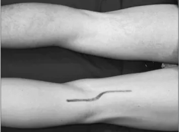
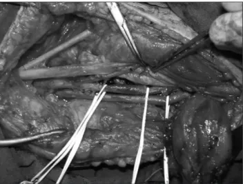
With the diagnosis of popliteal artery entrapment, the pa-tient underwent surgical treatment. he posterior approach, using the italic S incision was selected (Figure 2). he right limb was operated irst. he surgical indings were the same in both limbs, i.e., the popliteal artery was displaced medially of its usual course, not running along the popliteal vein and the posterior tibial nerve between the heads of the gastrocnemius muscles, but medially to the medial gastrocnemius muscle ten-don, as illustrated in Figure 3. he popliteal artery was dissected proximally and distally to the compression site and partial my-ectomy was performed in the medial gastrocnemius muscle, with popliteal artery release (Figure 4). Ater myectomy in the medial head of the gastrocnemius muscle and popliteal ar-tery release, intraoperative arteriography was performed, with
A
B
Figure 1. Preoperative arteriography showing bilateral medial bypass of popliteal arteries at dorsal foot lexion.
Figure 2. Preoperative italic S marks of the incision for posterior access.
he patient is under clinical follow-up with pulses pre-vent even at forced dorsal foot lexion. he same operation was performed on the let lower limb 90 days ater the irst surgical intervention.
Discussion
Popliteal artery entrapment syndrome is more frequent
in men, and most cases are unilateral1. Imaging exams are
decisive for the diagnosis of this syndrome 10-12, and may
in-clude Doppler ultrasonography10,11, computed axial
tomog-raphy12, magnetic resonance10 and angiography12. he latter
are more useful for allowing better surgery planning10,12.
he combined use of more than one imaging exam can be more efective than just one exam12.
he prevalence of PAES in the general population rang-es from 0.16 to 3.5%, and type III is the most common form, afecting 35% of all cases11. he patient PAES is young, with
a recent history of intense sport practice13.
A Brazilian study investigated the presence of this syn-drome in 42 asymptomatic individuals, half of whom were athletes and half were sedentary subjects. Popliteal com-pression was detected in 6 individuals (14.2%), 2 (4.7%) in athletes and 4 (9.5%) in non-athletes, with no statistically signiicant relation between PAES and physical activity14.
In this study, pocketDoppler exam of the posterior tibial artery and the ankle-brachial index were compared to the Doppler ultrasonography for the diagnosis of PAES. Both showed good sensitivity and speciicity and were suggested as diagnostic screening instruments in individuals suspect-ed of having the syndrome14.
he importance of correct diagnosis and treatment of PAES is relected on the diferential diagnosis and the re-sulting complications of the disease. he physician should distinguish PAES from arteritis, neurogenic claudication, muscle diseases, adventitial tumors in the popliteal artery, cystic disease of popliteal artery and traumatic causes of le-sion in the popliteal artery11. Non-treated popliteal artery
entrapment can lead, in the anatomical form, to complica-tions, such as stenosis, embolism, post-stenotic dilatation and arterial thrombosis, and in the functional form, to in-capacity to practice physical exercises9,15.
he most common symptoms are: intermittent claudication9,11,12,16, in 40% of the patients11, and feet
and calf pain, which appear ater physical exertion9,17.
Intermittent claudication - complaints of pain only while walking – can occur9. Our patient reported calf fatigue
and toe paresthesia during intense cycling, being asymp-tomatic in the let side, despite the popliteal artery com-pression at forced dorsal lexion.
It should be observed that the patient had no other diseases, such as diabetes and atherosclerosis9. Patients
without thrombotic complications have normal pulses with the lower limbs in neutral position and may have
Figure 4. Arteriography during the surgery showing absence of artery compression at dorsal foot lexion.
reduced or no pulses at forced dorsal foot lexion9. In the
case presented here, there were no posterior and dorsal tibial pulses in the right foot, without popliteal artery thrombosis; what probably happened was an intense ar-tery spasm, due to repeated trauma, as the intraoperative control arteriography ater partial myectomy in the medial head of the gastrocnemius muscle showed normal low in the posterior and dorsalis pedis arteries and pulses were detected at digital palpation.
In Brazil, the irst case reports on PAES were made by Ximenes and Ristow et al.19,20. Gallicchio and Schafer21 and
Castiglia22 were the irst to publish Brazilian book chapters
on the subject,
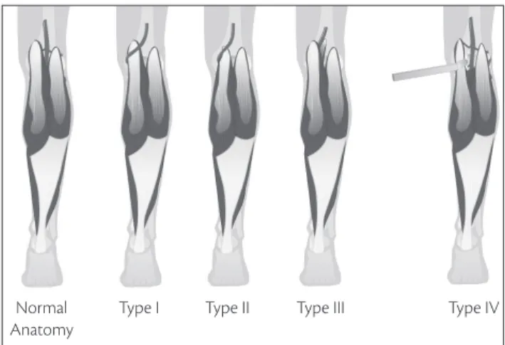
Created in 1971 by Delaney and Gonzáles, the classi-ication of the various types of popliteal artery entrapment initially included only types I, II, III and IV3,9. In 1979, type
V was added by Rich et al.18. In 1999, Levien and Veller8
stated that 50% of the population can have functional com-pressions and that, in these cases, the treatment should be
primarily clinical. hen, type VI was added8. herefore, the
most frequent popliteal artery entrapment classiication used today is:
TYPE I – Medial deviation of the popliteal artery, me-dially to the medial tendon of the gastrocnemius muscle that is inserted in the internal condyle of the femur.
TYPE II – he popliteal artery is normal, running an-teriorly to the medial tendon of the gastrocnemius muscle that, for having its insertion more laterally than usual, compresses the artery.
TYPE III – he gastrocnemius muscle has an addi-tional tendon, which is inserted more laterally, compress-ing the artery.
TYPE IV – he popliteal muscle compresses the popliteal artery, with normal anatomy of the gastrocne-mius muscle.
TYPE V – he popliteal artery and the popliteal vein are simultaneously compressed.
TYPE VI – Muscle hypertrophy with normal struc-ture, resulting in functional compression of the popliteal artery and the popliteal vein.
Figure 6 shows types I, II, II and IV of popliteal artery entrapment and the normal anatomical relation. he case reported was classiied as type I.
he treatment of PAES caused by muscle insertion anomalies has a formal surgical indication, even in the
asymptomatic patient12, thus preventing vascular
compli-cations that cause limb loss. In these cases, the operation eliminates the factor that causes entrapment and repairs the artery. We have two approached to the popliteal fossa:
medial and posterior accesses3,9,12. he posterior approach,
made through a S shaped incision, allows a better view of the structures, making it easier for the surgeon12. he
medial approach, which was used for the irst time in a functional entrapment case in 1974 by Darling et al.23,
is indicated in the presence of artery thrombosis, to al-low dissection of the great saphenous vein to be used as a grat9,12,15, as well long occlusions, in which a
femorop-opliteal bypass is required9. he question of surgical
ac-cess to the popliteal fossa is still not a consensus3. In the
case reported here, the posterior approach was used, with perfect view for treatment of the anomaly. We believe that, in cases of PAES without stenosis or post-aneurysmatic dilatation and with compression only, the posterior access is the best option, as we could explore the whole popliteal fossa without anatomical anomalies going unnoticed and exclude the presence of other factors for stenosis3.
References
1. Levien LJ. Popliteal artery entrapment syndrome. SeminVasc Surg.
2003;16(3):223-31.
2. Stuart TP. Note on a variation in the course of the popliteal artery. J. Anat Physiol. 1879;13(Pt 2):162–5.
3. Araújo JD, Araújo Filho JD, Ciorlin E, et al. Aprisionamento de vasos poplíteos: diagnóstico e tratamento e o conceito do aprisiona-mento funcional. J Vasc Bras. 2002;1(1):22-31.
4. Love JW, Whelan TJ. Popliteal artery entrapment syndrome. Am J Surg. 1965;109:620–4.
5. Collins PS, McDonald PT, Lim RC. Popliteal artery entrapment: an evolving syndrome. J Vasc Surg. 1989;10(5):484-90.
6. Wright LB, Matchett WJ, Cruz CP,et al. Popliteal artery disease: di-agnosis and treatment. Radiographics. 2004;24(2):467–79. Figure 6. Illustration of the normal anatomy and the various types of popliteal artery entrapment syndrome according to the modiied clas-siication of Delaney and Gonzáles.
Type I Normal
Anatomy
7. Schurmann G, Mattfeldt T, Hofmann W, et al. he popliteal artery entrapment syndrome: presentation, morphology and surgical treatment of 13 cases. Eur J Vasc Surg. 1990;4(3):223-31.
8. Levien LJ, Veller MG. Popliteal artery entrapment syndrome: more common than previously recognized. J Vasc Surg. 1999;30(4):587-98.
9. Almeida MJ, Yoshida WB, Melo NR. Síndrome do aprisionamento da artéria poplítea. J Vasc Bras. 2003;2(3):211-9.
10. Ikawa MH, Natour J, Jannini MG, et al. Síndrome do entrelaçamen-to da artéria poplítea: um diagnóstico diferencial de claudicação de membros inferiores: contribuição da ressonância magnética para diagnóstico e avaliação. Rev Bras Reumatol. 2007;47(6):442-5.
11. Oliveira FM, Santos ACB, Takito AM, et al. Bilateral popliteal artery entrapment syndrome: case report. J Vasc Bras. 2008;7(2):159-62.
12. Gourgiotis S, Aggelakas J, Salemis N, et al. Diagnosis and surgical approach of popliteal artery entrapment syndrome: a retrospec-tive study. Vasc Health Risk Manag. 2008;4(1):83-8.
13. Bonamigo TP, Becker M, Zylberstein S. Síndrome de aprisionamen-to da artéria poplítea: diagnóstico e tratamenaprisionamen-to. Relaaprisionamen-to de caso. Rev Bras Ortop. 1996;31(11):952-6.
14. Almeida MJ,Bonetti Yoshida W,Habberman D,et al. Extrinsic com-pression of popliteal artery in asymptomatic athlete and non-athlete individuals. A comparative study using duplex scan (color duplex sonography). Int Angiol. 2004;23(3):218-29.
15. Imtiaz N. Popliteal artery entrapment syndrome. J Coll Physicians Surg Pak. 2011;21(6):376-8.
16. Lamb CM, Davies CG, Whitbread T. Two cases of mis-diagnosed popliteal artery entrapment syndrome. Eur J VascEndovasc Surg. 2010;20(2):e16-8.
17. Yamaguchi H, Miura T, Eishi K, Tsuda N. A case of popliteal artery aneurysm associated with popliteal artery entrapment syndrome. Ann Vasc Dis. 2010;3:140-3.
18. Rich NM, Hughes CW. Popliteal artery and vein entrapment. Am J Surg. 1967;113:696-8.
19. Ximenes JOC. Síndrome de enlaçamento da artéria poplítea: relato de um caso e revisão da literatura. Rev Col Bras Cirur. 1975;2:74-77.
20. Ristow AVB, Arruda AM, Baptista H. Síndrome de compressão da artéria poplítea. Med Hoje. 1979;5:80-82.
21. Gallicchio VG, Schafer NT. Síndrome do aprisionamento da arté-ria poplítea. In: Bonamigo TP, Burihan E, Cinelli M, et al. Doenças da aorta e seus ramos: diagnóstico e tratamento. São Paulo: Fundo Editorial Byk; 1991. p. 324-44.
22. Castiglia V. Síndrome do aprisionamento da artéria poplítea. In Mafei FHA, Lastória S, Yoshida WB, et al. Doenças vasculares peri-féricas. Rio de Janeiro: Medsi; 1995. p. 701-15.
23. Darling RC, Buckley CJ, Abott WM, et al. Intermittent claudication in young athletes: popliteal artery entrapment syndrome. J Trauma 1974;14(7):543-52.
Correspondence
Nelson Mesquita Júnior Rua Deputado Heitor Alencar Furtado, 1.819 apto 1.302 – Mossunguê CEP 81200-110 – Curitiba (PR), Brazil E-mail: [email protected]
Author’s contributions