EDITORIAL
he interface between images and stent grafts for the
endovascular surgeon
Luiz Antonio Furuya
Vascular and endovascular surgeon j Vasc Bras. 2011;10(4):282-283.
Some years ago, vascular surgeons started receiv-ing in their oices a “new” type of exam, called Multislice Tomography or Angiotomography.
In the exam folder, there were tens of color images, re-constructions in several perspectives and sizes. he images were of amazing quality and beautiful, but of little use in making medical decisions. he folder also had a CD with a visualization sotware program and over 2000 axial images of thorax, abdomen and pelvis, with and without contrast.
Even with this vast amount of data, the images could not be manipulated, because the standard sotware program included was a simple image viewer, with few and limited resources. It was not possible do to any multi-planar or 3D reconstruction. he lack of a quality viewer (image post-processing sotware program) led to great dilemma: why request a modern, and obviously expensive, exam if it was not possible to make the best use of these data? he huge amount of information was not enough, on its own, for the exact planning, for instance, of endovascular treatment of aortic aneurysm: precise measurements and consequent se-lection of the best device to each patient.
Infrarenal aortic stent grat implantation is a relatively simple procedure that requires preoperative measurements of vessel length and diameter. However, the production and implantation of devices with branches to the visceral arteries is much more complex and involve several steps, including the evaluation of preoperative images, familiar-ity with the accesses to visceral vessels and similar skills to those required for infrarenal aortic stent grat implantation. he devices that involve visceral branches require, besides the sagittal images used in infrarenal devices, a 3D plan-ning and multiplanar reconstruction, options not available in simple visualization sotware programs.
he users that did not have access to the infrastructure of a modern workstation equipped with graphic resources needed new DICOM (Digital Imaging and Communications
in Medicine) that enable them to use the data contained in these CDs of multislice computed tomography or high-ield magnetic resonance.
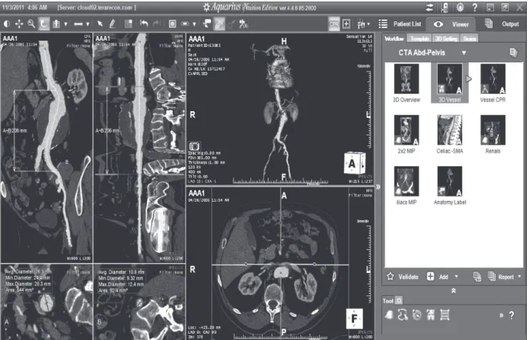
In a few years, several DICOM viewers with better in-terfaces to CDs sotware programs were launched in the market, enabling endovascular surgeons to inally plan their cases adequately. Among the several programs avail-able in the market, special attention will be now dedicated to iNtuition Cloud, of TeraRecon.
his sotware program ofers several advantages when compared to other viewers. First, it is compatible with many platforms, such as Windows, Mac OS X, Android or Iphone, and many browsers, such as Internet Explorer, Mozilla and Safari, among others. hrough a LAN (broad-band) or 3G mobile connection, the program transforms the user’s equipment into a complete workstation, with all resources, anytime and anywhere, either in the oice, at home or even in the operating room, thanks to the sotware program technology.
TeraRecon’s iNtuition Cloud ofers an automatic pre-processing system (AquariusAPS), which can be activated to automatically perform several image processing stages, such as: bone removal, centerline detection, anatomy detec-tion, parametric mapping applicadetec-tion, pulmonary nodule detection, and not only on 3D reconstructions, but espe-cially using multi-planar and 3D reconstructions.
TeraRecon’s iNtuition Cloud uses rendering hardware called VolumePro (render board for medical images, pro-prietary of TeraRecon), enabling the system to use the pow-erful mechanism of 3D volume rendering in real time.
he interface between images and stent grafts for the endovascular surgeon - Furuya LA J Vasc Bras 2011, Vol. 10, Nº 4 283
or in his oice, via browser, a few minutes ater the exam is completed by the tomograph or resonance equipment.
he possibility to easily access the images by a group of stu-dents and resident physicians, for instance, can expand the med-ical education boundaries to standards never imagined before.
As it is not a free access sotware program, investments are required to have access to such technology, which can be funded by an institution (hospital, university, college, etc.), a diagnostic medicine company or even an individual user (a physician, for instance), providing personalized op-tions to each type of client.
We can say for sure that the age of caliper and negato-scope has given way to the age of mouse and high-deinition displays. Precision and certainty replaced approximations
and unsupported evaluations. Much has yet to come, and the physician that gradually incorporates improvements in his clinical practice will surely discover ways to beneit from technology, generating more knowledge and higher quality in the treatment of his/her patients.
References
1. Lee WA. Endovascular abdominal aortic aneurysm sizing and case planning using the TeraRecon Aquarius workstation. Vasc Endovascular Surg. 2007;41(1):61-7.
2. Roy K. Greenberg, M.D. Endovascular aneurysm repair using branched or fenestrated devices [Internet]. Available from: http:// www.terarecon.com/downloads/news/casestudy_greenberg_ complexaneurysms.pdf.