Stress Resistance via DAF-16/FOXO in
Caenorhabditis
elegans
Yeu-Ching Shi1, Chan-Wei Yu1, Vivian Hsiu-Chuan Liao1*, Tzu-Ming Pan2*
1Department of Bioenvironmental Systems Engineering, National Taiwan University, Taipei, Taiwan,2Department of Biochemical Science and Technology, National Taiwan University, Taipei, Taiwan
Abstract
Background:Monascus-fermented products are mentioned in an ancient Chinese pharmacopoeia of medicinal food and herbs.Monascus-fermented products offer valuable therapeutic benefits and have been extensively used in East Asia for several centuries. Several biological activities ofMonascus-fermented products were recently described, and the extract of Monascus-fermented products showed strong antioxidant activity of scavenging DPPH radicals. To evaluate whether Monascus-fermented dioscorea products have potential as nutritional supplements, Monascus-fermented dioscorea’s modulation of oxidative-stress resistance and associated regulatory mechanisms in Caenorhabditis elegans were investigated.
Principal Findings: We examined oxidative stress resistance of the ethanol extract of red mold dioscorea (RMDE) in C. elegans, and found that RMDE-treated wild-type C. elegans showed an increased survival during juglone-induced oxidative stress compared to untreated controls, whereas the antioxidant phenotype was absent from adaf-16mutant. In addition, the RMDE reduced the level of intracellular reactive oxygen species inC. elegans. Finally, the RMDE affected the subcellular distribution of the FOXO transcription factor, DAF-16, inC. elegansand induced the expression of thesod-3 antioxidative gene.
Conclusions:These findings suggest that the RMDE acts as an antioxidative stress agent and thus may have potential as a nutritional supplement. Further studies inC. eleganssuggest that the antioxidant effect of RMDE is mediated via regulation of the DAF-16/FOXO-dependent pathway.
Citation:Shi Y-C, Yu C-W, Liao VH-C, Pan T-M (2012)Monascus-Fermented Dioscorea Enhances Oxidative Stress Resistance via DAF-16/FOXO inCaenorhabditis elegans. PLoS ONE 7(6): e39515. doi:10.1371/journal.pone.0039515
Editor:Robin Charles May, University of Birmingham, United Kingdom
ReceivedDecember 31, 2011;AcceptedMay 27, 2012;PublishedJune 22, 2012
Copyright:ß2012 Shi et al. This is an open-access article distributed under the terms of the Creative Commons Attribution License, which permits unrestricted use, distribution, and reproduction in any medium, provided the original author and source are credited.
Funding:The authors have no support or funding to report.
Competing Interests:The authors have declared that no competing interests exist.
* E-mail: [email protected] (VH-CL); [email protected] (T-MP)
Introduction
Red mold rice has long been used as a traditional food and dairy supplement. It was mentioned in an ancient Chinese pharmaco-poeia of medicinal food and herbs. Several biological activities of red mold rice were recently described, including inhibition of the biosynthesis of cholesterol for treating hyperlipidemia [1], improvements of the memory and learning ability in Ab-infused rats, and a reduction in blood glucose levels in diabetic rats [2,3]. In recent years, red mold dioscorea has been extensively studied. Red mold dioscorea contains various metabolites, including dimerumic acid, tannins, phenols, and polyketides (monacolins) with antioxidative properties and anti-inflammatory responses [4,5]. In particular, monacolins are believed to account for the majority of the cholesterol-controlling activity ofMonascus[1]. An extract ofM. ankawas shown to exhibit strong antioxidant action of scavenging the 1-1-diphenyl-2-picrylhydrazyl (DPPH) radical and inhibiting lipid peroxidation [6]. In our previous study, the ethanol extract of red mold rice (RMRE) was shown to have antioxidative activity of scavenging DPPH radicals [7]. In
addition, the constituents of the RMDE were complicated with monascin and ankaflavin being the major components [8].
Oxidative stress occurs in cells with excessive production of reactive oxygen species (ROS) such as a superoxide anion (O2-)
and hydrogen peroxide (H2O2) which can overwhelm a cell’s
natural antioxidant defense, leading to injury to biomolecules such as lipids, proteins, and DNA [9]. Oxidative stress is thought to contribute to the general decline in cellular functions associated with human diseases, such as Alzheimer’s disease [10,11], atherosclerosis [12], diabetes [13,14], Parkinson’s disease [15,16], and human cancers [17,18] as well as the aging [19,20] process itself.
The nematode Caenorhabditis elegans (C. elegans) has become a popular model to study molecular mechanisms of drug effects and disease pathogeneses. Many key findings with relevance for mammals were discovered in the well-characterized C. elegans. There is strong conservation of biological principles between
diseases, including Alzheimer’s [22,23] and diabetes [24,25]. In contrast to cell-culture systems and animal experiments,C. elegans
is easy to culture, and because of the transparent appearance of the worms, fluorescent markers, such as reporter genes, can be observed in living animals [26]. The insulin/insulin-like growth factor (IGF)-1 signal transduction pathway plays an important role in regulating both longevity and stress resistance [27]. Activity of DAF-16, a FOXO transcription factor, is influenced by the insulin pathway which is involved in antioxidative defense, stress resistance, and metabolism [28].
In order to evaluate whether Monascus-fermented dioscorea products have potential as nutritional supplements, modulation of thein vivoantioxidant activity of the ethanol extract of red mold dioscorea (RMDE) and its associated regulatory mechanism in
C. elegans were investigated. Herein, we analyzed the antioxidant activity of the RMDE by oxidative-stress assays and measured intracellular ROS levels in C. elegans. In addition, factors and genetic requirements that influence oxidative-stress resistance by the RMDE are dissected.
Results
The RMDE Enhanced Oxidative-stress Resistance in
C. elegans
The RMDE was prepared and analyzed as described previously [8]. To investigate whether RMDE has an antioxidant effect in
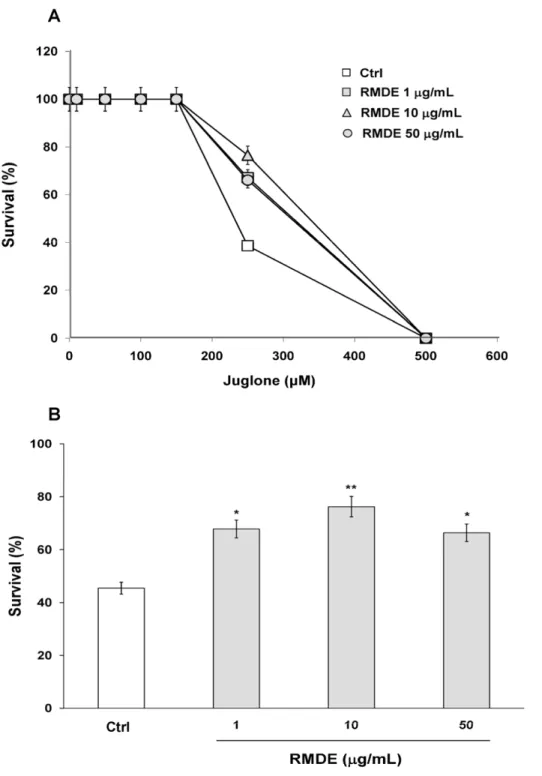
C. elegans, wild-type (WT) N2 worms were pretreated with RMDE, followed by exposure to juglone-induced oxidative stresses. WT N2 synchronized L1 larvae were pretreated with 1, 10, and 50mg/ mL RMDE and 0.1% DMSO as solvent control for 72 h at 20uC. Adult animals were then exposed to 0, 10, 50, 100, 150, 150, 250, and 500mM juglone, a redox cycler that generates intracellular oxidative stress [29] and then they were incubated for 3.5 h (Fig. 1A).
During pretreatment with the RMDE, no adverse effects on the worms, including survival, growth rate, progeny production, body length, or morphological changes, were observed. The dose response analysis for juglone showed that 250mM juglone was the optimal concentration to examine the antioxidant effect of RMDE inC. elegans(Fig. 1A). The results showed that pretreatment with 1, 10, and 50mg/mL RMDE significantly increased the survival of worms exposed to juglone-induced oxidative stress (Fig. 1A, B).
The oxidative stress assays were further evaluated by using H2O2 and paraquat. After RMDE (10mg/mL) treatment, adult WT N2 worms were exposed to 1 mM H2O2 [30–32] and
150 mM paraquat [33–35] for 4.5 h and 12 h, respectively and then the viability of worms was scored. In agreement with the observation by juglone, RMDE (10mg/mL) significantly increased the survival of WT N2 worms upon H2O2and paraquat exposure
(Fig. S1).
The RMDE Decreased Intracellular ROS Levels in
C. elegans
Next, we examined whether RMDE-enhanced oxidative-stress resistance was due to its ROS-scavenging ability. Non-fluorescent DCF-DA is a freely cell-permeable dye, which is readily converted to the fluorescent 2979-dichlorofluorescein (DCF) due to an interaction with intracellular H2O2. Figure 2A shows that the
RMDE significantly decreased intracellular ROS when WT N2 worms were treated with 1, 10, and 50mg/mL RMDE. Since as low as 10mg/mL RMDE was able to significantly alleviate the amount of intracellular ROS as well as significantly enhance the antioxidant effect, 10mg/mL RMDE was chosen as the working concentration for further experiments.
We next examined RMDE’s ROS-scavenging ability using the
mev-1 mutant. mev-1 is a defect of the mitochondrial complex which exhibits increased ROS levelsin vivo. Thus,mev-1mutant is hypersensitive to elevated oxygen levels [36]. After RMDE treatment, adultmev-1animals were exposed to 250mM juglone for 3.5 h. The survival ofmev-1 mutants significantly increased with 10mg/mL RMDE treatment compared to that of untreated ones (Fig. 2B). Taken together, RMDE may act against oxidative stress through its intracellular ROS-scavenging ability and decreases mitochondrial ROS toxicity.
The RMDE Enhanced Oxidative-stress Resistance in
C. elegansVia DAF-16
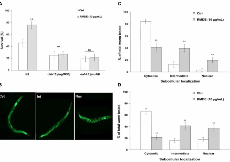
To identify RMDE’s mechanism of action, we examined its effect on DAF-16. DAF-16 is a forkhead (FOXO) transcription factor with crucial functions in controlling stress responses and aging in C. elegans and mammals [37]. To analyze the involvement of DAF-16 in RMDE-mediated oxidative-stress resistance, we performed a juglone-induced oxidative stress test withdaf-16(mgDf50)anddaf-16(mu86)deletion mutants. Both daf-16(mgDf50) and daf-16(mu86) are null mutants [38,39]. For the 250mM juglone-induced oxidative stress tests, unlike WT N2 worms, the survival of both RMDE-treated daf-16(mgDf50) and
daf-16(mu86) mutants did not show a significant difference compared to that of untreated ones (Fig. 3A).
Since thedaf-16mutant is more sensitive to juglone, we further evaluated how the daf-16 mutant behaves at a lower juglone concentration, and whether RMDE has an effect at this lower concentration. We used 150mM juglone at which WT N2 worms did not show significantly different survival between RMDE untreated and treated conditions (Fig. 1A) to evaluate the effect of RMDE on daf-16 mutant. The result showed that survival of RMDE treated and untreated control daf-16(mu86) mutant was not significantly different upon 150mM juglone exposure (Figure S2). This is in agreement with the effects of RMDE on daf-16(mgDf50)anddaf-16(mu86)mutants by using higher concentra-tion juglone (250mM) (Fig. 3A, Fig. S2). Taken together, the RMDE may provide oxidative-stress resistance in C. elegans via DAF-16.
To further investigate the role of DAF-16 in modulating RMDE-induced oxidative-stress resistance, we examined the translocation of DAF-16. Localization of DAF-16 in nuclei is an essential prerequisite for its ability to activate target gene transcription [40–43]. Localization of DAF-16 was studied under normal culture conditions and in cultures exposed to juglone. The subcellular distribution of DAF-16 was classified into the categories of ‘‘cytosolic’’, ‘‘intermediate’’, and ‘‘nuclear’’ accord-ing to their localization phenotypes (Fig. 3B). Control untreated worms incubated under normal culture conditions revealed a predominant cytosolic localization of DAF-16 (Fig. 3 C). Treatment of transgenic worms with 10mg/mL RMDE for 3 days resulted in increases in the fractions of worms showing nuclear localization of DAF-16 with cytosolic (41.0% 67.7%), intermediate (39.5% 66.6%), and nuclear (19.5% 64.3%) localization phenotypes (Fig. 3C). Following exposure to 50mM juglone for 5 min, fractions of intermediate and nuclear localization of DAF-16 were greater in RMDE-treated transgenic worms (Fig. 3D). Therefore, RMDE affected the subcellular distribution of DAF-16 and caused translocation of DAF-16 from the cytoplasm to nuclei. Taken together, the results suggest that RMDE enhanced oxidative-stress resistance in C. elegans via DAF-16.
The RMDE Upregulated SOD-3 Expression
To elucidate whether the above-described increase in oxidative-stress resistance was due to the RMDE regulating a specific oxidative- stress-response gene, we examined the responsiveness of the antioxidant enzyme, SOD, to RMDE treatment. SOD is a major enzyme that protects against oxidative stress by catalyzing the removal of O2
-[44]. InC. elegans,sod-3encodes manganese (Mn)SOD, andsod-3is known to be a target gene of DAF-16 [37].
The CF1553 transgenic strain which contains a reporter gene of
sod-3fused to GFP was treated with 10mg/mL RMDE for 3 days. Figure 4A shows that 10mg/mL RMDE treatment alone significantly upregulated SOD-3::GFP expression compared to Figure 1. Effects of the ethanol extract of red mold dioscorea (RMDE) on oxidative-stress resistance of wild-type (WT)Caenorhabditis elegansN2.(A) Dose response for juglone exposure. Synchronized WT L1 larvae were pretreated with the RMDE (1, 10, and 50mg/mL) or 0.1% DMSO as the solvent control for 72 h at 20uC. Subsequently, adult worms were subjected to oxidative-stress assays. For the oxidative-stress assays, RMDE-treated (n =100) and control (0.1% DMSO,n =100) adult worms were exposed to 0, 10, 50, 100, 150, 150, 250, and 500mM juglone for 3.5 h at 20uC and then scored for viability. (B) Survival of RMDE-treated and control adult worms exposed to 250mM juglone for 3.5 h at 20uC. The test was performed three times. Error bars represent the standard error, and differences compared to the control (0mg/mL, 0.1% DMSO) were considered significant atp,0.05 (*),p,0.01 (**) by one-way ANOVA and the LSD post-hoc test.
untreated worms. Additionally, when strain CF1553 was further challenged with 150mM juglone exposure for 1 h, the results showed that the expression level of SOD-3::GFP was also
significantly increased in CF1553 worms treated with RMDE compared to that untreated ones (Fig. 4A).
To further confirm SOD-3 expression is regulated by RMDE, we measured SOD-3 mRNA by qRT-PCR (Fig. 4B). In addition to Figure 2. Effects of the ethanol extract of red mold dioscorea (RMDE) on reactive oxygen species (ROS) accumulation in Caenorhabditis elegans.(A) Synchronized WT L1 larvae were pretreated with the RMDE or 0.1% DMSO as the solvent control for 72 h at 20uC. Subsequently, intracellular ROS were analyzed and the results are presented as relative fluorescence units (RFU) of 20 individual worms (n =160). (B) Oxidative-stress resistance of themev-1mutant. Synchronizedmev-1L1 larvae were pretreated with the RMDE or 0.1% DMSO as the solvent control for 72 h at 20uC. Subsequently, mutant worms were subjected to oxidative-stress assays. RMDE-treated (n =76) and control (0.1% DMSO,n =101) worms were exposed to 250mM juglone for 3.5 h at 20uC and then scored for viability. The test was performed three times. Error bars represent the standard error, and differences compared to the control (0mg/mL, 0.1% DMSO) were considered significant atp,0.05 (*),p,0.01 (**), andp,0.001 (***) by one-way ANOVA and the LSD post-hoc test.
doi:10.1371/journal.pone.0039515.g002
examine SOD-3 mRNA level in WT N2 nematodes in response to RMDE, SOD-3 mRNA levels in response to RMDE were also investigated in daf-16(mgDf50) mutant and CF1553 strain. The results showed that RMDE caused a 4.9-fold and 1.7-fold increase of SOD-3 mRNA level in WT N2 and CF1553, respectively comparing with that untreated ones (Fig. 4B). In contrast, SOD-3 mRNA level was diminished indaf-16(mgDf50)mutant showing no significant difference between RMDE-treated and untreated worms (Fig. 4B). Therefore, the results further confirmed the involvement of SOD-3 expression and DAF-16 in regulating RMDE antioxidant properties. Taken together, these findings suggest that RMDE may act against oxidative stress via DAF-16/FOXO.
Discussion
The RMDE was studiedin vitroandin vivo, and several biological effects were reported in mammalian systems [4,5,45]. However, its potential as a nutritional supplement and the mechanisms by which
it exerts its actionin vivostill remain to be further elucidated. Herein, we usedC. elegans as anin vivomodel to examine the protective potential and mode of action of the RMDE. There are several studies in which the protective actions of flavonoids and polyphenols inC. eleganswere mainly attributed to their antioxidative activities [26,46]. In the present study, we showed that the survival of WT worms was significantly increased with RMDE treatment under oxidative stress agents (juglone, H2O2, and paraquat)-induced
To identify RMDE’s mechanism of action, we examined its effect on DAF-16/FOXO. FOXO transcription factors are conserved from worms to human and regulated by insulin signaling pathway [37]. DAF-16 is the only FOXO protein in
C. elegans[37]. InC. elegans, the DAF-16 transcription factor in the insulin signaling pathway is considered a key regulator of many important biological processes including lifespan, metabolism, and stress responses [47]. We examined the ability of loss-of-function Figure 4. Effects of the ethanol extract of red mold dioscorea (RMDE) on the expression of superoxide dismutase (SOD).(A). Effects of RMDE on the expression of SOD-3::green fluorescent protein (GFP). Immediately after hatching, age-synchronized, transgenic L1 worms of the CF1553 strain (sod-3:: GFP) were treated with 10mg/mL RMDE or 0.1% DMSO as the solvent control for 72 h at 20uC, followed by 150mM juglone exposure for 1 h. Subsequently, the total GFP fluorescence of each whole worm was quantified by Image-Pro Plus software. Data shown are the average number of pixels in transgenicCaenorhabditis elegans(n= 20) at each indicated treatment. (B) Effects of RMDE on the SOD-3 mRNA level. Synchronized WT N2, daf-16(mgDf50)mutant, and CF1553 strain L1 larvae were pretreated with 10mg/mL RMDE or 0.1% DMSO as the solvent control for 72 h at 20uC and then total RNA was extracted. mRNA level of SOD-3 was determined by quantitative real-time RT-PCR. SOD-3 mRNA levels were normalized to the expression of AMA-1. The fold change was normalized to that observed in untreated controlC. eleganssamples. The test was performed three times. Error bars represent the standard error, and differences compared to the control (0.1% DMSO) were considered significant atp,0.05 (*),p,0.01 (**) by one-way ANOVA and the LSD post-hoc test. ns, no significant.
doi:10.1371/journal.pone.0039515.g004
daf-16 deletion mutants to resist oxidative stress during RMDE exposure. The premise was that if daf-16 gene is required for oxidative-stress resistance by the RMDE, then the RMDE would be unable to enhance oxidative-stress resistance in that mutant. Results showed that daf-16 null mutants did not exhibit a significantly enhanced survival after RMDE treatment followed by juglone exposure compared to untreated worms (Fig. 3A). This indicated that the RMDE mediates oxidative-stress resistance via DAF-16.
To further validate that DAF-16 is required for the protective effect of the RMDE, the effect of the RMDE on the translocation of DAF-16 from the cytoplasm to nuclei was examined (Fig. 3C, D). In addition, the gene expression of SOD-3, a target gene of DAF-16, was examined after RMDE exposure (Fig. 4A, B). Results showed that the RMDE was able to enhance DAF-16 translocation from the cytoplasm to nuclei (Fig. 3C, D). Moreover, SOD-3 expression was induced by RMDE exposure (Fig. 4A). Furthermore, RMDE caused a significant increase of SOD-3 mRNA level in WT N2 and CF1553 comparing with the untreated ones (Fig. 4B). In contrast, SOD-3 mRNA level was diminished in daf-16 mutant showing no significant difference between RMDE-treated and untreated worms (Fig. 4B). Nuclear localization of DAF-16 is a prerequisite for transcriptional activation of its target genes such as genes for antioxidative enzymes like MnSOD (sod-3) and catalases (ctl-1 andctl-2) [28]. Therefore, the antioxidant effect of the RMDE is likely mediated via regulation of a DAF-16-dependent pathway. It is noted that there are conflicting data regarding the response of SOD-3 to juglone exposure. Van Raamsdonk and Hekimi [48] found that a
sod-3deletion does not affect juglone resistance significantly. They found a small effect with paraquat but this is not found by Doonan et al [49]. In contrast, Heidler et al [50] showed that SOD-3 expression and enzymatic activity were increased upon juglone exposure. Therefore, although our results showed that RMDE enhances SOD-3 upregulation and support the activation of DAF-16 by RMDE, it is not necessary that SOD-3 mediates the stress resistance as other DAF-16 target genes might also involved in RMDE-induced resistance.
RMDE itself can lead to nuclear localization of DAF-16 (Fig. 3C) and induce expressions of SOD-3 in transgenic strain carrying SOD-3::GFP (Fig. 4A) and mRNA level (Fig. 4B). This suggests that RMDE has similar effect as mild stress stimulus. In spite of the deleterious effects of ROS, recent research has indicated that ROS serve many important and non-damaging roles in both intracellular and extracellular signal transduction that involves diverse functions from vascular health to host defense [51]. Stress hormesis occurs when a low level stress elicits adaptive beneficial responses that protect against subsequent exposure to severe stress [52]. Recent findings suggest that mild oxidative stress from low concentrations of juglone can extendC. eleganslifespan by hormetic mechanisms [50]. Therefore, it is possible that by acting as mild stress stimulus, RMDE activates an adaptive response leading to an increase resistance inC. elegansto a subsequent severe stress (such as high concentration, 250mM juglone exposure).
In conclusion, our study is the first to demonstrate that the RMDE has the capacity to increase in vivo oxidative-stress resistance. This finding is supported by recent studies showing that the RMDE exerts protection against oxidative stress and diabetic disease-associated inflammation [3,4,7,45,53]. Further study in C. elegans suggests that the antioxidative effect of the RMDE is mediated via regulation of DAF-16/FOXO-dependent pathway. These findings indicate that the RMDE acts as an antioxidative stress agent and thus may have potential as a nutritional supplement. Additionally, the RMDE may have
therapeutic potential for preventing oxidative stress-associated diseases like diabetes and Alzheimer’s disease as well as aging.
Materials and Methods
Caenorhabditis ElegansStrains and Handling Procedures
Strains used in this study were: Bristol N2 (wild-type; WT); GR1307, daf-16(mgDf50); CF1038, daf-16(mu86); TK22, mev-1
(kn1); CF1553, muIs84[pAD76(sod-3::GFP)], and TJ356, zIs356[-DAF-16::GFP]. AllC. elegans strains as well as theEscherichia coli
OP50 strain were obtained from theCaenorhabditisGenetics Center (CGC), University of Minnesota, MN, USA). Worms were maintained (unless otherwise stated) at 20uC on nematode growth medium (NGM) agar plates carrying a lawn of E. coli OP50 according to Brenner [54]. Synchronization of worm cultures was achieved by hypochlorite treatment of gravid hermaphrodites [55].
Stress-resistance Assays
Synchronized WT or mutant strain L1 larvae were incubated in liquid S-basal containingE. coliOP50 bacteria at 109cells/mL and various concentrations of the RMDE or 0.1% dimethyl sulfoxide (DMSO) as the solvent control for 72 h. Subsequently, adult worms were subjected to stress assays. For the oxidative-stress assays, 5-hydroxy-1,4-naphthoquinone (juglone), hydrogen peroxide (H2O2), and paraquat (Sigma, St. Louis, MO, USA) were
used to induce oxidative stress in worms. RMDE-treated and control adult worms were transferred to S-basal containing various concentrations of juglone, incubated for 3.5 h at 20uC, and then scored for viability. The survival of worms was determined by touch-provoked movement. Worms were scored as dead when they failed to respond to repeated touching with a platinum wire pick. The test was performed at least three times.
Measurement of ROS
Intracellular ROS in C. elegans were measured using 29,79 -dichlorodihydrofluoroscein diacetate (H2DCFDA) (Sigma).
Syn-chronized WT L1 larvae were incubated in liquid S-basal containing E. coli OP50 bacteria at 109 cells/mL and various concentrations of the RMDE or 0.1% DMSO as the solvent control for 72 h. The adult worms were then washed three times with phosphate buffered saline (PBS), followed by incubation in 250mL PBS containing 100mM H2DCFDA for 3 h.
Subsequent-ly, worms were washed twice with PBS and individual worms of each population were transferred into the wells of a 96-well microtiter plate containing 100mL PBS. The ROS measurement was carried out in an FLx800 Microplate Fluorescent Reader (Bio-Tek Instruments, Winookski, VT, USA) for quantification of fluorescence with excitation at 485 nm and emission at 530 nm. The results are presented as relative fluorescence units (RFU) of 20 individual worms. The test was performed at least three times.
Induction of a Stress-response Reporter
sodium azide, and capped with coverslips. Epifluorescence images were captured with a Leica epifluorescence microscope (Leica, Wetzlar, Germany) using a suited filter set (with excitation at 480620 nm and emission at 510620 nm) with a cooled charge-coupled device (CCD) camera. Adult worms were examined, and total GFP fluorescence for each whole worm was quantified by Image-Pro Plus software (Media Cybernetics, Bethesda, MD, USA). The test was performed at least three times.
Subcellular DAF-16 Localization
Synchronized L1 larvae of the TJ356 transgenic strain stably expressing a DAF-16::GFP fusion protein as a reporter [37] were incubated in liquid S-basal containingE. coliOP50 bacteria at 109 cells/mL and a final concentration of 10mg/mL RMDE or 0.1% DMSO as the solvent control for 72 h. The effect of the RMDE on oxidative stress was also examined by treatment with 50mM juglone for 5 min. Subsequent to this treatment, worms were placed on microscope slides and capped with coverslips, and the subcellular DAF-16 distribution was analyzed by fluorescence microscopy on an epifluorescence microscope (Leica). Expression patterns of TJ356 worms were classified into three categories (cytosolic, intermediate, and nuclear) with respect to major localization of the DAF-16::GFP fusion protein. Subcellular DAF-16 localization was examined in approximately 20 animals per condition. The test was performed at least three times.
RNA and Real-time Quantitative Reverse-transcription Polymerase Chain Reaction (qRT-PCR) Analysis
Synchronized L1 larvae were incubated in liquid S-basal containing E. coli OP50 bacteria at 109 cells/mL and a final concentration of 10mg/mL RMDE or 0.1% DMSO as solvent control for 72 h. Total RNA from adult worms was isolated using TRIzol according to manufacturer’s instructions (Invitrogen, Carlsbad, CA, USA) and cDNA was synthesized using Super-Script III First-strand synthesis super-Mix for qRT-PCR (Invitro-gen). The qRT-PCR was performed on a Plus One real-time cycler (Applied Biosystems, Carlsbad, CA, USA) using a SYBR Green PCR core kit (Applied Biosystems). RT-PCR levels were normalized to the expression ofama-1, which encodes the large subunit of RNA polymerase II [57,58]. The fold change was normalized to that observed in untreatedC. eleganssamples. The qRT-PCR primers for AMA-1 are: forward primer: 59 -CTGACCCAAAGAACACGGTGA-39; reverse primer: 59 -TCCAATTCGATCCGAAGAAGC-39. Primers for SOD-3 are: forward primer: 59-AGCATCATGCCACCTACGTGA-39; re-verse primer: 59-CACCACCATTGAATTTCAGCG-39. The test was performed three times.
Statistical Analyses
Statistical analyses were performed using SASH 9.2 Software (SAS Institute, Cary, NC, USA). Results are presented as the
mean6standard error. The statistical significance of differences between populations was demonstrated by a one-way analysis of variance (ANOVA) and least significant difference (LSD) post-hoc test. Differences were considered significant atp,0.05,p,0.01, or
p,0.001 (see Figures).
Supporting Information
Figure S1 Effects of the ethanol extract of red mold dioscorea (RMDE) on oxidative-stress resistance of wild-type (WT)Caenorhabditis elegansN2 using differ-ent oxidative stress agdiffer-ents.Synchronized WT L1 larvae were pretreated with the RMDE 10mg/mL or 0.1% DMSO as the solvent control for 72 h at 20uC. Subsequently, adult worms were subjected to stress assays. Worms used for oxidative-stress assays were: RMDE-treated (juglone,n= 100; H2O2,n= 75;
paraquat, n= 45) and 0.1% DMSO control (juglone, n= 100; H2O2, n= 75; paraquat, n= 45). Adult worms were exposed to
250mM juglone, 1 mM H2O2, and 150 mM paraquat for 3.5 h,
4.5 h, and 12 h at 20uC, respectively and then scored for viability. The test was performed three times. Error bars represent the standard error, and differences compared to the control (0mg/mL, 0.1% DMSO) were considered significant atp,0.05 (*),p,0.01 (**) by one-way ANOVA and the LSD post-hoc test.
(TIF)
Figure S2 Effects of the ethanol extract of red mold dioscorea (RMDE) on DAF-16 by using different concen-trations of juglone. Synchronized daf-16 (mu86) mutant L1 larvae were pretreated with the RMDE (10mg/mL) (150mM juglone,n= 116; 250mM juglone,n= 118) or 0.1% DMSO as the solvent control (150mM juglone, n= 111; 250mM juglone, n= 143) for 72 h at 20uC. Subsequently, worms were subjected to oxidative-stress assays. RMDE-treated worms were exposed to 150 and 250mM juglone for 3.5 h at 20uC and then scored for viability. The test was performed three times. Error bars represent the standard error, and differences compared to the control (0mg/ mL, 0.1% DMSO) were considered significant at p,0.05 (*),
p,0.01 (**) by one-way ANOVA and the LSD post-hoc test. ns, no significant.
(TIF)
Acknowledgments
Caenorhabditis elegansstrains were provided by theCaenorhabditis Genetics Center funded by the NIH National Center for Research Resources.
Author Contributions
Conceived and designed the experiments: VHL. Performed the experi-ments: YCS CWY. Analyzed the data: YCS CWY. Contributed reagents/ materials/analysis tools: VHL TMP. Wrote the paper: VHL YCS.
References
1. Shepherd JC, Cobbe SM, Ford I (1995) Prevention of coronary heart disease with pravastatin in men with hypercholesterolemia. West of Scotland Coronary Prevention Study Group. New Engl J Med 333: 1301–1307.
2. Lee CL, Kuo TF, Wu CL, Wang JJ, Pan TM (2010) Red mold rice promotes neuroprotective sAPPalpha secretion instead of Alzheimer’s risk factors and amyloid beta expression in hyperlipidemic Abeta40-infused rats. J Agric Food Chem 58: 2230–2238.
3. Shi YC, Pan TM (2010) Anti-diabetic effects ofMonascus purpureusNTU 568 fermented products on streptozotocin-induced diabetic rats. J Agric Food Chem 58: 7634–7640.
4. Lee CL, Wang JJ, Kuo SL, Pan TM (2006)Monascusfermentation of dioscorea for increasing the production of cholesterol-lowering agent-monacolin K and anti-inflammation agent-monascin. Appl Microbiol Biotechnol 72: 1254–1262.
5. Kuo CF, Chyau CC, Wang TS, Li CR, Hu TJ (2009) Enhanced antioxidant and anti-inflammatory activities ofMonascus pilosusfermented products by addition of turmeric to the medium. J Agric Food Chem 57: 11397–11405.
6. Aniya Y, Ohtani II, Higa T, Miyagi C, Gibo H, et al. (2000) Dimerumic acid as an antioxidant of the mold,Monascus anka. Free Radic Biol Med 28: 999–1004. 7. Lee CL, Hung HK, Wang JJ, Pan TM (2007) Red mold dioscorea has greater hypolipidemic and antiatherosclerotic effect than traditional red mold rice and unfermented dioscorea in hamsters. J Agric Food Chem 55: 7162–7169. 8. Hsu YW, Hsu LC, Liang YH, Kuo YH, Pan TM (2010) Monaphilones A-C,
three new antiproliferative azaphilone derivatives fromMonascus purpureusNTU 568. J Agric Food Chem 58: 8211–8216.
9. Fox RB (1984) Prevention of granulocyte-mediated oxidant lung injury in rats by a hydroxyl radical scavenger, dimethylthiourea. J Clin Invest 74: 1456–1464.
10. Perry G, Cash AD, Smith MA (2002) Alzheimer disease and oxidative Stress. J Biomed Biotechnol 2: 120–123.
11. Markesbery WR (1997) Oxidative stress hypothesis in Alzheimer’s disease. Free Radic Biol Med 23: 134–147.
12. Singh U, Jialal I (2006) Oxidative stress and atherosclerosis. Pathophysiology 13: 129–142.
13. Maritim AC, Sanders RA, Watkins JB (2003) Diabetes, oxidative stress, and antioxidants: a review. J Biochem Mol Toxicol 17: 24–38.
14. Monnier L, Mas M, Ginet C, Michel F, Villon L, et al. (2006) Activation of oxidative stress by acute glucose fluctuations compared with sustained chronic hyperglycemia in patients with type 2 diabetes. JAMA 295: 1681–1687. 15. Jenner P (2003) Oxidative stress in Parkinson’s disease. Ann Neurol 53: S26–36. 16. Jenner P, Olanow CW (1996) Oxidative stress and the pathogenesis of
Parkinson’s disease. Neurology 47: 161S–170S.
17. Brown NS, Bicknell R (2001) Hypoxia and oxidative stress in breast cancer: Oxidative stress -its effects on the growth, metastatic potential and response to therapy of breast cancer. Breast Cancer Res 3: 323–327.
18. Hileman EO, Liu J, Albitar M, Keating MJ, Huang P (2004) Intrinsic oxidative stress in cancer cells: a biochemical basis for therapeutic selectivity. Cancer Chemother Pharmacol 53: 209–219.
19. Kregel KC, Zhang HJ (2007) An integrated view of oxidative stress in aging: basic mechanisms, functional effects, and pathological considerations. AJP-Regu Physiol 292: R18–R36.
20. Finkel T, Holbrook NJ (2000) Oxidants, oxidative stress and the biology of ageing. Nature 408: 239–247.
21. Kaletta T, Hengartner MO (2006) Finding function in novel targets:C. elegansas a model organism. Nature Rev Drug Discov 5: 387–399.
22. Wu Y, Wu Z, Butko P, Christen Y, Lambert MP, et al. (2006) Amyloid-b -induced pathological behaviors are suppressed byGinkgo bilobaextract EGb 761 and ginkgolides in transgenicCaenorhabditis elegans. J Neurosci 26: 13102–13113. 23. Luo Y (2006) Alzheimer’s disease, the nametodeCaenorhabditis elegans, andGinkgo
bilobaleaf extract. Life Sci 78: 2066–2072.
24. Forsythe ME, Love DC, Lazarus BD, Kim EJ, Prinz WA, et al. (2006)
Caenorhabditis elegans ortholog of a diabetes susceptibility locus: oga-1 (O -GlcNAcase) knockout impacts O-GlcNAc cycling, metabolism, and dauer. PNAS 103: 11952–11957.
25. Schlotterer A, Kukudov G, Bozorgmehr F, Hutter H, Du X, et al. (2009)C. elegansas model for the study of high glucose- mediated life span reduction. Diabetes 58: 2450–2456.
26. Kampkotter A, Nkwonkam CG, Zurawski RF, Timpel C, Chovolou Y, et al. (2007) Investigations of protective effects of the flavonoids quercetin and rutin on stress resistance in the model organismCaenorhabditis elegans. Toxicology 234: 113–123.
27. Partridge L, Gems D (2002) Mechanisms of aging; public or private? Nat Rev Genet 3: 165–175.
28. Murphy GT, McCarroll SA, Bargmann CI, Fraser A, Kamath RS, et al. (2002) Genes that act downstream of DFA-16 to influence lifespan ofCaenorhabditis elegans. Nature 424: 277–284.
29. Blum J, Fridovich I (1983) Superoxide, hydrogen peroxide, and oxygen toxicity in two free-living nematode species. Arch Biochem Biophys 222: 35–43. 30. Jansen WTM, Bolm M, Balling R, Chhatwal GS, Schnabel R (2002) Hydrogen
peroxide-mediated killing ofCaenorhabditis elegansbyStreptococcus pyogenes.Infect Immun 70: 5202–5207.
31. Wolf M, Nunes F, Henkel A, Heinick A, Paul RJ (2008) The MAP Kinase JNK-1 ofCaenorhabditis elegans: location, activation, and influences over temperature-dependent insulin-like signaling, stress responses, and fitness. J Cell Physiol 214: 721–729.
32. Hudson AL, Sotirchos IM, Davey MW (2011) The activity and hydrogen peroxide sensitivity of the peroxiredoxins from the parasitic nematode
Haemonchus contortus.Mol Biochem Parasitol 176: 17–24.
33. Hertweck M, Go¨bel C, Baumeister R (2004)C. elegansSGK-1 is the critical component in the Akt/PKB kinase complex to control stress response and life span. Dev Cell 6: 577–588.
34. Ayyadevara S, Alla R, Thaden JJ, Reis RJS (2008) Remarkable longevity and stress resistance of nematode PI3K-null mutants. Aging Cell 7: 13–22.
35. Sa¨mann J, Hegermann J, von Gromoff E, Eimer S, Baumeister R, et al. (2009)
Caenorhabditits elegansLRK-1 and PINK-1 act antagonistically in stress response and neurite outgrowth. J Biolo Chem 284: 16482–16491.
36. Ishii N (2000) Oxidative stress and aging inCaenorhabditis elegans. Free Radic Res 33: 857–864.
37. Henderson ST, Johnson TE (2001) daf-16 integrates developmental and environmental inputs to mediate aging in the nematodeCaenorhabditis elegans. Current Biol 11: 1975–1980.
38. Lin K, Dorman JB, Rodan A, Kenyon C (1997)daf-16: An HNF-3/forkhead family member that can function to double the life-span ofCaenorhabditis elegans. Science 278: 1319–1322.
39. Ogg S, Paradis S, Gottlieb S, Patterson GI, Lee L, et al. (1997) The fork head transcription factor DAF-16 transduces insulin-like metabolic and longevity signals inC. elegans. Nature 389: 994–999.
40. Furuyama T, Nakazawa I, Mori N (2000) Identification of the differential distribution patterns of mRNAs and consensus binding sequences for mouse DAF-16 homologues. Biochem J 349: 629–634.
41. Kuningas M, Ma¨gi R, Westendorp RG, Slagboom PE, Remm M, et al. (2007) Haplotypes in the human Foxo1a and Foxo3a genes; impact on disease and mortality at old age. Eur J Hum Genet 15: 294–301.
42. Vijg J, Campisi J (2008) Puzzles, promises and a cure for aging. Nature 454: 1065–1071.
43. Keowkase R, Aboukhatwa M, Luo Y (2010) Fluoxetine protects against amyloid-beta toxicity, in part viadaf-16mediated cell signaling pathway, inCaenorhabditis elegans. Neuropharmacology 59: 358–365.
44. Fridovich I (1995) Superoxide radical and superoxide dismutases. Annu Rev Biochem 64: 97–112.
45. Hsu WH, Lee BH, Pan TM (2010) Red mold dioscorea-induced G2/M arrest and apoptosis in human oral cancer cells. J Sci Food Agric 90: 2709–2715. 46. Wilson MA, Shukitt-Hale B, Kalt W, Ingram DK, Joseph JA, et al. (2006)
Blueberry polyphenols increase lifespan and thermotolerance inCaenorhabditis elegans. Aging Cell 5: 59–68.
47. Mukhopadhyay A, Oh SW, Tissenbaum HA (2006) Worming pathways to and from DAF-16/FOXO. Exp Gerontol 41: 928–934.
48. Van Raamsdonk JM, Hekimi S (2009) Deletion of the mitochondrial superoxide dismutase sod-2 extends lifespan in Caenorhabditis elegans. PLoS genetics 5: e1000361.
49. Doonan R, McElwee JJ, Matthijssens F, Walker GA, Houthoofd K, et al. (2008) Against the oxidative damage theory of aging: superoxide dismutases protect against oxidative stress but have little or no effect on life span inCaenorhabditis elegans. Genes Dev 22: 3236–3241.
50. Heidler T, Hartwig K, Daniel H, Wenzel U (2010)Caenorhabditis eleganslifespan extension caused by treatment with an orally active ROS-generator is dependent on DAF-16 and SIR-2.1. Biogerontology 11: 183–195.
51. Bartz RR, Piantadosi CA (2010) Clinical review: oxygen as a signaling molecule. Crit Care 14: 234–242.
52. Calabrese EJ, Bachmann KA, Bailer AJ, Bolger PM, Borak J, et al. (2007) Biological stress response terminology: Integrating the concepts of adaptive response and preconditioning stress within a hormetic dose-response framework. Toxicol Appl Pharmacol 222: 122–128.
53. Shi YC, Pan TM (2010) Antioxidant and pancreas-protective effect of red mold fermented products on streptozotocin-induced diabetic rats. J Sci Food Agric 90: 2519–2525.
54. Breneer S (1974) The genetics ofCaenorhabditis elegans. Genetics 77: 71–94. 55. Sulston J, Hodgkin J (1988) Methods. In: Wood, W., Editor. The Nematode
Caenorhabditis elegans, Cold Spring Harbor Laboratory, Cold Spring Harbor, New York, pp 587–606.
56. Rea SL, Wu D, Cypser JR, Vaupel JW, Johnson TE (2005) A stress-sensitive reporter predicts longevity in isogenic populations ofCaenorhabditis elegans. Nat Genet 37: 894–898.
57. Johnstone IL, Barry JD (1996) Temporal reiteration of a precise gene expression pattern during nematode development. EMBO J 15: 3633–3639.