ISJ 4: 92-94, 2007
ISSN 1824-307X
TECHNICAL REPORT
A full-length protocol to test hemolytic activity of palytoxin on human erythrocytes
D Malagoli
Department of Animal Biology, University of Modena and Reggio Emilia, Modena, Italy
Accepted September 18, 2007
Abstract
The hemolytic assay protocols currently utilized to test the presence of the marine biotoxin
palytoxin (PTX) are deeply analyzed. In some points, slight modifications and rearrangements have
been realized, to obtain an exhaustive protocol suitable to test PTX activity on human erythrocytes.
Key words: palytoxin; hemolysis; method
Introduction
Palytoxin (PTX) is a non-protein toxin firstly isolated from the soft coral of the genus Palythoa
more than 30 years ago (Moore and Scheuer, 1971). PTX is considered one of the most toxic molecules occurring in nature (Lenoir et al., 2004), and it has also recently been studied also as a skin tumor promoter (Wattenberg, 2007). Since marine animals display a certain degree of resistance to its action (Gleibs and Mebs, 1999), PTX can accumulates in the food chain therefore provoking severe, or even lethal, intoxications to humans (Taniyama et al., 2003). Even though PTX is not an hemolysin (Habermann, 1989), its interaction with Na+/K+ ATPase on erythrocyte membranes (Artigas and Gadsby, 2003; Hilgemann, 2003) provokes a delayed release of hemoglobin. This activity is the base of the hemolytic assay, proposed by Bignami (1993) as a more sensitive alternative to enzyme-linked immunoassay. Bignami (1993) realized his protocol taking advantage from the already known hemolytic activity registered for PTX in mammalian erythrocytes (Habermann et al., 1981). The text based its specificity on the capability of the glycoside ouabain to inhibit PTX activity (Habermann and Chhatwal, 1982; Habermann, 1989). Since then, several studies indirectly identified PTX in invertebrate (Gleibs and Mebs, 1999) and vertebrate (Taniyama et al., 2001, 2003) extracts by mean of the hemolytic assay. However, some considerations have to introduced before analyzing the numerous report concerning the use of PTX in hemolytic assays.
___________________________________________________________________________ Corresponding author:
Davide Malagoli
Department of Animal Biology Via Campi, 213/D, 41100 Modena Italy
E-mail: [email protected]
Different mammalian species exhibit a specific degree of sensitivity to PTX (Habermann, 1989) and different modalities are utilized by the researcher for quantifying hemolytic activity of the samples, thus introducing slight modifications in the method described in each report (Bignami, 1993; Gleibs et al. 1995; Gleibs and Nebs, 1999; Taniyama et al., 2001, 2003; Lenoir et al., 2004; Riobó et al., 2006). On these basis, it is difficult to obtain a complete and detailed protocol for evaluating the hemolytic activity of PTX, especially for human erythrocytes, from a single report. The aim of this Technical Report, is to present an exhaustive and detailed protocol to evaluate the hemolytic activity of PTX on human A+ erythrocytes and to confront the collected results with those already present in literature.
Materials and methods
Hemolysis assay
Human A+ whole blood was used within 24 h after bleeding and washed three times (Gleibs et al., 1995) in 9 volumes of sterile 0.9 % NaCl saline solution. After each washing, cells were pelleted by centrifugation at 150xg for 5 min and the supernatant was discarded. The final pellet was diluted 1:9 (v/v) in sterile 0.9 % NaCl saline solution than 1:24 (v/v) in sterile Dulbecco’s phosphate buffer saline (D-PBS), pH 7.0 (Bignami, 1993) containing 0,5 mM boric acid and 1 mM calcium chloride (Taniyama et al., 2001). Red cell suspensions (1 ml of final volume) were incubated with an aqueous solution (Taniyama et al., 2003) of PTX standard, from 10-3 to 103 ng/ml. The following times of incubations were tested: 4, 6, 8 and 24 h. However, since no differences were observed from samples incubated for 6 h or more, all the repetitions have been performed with incubation
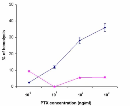
Fig. 1 Dose-dependent hemolytic effects of PTX standard (blue) on human erythrocytes and their reversal in presence of 100 μM ouabain (purple). Dose-ranging experiments were performed from 10-3
to 103 ng/ml of PTX. However, since for concentrations ≤ 100 ng/ml, no effects were observed, the 100 ng/ml concentration is represented as the first value considered. Error bars represent SD.
time of 6 h. Incubation temperature was set at 37 °C (Taniyama et al., 2001), and during the incubation the samples were occasionally (maximum once per h) resuspended by inversion. For negative controls, red blood cell suspension was added with 100 μM ouabain and incubated for 30 min at 37 °C, prior to the addition of PTX (Gleibs et al., 1995). In order to avoid any possible differences due to a diverse manipulation, also samples that did not contain the glycoside ouabain were incubated for 30 min at 37 °C before the addition of PTX. Total lysis of erythrocyte suspension was obtained by incubating the cells with 0,1 % v/v Tween 20. In order to evaluate the degree of spontaneous lysis, also tubes containing exclusively the red blood cell suspension in D-PBS were set. For each concentration and control, the experiments were set in triplicate. A total of eight independent experiments have been performed.
Hemolysis evaluation
After the incubation, the cell suspensions were centrifuged at 900xg for 10 min (Taniyama et al., 2003) and the supernatant was carefully collected, by paying attention not to disturb the pellet. The absorbance at 405/540 nm of supernatant was measured with a “Helios β" spectrophotometer (Spectronic, Unicam, Cambridge, UK). The value of absorbance of erythrocytes maintained exclusively in D-PBS has been utilized to set the 0 value before reading the samples that contained PTX. Hemolytic levels were expressed by percentage of hemolysis, calculated with the ratio between the value measured for each sample and that registered for the total hemolysis (Taniyama et al., 2003).
Chemical Reagents
Human erythrocytes were obtained from Laboratorio Medicina Trasfusionale (Policlinico, Azienda Ospedaliero Universitaria, Modena, Italy), PTX standard was from Wako Chemicals (Neuss, Germany). A second PTX standard solution was generously gifted by Centro di Ricerche Marine (Cesenatico, FC, Italy). All the other chemicals, including D-PBS and ouabain, came from Sigma-Aldrich (St Louis, CA, USA). All the plastics used were sterile and cell-culture tested (Sarstedt, Nümbrecht, Germany).
Results and Discussion
The applied protocol confirms that PTX is able to induce hemolysis in human erythrocytes (Fig. 1), and the levels of the lysis are comparable with those observed by other groups using the same source for red blood cells (Taniyama et al., 2001, 2003). It is worth noting that the value here presented indicated a discrepancy with those already published, since the effects of PTX were detectable only from 101 ng/ml, while at this value Taniyama and collaborators found a clear activity of PTX (Taniyama et al., 2001). In order to verify if present data could be due to an erroneous preparation of the PTX standard, experiments were repeated with a second PTX solution coming from Centro di Ricerche Marine. However, data collected with this second standard substantially confirmed those collected with the first one. Differently from this first result, there is a substantial agreement between data presented here and those collected by Taniyama et al (2001) for what concerns the
maximum levels of hemolysis detectable in human erythrocytes.
As it has been observed from a long time (Habermann and Chhatwal, 1982), PTX effects on human erythrocytes are completely reverted by the glycoside ouabain (Taniyama et al., 2001). The results obtained by using 100 μM ouabain confirm this indication even if a small percentage of erythrolysis have been observed in the presence of the glycoside (Fig. 1). Also other concentrations have been suggested for ouabain (Habermann and Chhatwal 1982, Taniyama et al., 2001), but that adopted here was the most effective and did not significantly influence the basal levels of erythrolysis.
As far as data analysis is concerned, Fig. 1 presents data collected by using the absorbance wavelength of 405 nm, following Gleibs and collaborators (1995). Different values of wavelength, such as 540 nm, have been utilized to evaluate the hemolysis of mouse erythrocytes (Lenoir et al., 2004). However, even if also the latter wavelength has been utilized in the present research, the most repeatable data were obtained at 405 nm, after imposing to the spectrophotometer the value 0 for the samples maintained only in D-PBS.
If some differences in the protocols used to test the hemolytic activity of PTX can be retrieved, probably the most notable ones concern the unity chosen to measure the levels of hemolysis. Gleibs
et al (1995), working on Palythoa and other marine specimen, describe the hemolytic unit (HU) as the amount of material necessary to produce 50 % hemolysis in human erythrocytes within 4 h incubation PTX. A similar definition of the HU, without references to the time of incubation, is given also by Lenoir et al (2004), that worked with mouse red cells. Working with human erythrocytes, Gleibs and Mebs (1999) quantified one hemolytic unit (HU) in 0,4 μg of PTX, but due to the different sensitivity of mammalian erythocytes to PTX (Habermann, 1989), this value should probably be referred only to human red cells. Taniyama et al. (2001) quantify PTX effects with the percentage of hemolysis, calculated with the ratio between the value measured for a sample and that registered for the total hemolysis. The percentage is a pure number that can be applied independently from the source of the erythrocytes, therefore it has been chosen here to evaluate PTX activity from commercially available standards. However, since the percentage of hemolysis should be properly calculated as follows: [absorbance in test - absorbance in D-PBS]/[absorbance in 100 % hemolysis - absorbance in D-PBS] (Hubert et al., 1997), the value of absorbance in D-PBS has been utilized to set the 0 value on the spectrophotometer. Only after this procedure, the ratio proposed by Taniyama et al. (2001) can be properly applied.
Acknowledgements
The author wish to thank Laboratorio Medicina Trasfusionale (Policlinico, Modena, Italy) that kindly supplied the human blood and Centro di Ricerche
Marine (Cesenatico, FC, Italy) for the PTX solution. The author gratefully acknowledges also Prof. Enzo Ottaviani (University of Modena and Reggio Emilia, Italy) for the critical reading of the manuscript.
References
Artigas P, Gadsby DC. Na+/K+-pump ligands modulate gating of palytoxin-induced ion channels. Proc. Natl. Acad. Sci. USA 100: 501-5055, 2003.
Bignami GS. A rapid and sensitive hemolysis neutralization assay for palytoxin. Toxicon 31: 817-820, 1993.
Gleibs S, Mebs D, Werding B. Studies on the origin and distribution of palytoxin in a Caribbean coral reef. Toxicon 33: 1531-1537, 1995. Gleibs S, Mebs D. Distribution and sequestration of
palytoxin in coral reef animals. Toxicon 37: 1521-1527, 1999.
Habermann E. Palytoxin acts through Na+,K+ -ATPase. Toxicon 27: 1171-1187, 1989.
Habermann E, Ahnert-Hilgert G, Chhatwal GS, Béress L. Delayed hemolytic action of palytoxin. General characteristics. Biochim. Biophys. Acta 649: 481-486, 1981.
Habermann E, Chhatwal GS. Ouabain inhibits the increase due to palytoxin of cation permeability of erythrocytes. Naunyn Schmiedebergs Arch. Pharmacol. 319: 101-107, 1982.
Hilgemann DW. From a pump to a pore: how palytoxin opens the gates. Proc. Natl. Acad. Sci. USA 100: 386-388, 2003.
Hubert F, Cooper EL, Roch P. Structure and differential target sensitivity of the stimulable cytotoxic complex from hemolymph of the Mediterranean mussel Mytilus galloproÍincialis. Biochim. Biophys. Acta 1361: 29–41, 1997. Lenoir S, Ten-Hage L, Turquet J, Quod JP, Bernard
C, Hennion MC. Firstevidence of palytoxin analogues from an Ostreopsis mascarenensis
(Dinophyceae) benthic bloom in southwestern Indian Ocean. J. Phycol. 40: 1042-1051, 2004. Moore RE, Scheuer PJ. Palytoxin: a new marine
toxin from a coelenterate. Science 1971 172: 495-498, 1971.
Riobó P, Paz B, Franco JM. Analysis of palytoxin-like in Ostreopsis cultures by liquid chromatography with precolumn derivatization and fluorescence detection. Anal. Chim. Acta 566: 217-223, 2006.
Taniyama S, Mahmud Y, Tanu MB, Takatani T, Arakawa O, Noguchi T. Delayed hemolytic activity by the freshwater puffer Tetraodon sp.
toxin. Toxicon 39: 725-727, 2001.
Taniyama S, Arakawa O, Terada M, Nishio S, Takatani T, Mahmud Y et al.. Ostreopsis sp., a possible origin of palytoxin (PTX) in parrotfish
Scarus ovifrons. Toxicon 42: 29-33, 2003. Wattenberg EV. Palytoxin: exploiting a novel skin
tumor promoter to explore signal transduction and carcinogenesis. Am. J. Physiol. Cell Physiol. 292: C24-C32, 2007.