J of Evolution of Med and Dent Sci/ eISSN- 2278-4802, pISSN- 2278-4748/ Vol. 3/ Issue 47/Sept 25, 2014 Page 11331
USG AND MRI CORRELATION IN THE EVALUATION OF MENISCAL LESIONS
OF KNEE
Ravichandra G1, Aravinda M2, Sajeer Mohammed Usman3, Vivek S4
HOW TO CITE THIS ARTICLE:
Ravichandra G, Aravinda M, Sajeer Mohammed Usman, Vivek S. USG and MRI Correlation in the Evaluation of Meniscal Lesions of Knee. Journal of Evolution of Medical and Dental Sciences 2014; Vol. 3, Issue 47, September 25; Page: 11331-11337, DOI: 10.14260/jemds/2014/3482
ABSTRACT: AIMS AND OBJECTIVE: The objective of this study was to evaluate the role of a handheld USG of 7-13 MHz in evaluating meniscal lesions of knee and comparing it to the results obtained with magnetic resonance imaging. MATERIAL AND METHODS: A comparative double blind study was done between ultrasonography and MRI of fifty patients with a history of knee trauma or with suspected knee meniscal lesions, who were referred to the department of Radiodiagnosis. MRI and USG results were finally correlated with arthroscopy findings. All these patients had a significant history and clinical evaluation suggested meniscal lesion of the knee joint. Cases which have been operated previously on the same knee were excluded from this study. RESULTS: Out of the 50 patients, a total of 100 menisci were examined, out of which after final arthroscopy confirmation, 34 had meniscal tears, 3 degenerative tears, and two patients had meniscal cysts both of which were not associated with a tear of the meniscus. CONCLUSION: The specificity of USG matched that of MRI and it can reasonably be applied to confirm the clinical diagnosis before undertaking arthroscopy. However, the lower sensitivity suggests that there is still a need to improve the technique to reduce the number of false-negative diagnoses and thus to avoid unnecessary arthroscopy. USG may be used as a screening tool prior to arthroscopy in selected cases where MRI is a contraindication or is not available or if the patient is not affording.
KEYWORDS: Meniscal lesions, knee, MRI, USG.
INTRODUCTION: MRI scanning of the knee joint is the noninvasive procedure which is widely used to diagnose traumatic knee injuries. MRI scanning is routinely performed before arthroscopic management. USG is also helpful in diagnosing traumatic knee injuries. Our objective was to correlate MRI and USG findings with arthroscopy in meniscal injuries and to review which modality offers better sensitivity and specificity.
Approximately two-thirds of all derangements of the knee joint are due to lesions or degenerative changes of the menisci.1,2,3
MRI has revolutionized cross sectional imaging of the musculoskeletal system and has become the most widely used technique for a wide variety of pathologic conditions. However, while MRI was gaining its ascendancy, ultrasonography was also being used for musculoskeletal imaging, which is an important complementary tool, and there is now a large body of literature documenting the effectiveness of musculoskeletal sonography.4
J of Evolution of Med and Dent Sci/ eISSN- 2278-4802, pISSN- 2278-4748/ Vol. 3/ Issue 47/Sept 25, 2014 Page 11332 Traumatic injuries to the meniscal cartilage in the knee have always been a diagnostic challenge.6 The combination of clinical and MRI findings would reduce the number of arthroscopies to
5%.7
MATERIAL AND METHODS:A comparative double blind study was done between ultrasonography and MRI of fifty patients with a history of knee trauma or with suspected knee meniscal lesions, who were referred to the department of Radiodiagnosis. All these patients had a significant history and clinical evaluation suggested meniscal lesion of the knee joint. Cases which have been operated previously on the same knee were excluded from this study.
Magnetic resonance imaging protocol.
Magnetic resonance imaging performed with a 0.4 tesla machine Aperto from Hitachi. Imaging sequences obtained:
STIR coronal,
Proton density Coronal,
T1 Coronal,
T2 Axial,
T2 Sagittal,
Gradient T2 Sagittal (Sagittal taken with a 15 degree internal rotation axis).
ULTRASONOGRAPHY PROTOCOL: Ultrasonography was performed with a standard hand held ultrasound machine Envisor from Philips, 7-13 MHz machine, using a linear transducer. Images were obtained by placing the transducer longitudinally in relation to the knee joint on the medial and lateral side with the patient in prone position visualizing the femoral and tibial condyles with the meniscus in between. All USGs were performed by the same senior radiologist with an interest in musculoskeletal ultrasound. The radiologist was blinded of the clinical and magnetic resonance imaging findings of the patient prior to ultrasonography. Reporting was done immediately. Images were obtained for each portion of the meniscus and saved for reference
ARTHROSCOPY PROTOCOL: All patients underwent arthroscopy under spinal anaesthesia or epidural anaesthesia under complete sterile precautions. Arthroscopy was done using standard anteromedial and anterolateral portals, visualized with a camera with a 30 degree angle.
All patients underwent diagnostic arthroscopy in a step wise manner: 1. Suprapatellar pouch and patellofemoral joint.
2. Medial gutter.
3. Medial compartment. 4. Intercondylar notch.
5. Posteromedial compartment. 6. Lateral compartment.
7. Lateral gutter and posterolateral compartment.
J of Evolution of Med and Dent Sci/ eISSN- 2278-4802, pISSN- 2278-4748/ Vol. 3/ Issue 47/Sept 25, 2014 Page 11333
RESULTS: All the 50 patients who were referred to department of radio diagnosis underwent ultrasonography and magnetic resonance imaging. All patients were taken through the same study protocol strictly.
All patients included in the study, completed the study protocol since there was no follow up required. All parts of the protocol were completed in a single hospital admission. There were no complications in the study.
Medial menisci and lateral menisci were analyzed separately. Grade 1 and grade 2 lesions detected on MRI and ultrasound were considered as negative scans. The higher grade of the reported tear was taken into consideration when a range of grades were reported.
Out of the 50 patients, a total of 100 menisci were examined, out of which after final arthroscopy confirmation, 34 had meniscal tears, 3 degenerative tears, and two patients had meniscal cysts both of which were not associated with a tear of the meniscus.
Comparing the sensitivity, specificity, PPV, NPV and accuracy of ultrasonography with MRI was done taking the findings at arthroscopy as a positive finding and grade 3 tears on MRI and USG as positive, although grade 1 and grade 2 tears were reported on MRI and USG they were considered as
No Tear and taken as a negative. The true negative, true positive, false negative, false positive were
applied to the formula and tables were drawn from which charts were drawn separate for medial meniscus, lateral meniscus and combined total.
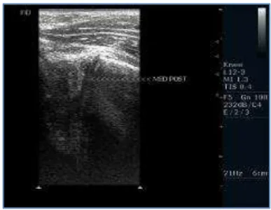
Fig. 1: 28 years old male with traumatic injury to the knee underwent USG, MRI and arthroscopy.
J of Evolution of Med and Dent Sci/ eISSN- 2278-4802, pISSN- 2278-4748/ Vol. 3/ Issue 47/Sept 25, 2014 Page 11334
Fig. 2: 22 yr old male underwent USG, MRI and arthroscopy following injury
Fig. 1B: Sagittal T2 WI shows double PCL sign indicating bucket
handle tear of medial meniscus
Fig. 1C: Arthroscopy showing the displaced bucket handle tear
Fig. 2B: Utrasonography of posterior horn of lateral meniscus showing a grade 3 tear Fig. 2A: Sagittal T2 WI showing
grade 3 changes in posterior horn of lateral meniscus
J of Evolution of Med and Dent Sci/ eISSN- 2278-4802, pISSN- 2278-4748/ Vol. 3/ Issue 47/Sept 25, 2014 Page 11335
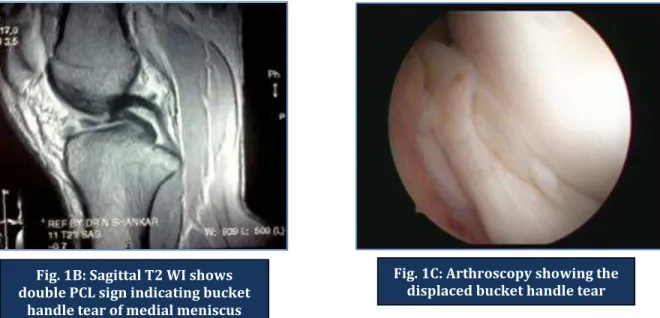
Fig. 3: 33 years old male with insidious onset of knee pain underwent USG, MRI and arthroscopy.
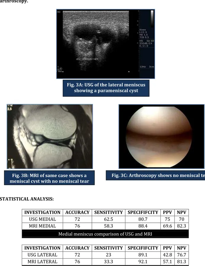
STATISTICAL ANALYSIS:
INVESTIGATION ACCURACY SENSITIVITY SPECIFIFCITY PPV NPV
USG MEDIAL 72 62.5 80.7 75 70
MRI MEDIAL 76 58.3 88.4 69.6 82.3
Medial meniscus comparison of USG and MRI
INVESTIGATION ACCURACY SENSITIVITY SPECIFIFCITY PPV NPV
USG LATERAL 72 23 89.1 42.8 76.7
MRI LATERAL 76 33.3 92.1 57.1 81.3
Lateral meniscus comparison of USG and MRI Fig. 3A: USG of the lateral meniscus
showing a parameniscal cyst
Fig. 3B: MRI of same case shows a meniscal cyst with no meniscal tear
J of Evolution of Med and Dent Sci/ eISSN- 2278-4802, pISSN- 2278-4748/ Vol. 3/ Issue 47/Sept 25, 2014 Page 11336
INVESTIGATION ACCURACY SENSITIVITY SPECIFIFCITY PPV NPV
USG 72 48.6 85.7 66.6 73.9
MRI 76 50 90.6 75 76.3
Total medial and lateral meniscus comparison ofUSG and MRI
CONCLUSION: Given that the specificity matched that of MRI, ultrasonography can reasonably be applied to confirm the clinical diagnosis before undertaking arthroscopy. However, the lower sensitivity suggests that there is still a need to improve the technique to reduce the number of false-negative diagnoses and thus to avoid unnecessary arthroscopy.
Ultrasonography may be used as a screening tool prior to arthroscopy in selected cases where MRI is a contraindication or is not available or if the patient is not affording. Ultrasonography shows a dynamic image of the meniscus and thus may prove useful if studied in conjunction with a proper clinical examination.
It is difficult to comment on the type of tear with an ultrasonography evaluation alone. Ultrasonography can easily identify a meniscal cyst, its size and locations. Ultrasonography cannot differentiate a traumatic tear from a degenerative tear.
REFERENCES:
1. Raunest J et al. The clinical value of magnetic resonance imaging in the evaluation of meniscal disorders. The Journal of bone and joint surgery, July 1991;73-A-1:11-16.
2. Martin E et al. Incidental meniscal findings on knee MRI in middle aged and elderly persons. New England Journal of Medicine 2008, September;359:1108-15.
3. Ian Douglas. Meniscal tears. Current Orthopaedics 2006;20:85-94.
4. Levon N. The top 10 reasons musculoskeletal sonography is an important complementary or alternative technique to MRI. American Journal of Roentgenology June 2008;190:1621-1626. 5. Park G Y, Min J, Moon S, Michael Y. The value of ultrasonography in the detection of meniscal
tears diagnosed by magnetic resonance imaging. American Journal of Physical Medical Rehabilitation. 87;1:14-20.
6. Douglas H, Arthur A, Martha Norris. Bucket handle tears of the medial and lateral menisci of the knee; value of MR Imaging in detecting displaced fragments. American Journal of Roentgenology: September 1995;165:621-625.
7. Kush singh, Clyde A, M Todd, Laurenec A. MRI appearance of variant of discoid lateral meniscus. American Journal of Roentgenology:2006;187:384-387.
8. M Schurz, J T Erdoes, P Platzer, NPetras, J T Hausmann, V Vécsei. The value of clinical examination and MRI versus intraoperative findings in the Diagnosis of meniscal tears.Scriptamedica (brno) April 2008;81-1:3–12.
9. David A et al. Current concepts and controversies in meniscal imaging. MRI clinics of North America, May 2000;8-2:243-270.
10.Michael F, Van Trommel. The use of Non contrast MRI in evaluating meniscal repair comparison with arthrography. Arthroscopy, 1998;14-1:2-8.
J of Evolution of Med and Dent Sci/ eISSN- 2278-4802, pISSN- 2278-4748/ Vol. 3/ Issue 47/Sept 25, 2014 Page 11337 12.R Mackenzie et al. Errors at knee MRI-True or False. The British Journal of Radiology,
1995;68:1045-1051.
13.Arthur A, Rajat M, Clinical MRI and Arthroscopic findings associated with failure to diagnose a lateral meniscal tear on knee MRI. American Journal of Roentgenology, 2008;190:22-26.
14.Radial tears of the medial meniscal root reliability and accuracy of MRI for diagnosis, American Journal of Roentgenology July 2008;191:81-85.
15.Jeffrey W, Arthur A, Kazohiko S, Comparison of the accuracy rates of 3-T and 1.5 T MRI of the knee in the diagnosis of meniscal tears, American Journal of Roentgenology, 2009 august;193: 509-514.
AUTHORS:
1. Ravichandra G. 2. Aravinda M.
3. Sajeer Mohammed Usman 4. Vivek S.
PARTICULARS OF CONTRIBUTORS:
1. Associate Professor, Department of Radiodiagnosis, Yenepoya Medical College, Yenepoya University, Mangalore.
2. Assistant Professor, Department of Orthopaedics, Yenepoya Medical College, Yenepoya University, Mangalore. 3. Resident, Department of Orthopaedics,
Yenepoya Medical College, Yenepoya University, Mangalore.
4. Resident, Department of Radiodiagnosis Yenepoya Medical College, Yenepoya University, Mangalore.
NAME ADDRESS EMAIL ID OF THE CORRESPONDING AUTHOR:
Dr. Vivek S,
Department of Radio Diagnosis, Yenepoya Medical College, Yenepoya University, Mangalore-575018, Karnataka, India.
Email: [email protected]