An interview with
• Graduated from the School of Dentistry, Rio de Janeiro Federal University (UFRJ) – Praia Vermelha, Brazil, 1978. • Specialist in Oral and Maxillofacial Surgery, UERJ, 1979.
• Residency in Oral and Maxillofacial Surgery, University of Texas-Dallas – USA, 1981-1984. • MSc and PhD, School of Dentistry, UFRJ – 1991 and 2001.
• Professor at UFRJ and Rio de Janeiro State University (UERJ) since 1979 to date. • Head Professor, Oral Surgery, School of Dentistry, UERJ, since 1995.
• Author of the following books: “Orthognathic Surgery for Orthodontists” (Ed. Santos), “Retained Tooth Surgery” (Ed. San-tos), “Update on Oral and Maxillofacial Surgery and Traumatology” (Ed. Santos).
• In private practice in Rio de Janeiro, Brazil, since 1979. • Renowned lecturer in Brazil and abroad.
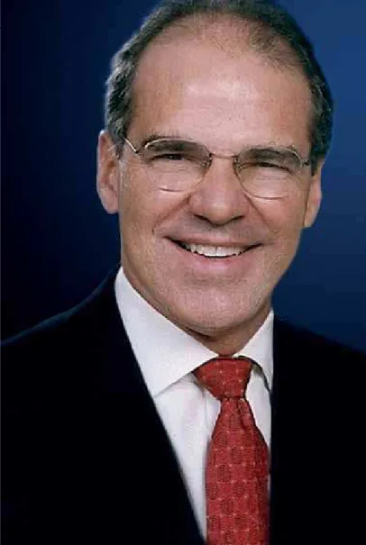
Prof. Paulo José d’Albuquerque Medeiros was born to Paulo Pinho de Medeiros and Conceição Rosário d’Albuquerque Me-deiros in the city of Rio de Janeiro, Brazil on March 7, 1957. He has been married for 31 years to Patricia Leo MeMe-deiros and they have two children: Alessandra Leão Medeiros Parente, 30, a law judge, and Leo Bruno Medeiros, 28, economist. He is fond of music and the movies, and is a ine singer. He has developed a reined taste for good wines, his favorite being Chateau Palmer. He is currently reading a pocket book titled “1001 wines to drink before you die”. His most daunting challenge in life: “Keeping up the motivation to teach, which is my true calling”.
Marco Antonio Almeida
How to cite this interview: Medeiros PJD. Interview. Dental Press J Orthod. 2012 Sept-Oct;17(5):8-23.
Submitted: July 2, 2012 - Revised and accepted: August 7, 2012
» Patients displayed in this article previously approved the use of their facial and intraoral photographs.
Paulo José
AEStHEticS
What is the greatest demand of patients: Esthet-ics of function? (Antenor Araújo)
Most patients expect improved esthetics. Don’t forget that - if the patient’s facial esthetics is not compromised - it is quite a challenge for the orthodontist to persuade the patient to agree to ortho-surgical treatment, even if they have some sort of malocclusion.
Which parameters are most important in the analysis of facial esthetics?
(Carlos Estevanell Tavares)
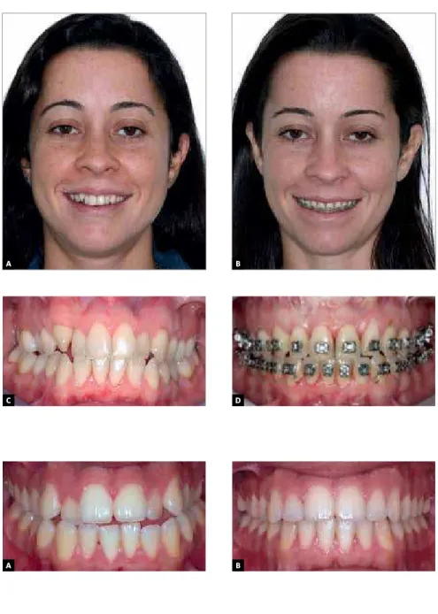
I could cite several variables, but I prefer to high-light the smile, which is our “calling card.” I’ve had several patients who, after correction of vertical maxillary excess, came to be questioned by friends who wanted to know if they were wearing contact lenses (Fig 1). It seems that an unsightly smile can “erase” other interesting facial features. The “buc-cal corridor”, which is sometimes treated by ortho-surgical expansion (Fig 2), and in other situations by simply improving the shape of dental arches through tooth movement, also greatly affects the smile and facial esthetics (Fig 3).
Figure 1 - Class II patient with vertical maxillary excess and anteroposterior mandibular deficiency (A). After max-illary repositioning and mandibular advancement. The surgical procedure highlighted the eyes (B).
Figure 3 - Class III malocclusion and maxil-lary constriction before ortho-surgical treat-ment. Occlusion after osteotomies of maxilla and mandible without segmentation. The up-per arch was expanded and its form improved through tooth movement.
Figure 2 - Before (A) and after (B) surgically assisted rapid maxillary expansion. “Buccal corridor” shows improvement. Initial occlu-sion (C) and after intervention (D).
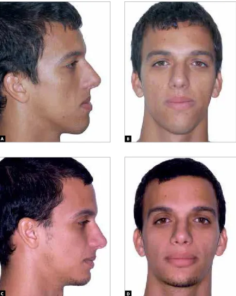
Figure 4 - Note nasal base tapering toward the tip. Increased nose harmony after slight base enlargement as a result of maxillary ad-vancement.
A
A
A
B
B
B
What steps do you take in cases of advancement or repositioning orthognathic surgery in the maxilla to avert undesirable efects on facial es-thetics? (Carlos Estevanell Tavares)
Nasal esthetics is a major concern of surgeons in planning and performing maxillary orthogna-thic surgery. Le Fort I osteotomy could cause nasal enlargement due to muscle detachment, but this
issue is particularly challenging in the aforesaid movements, i.e., maxillary advancement/reposi-tioning. In rare, select cases enlargement may even be desirable (Fig 4). The vast majority of patients, however, have the base of their nose sutured after maxillary repositioning, which is intended to pre-vent enlargement. This procedure is called “Nasal plication” (Fig 5).
A B
Figure 5 - A, B) This patient is a good candi-date for maxillary advancement combined with maxillary repositioning. C, D) Good con-trol of nasal base although the movement performed can be considered potentially det-rimental to the esthetics of the nose.
How do you address cases of closed mandibu-lar plane and decreased lower face?
(Carlos Estevanell Tavares)
In the past, this condition was mistakenly called “Short Face Syndrome”. Although not a true syn-drome, its expression tends to recur at different levels. The starting point for treatment is determining how many millimeters the maxilla should be lowered, and whether this lowering will have one single magnitude or different magnitudes, considering the incisal and
molar regions. A predictive tracing should indicate whether the mandible is likely to undergo a clockwise rotation only, and require surgery, or whether it will have to undergo advancement or setback osteotomy, depending on the initial malocclusion. The height of the mandibular symphysis in these individuals is usually short. Therefore, performing a genioplasty to increase the vertical dimension may be beneicial (Fig 6). These procedures elongate the face and im-prove the mandibular plane at the same time.
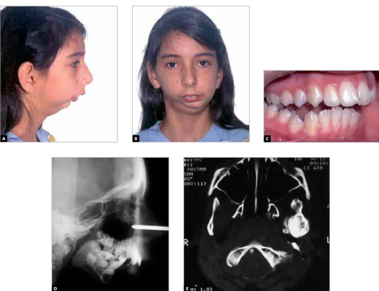
Figure 6 - Patient presented with anteroposterior and vertical maxillary hypoplasia in addition to anteroposterior mandibular excess. Lateral cephalometric radiograph highlighting the discrepancy between maxilla and mandible. Lateral cephalogram with mouth slightly open enabling visualization and quantification of vertical maxillary deficiency in relation to the upper lip. Patient after lowering and maxillary advancement combined with mandibular setback.
A
C
B
Figure 8 - Note unfavorable aspect of submental region (A). Note beneficial effect in the submental region after mandibular advancement (B).
Figure 7 - Note the unsightly shape of the nasal dorsum (A). Improved nasal esthetics as a result of orthognathic surgery (B).
When do you recommend plastic surgery as a com-plement to facial esthetics, concurrently with or-thognathic surgery? (Carlos Estevanell Tavares)
The plastic surgery procedures more commonly performed as adjuncts to orthognathic surgery are rhinoplasty, submental liposuction and plication of the platysma muscle. As regards indication and tim-ing, one should bear in mind that surgical movement of the maxilla and/or mandible can effect favorable changes in these anatomical regions. There are cas-es of individuals who wished to undergo rhinoplasty
and who gave it up after deciding that the effect of orthognathic surgery on the nose had delivered the desired esthetic outcome (Fig 7). Currently, most maxillofacial surgeons only indicate rhinoplasty when it is combined with isolated osteotomies of the mandible. When the patient is undergoing maxillary osteotomy, rhinoplasty is planned for 6 months af-ter orthognathic surgery, but only if at that time the patient still wishes it. The same applies in the case of mandibular advancement, which in itself produces esthetic effects in the submental region (Fig 8).
A
A
B
Conversely, in a patient undergoing mandibular set-back who presents with excess submental tissue, lipo-suction and/or plication of the platysma at the time of orthognathic surgery seems to be the best approach.
FUNctiON
Surgical mandibular retrusions can impair the airspace and compromise breathing. currently, how does surgical planning deal with this issue? (Arno Locks)
Although decreases in airspace have been ob-served in mandibular setback samples, there are
no studies demonstrating functional impairment in these individuals. This may be because man-dibular setbacks in excess of 7 mm or 8 mm are extremely rare. In treating severe Class III mal-occlusions, with overjets in excess of 10 mm, pro-fessionals tend to divide movements between the maxillary advancement and mandibular setback movements (Fig 9). Our group published an ar-ticle in the September/October 2011 issue of this journal which analyzed 17 patients who had un-dergone isolated mandibular setbacks and other setbacks associated with maxillary advancement. Figure 9 - Patient had a -17 mm overjet (A), Pro-nounced disharmony between maxilla and man-dible (B). After 8 mm maxillary advancement and 9 mm mandibular setback, an insignificant decrease in the airways can be seen. Patient reported that there was no respiratory impair-ment (C). Final occlusion (D, E).
A
D
B
E
The average mandibular setback was of about 7 mm, and we noted an average reduction of about 1 mm in oropharynx, and about 3.5 mm in the hy-popharynx. Despite these findings, none of the patients in the sample reported impaired breath-ing after surgery. In over 30 years of practice in orthognathic surgery I cannot recall having had any patients who returned with respiratory com-plaints after this procedure. I would only be reluc-tant to indicate mandibular setback for individuals with prior respiratory disorders or obese patients.
What scientific evidence is there regarding a cause-and-effect relationship between orthog-nathic surgery and snoring or sleep apnea? (Marco Antonio Almeida)
Orthognathic surgery has been used to treat sleep apnea in cases that prove refractory to more conservative, less invasive therapies. The literature reports that maxillary advancement performed si-multaneously with mandibular advancement has yielded favorable results in a significant number of patients. There are researchers out there who have been conducting reliable, well-designed research that can attest to this fact, but I’ve had no experi-ence with these patients.
A patient with no tMD symptoms before treat-ment starts presenting with tMD signs and symptoms after orthognathic surgery, such as clicking joints, pain in the tMJ region and re-stricted opening. What are the possible causes of this condition? Are the causes somehow re-lated to orthodontic treatment, orthognathic surgery or attributable to chance? (Weber Ursi)
Joint dysfunction hardly ever occurs after or-thognathic surgery. The most frequently occur-ring symptoms are muscle pain and discomfort, which the literature refers to as Myofascial Pain and Dysfunction Syndrome. This condition tends to be transient and is treated through measures to mitigate the symptoms and increase patient com-fort. The rare cases of patients who experienced joint noises, which by the way were not painful, the incident occurred in the first weeks of the postop-erative period and resolved spontaneously. This adaptability of patients might be due to the fact
that they are young and without prior dysfunction. I don’t believe either orthognathic surgery or or-thodontics is responsible for the onset of joint dys-function in this particular population.
tEcHNiQUE
What aspects of orthodontic preparation are you most often confronted with, which poten-tially compromise the outcome and stability of orthognathic surgery? How can these prob-lems be avoided? (Weber Ursi)
Instability of movements during dental orth-odontic preparation usually manifests itself in the medium or long term. After surgery the patient is still undergoing orthodontic treatment for about 1 year, after which a retainer tends to maintain the teeth in their correct position. In reassessing pa-tients 10, 20 and even 25 years after ortho-surgical treatment I have noticed that the most significant losses occur in the transverse direction, as is usu-ally the case with surgical relapses. When the pa-tient needs significant transverse gains, either orthopedic or surgically assisted maxillary expan-sion seems to offer better results.
Do you believe that evaluation by a psycholo-gist experienced with this type of patient in order to detect specific responses regarding acceptance of changes in appearance, actual motivation toward surgery, anxiety level, etc, would go a long way towards averting dissatis-faction with the outcome? Or is it your belief that this sort of detection can always and eas-ily be carried out by the orthodontist and/or surgeon? (Carlos Elias Ferreira de Freitas)
I see no need for ortho-surgical patients to be routinely evaluated by a psychologist. In specific cases I do think such an evaluation would be highly advisable. On occasion, I have obviously had a few unsuccessful cases, and have also encountered dif-ficulties in treating a handful of patients.
to what extent have the new distraction osteo-genesis techniques contributed to resolve large mandibular advancements—including mandibu-lar ramus lengthening—causing counterclockwise mandibular rotation? (Arno Locks)
Distraction osteogenesis, unlike what was ini-tially believed, has not replaced conventional or-thognathic surgery in conventional cases. Distrac-tion osteogenesis is optimally indicated in the first decade of the patient’s life to treat mandibular growth deficiencies. Cases of hemifacial microso-mia or retrognathia resulting from TMJ ankylosis have been successfully treated by this method. The discomfort caused by prolonged use of a distractor in addition to difficulty in controlling the tion vector are some of the disadvantages of distrac-tion osteogenesis vs. orthognathic surgery starting in the second decade of the patient’s life.
the conduct advocated by U.S. professor Lar-ry Wolford, who often indicates surgical in-terventions in the temporomandibular joint concurrent with orthognathic surgery when the patient presents with intra-joint changes, has met with widespread acceptance in Brazil. What is your opinion on the subject?
(Carlos Elias Ferreira de Freitas)
I worked with Dr. Wolford for 2 years during my residency program and I know for a fact that he is a highly judicious professional. I believe this is not a routine approach, and the numerous joint interven-tions that he performs simultaneously with orthog-nathic surgery are due to, irstly, the large volume of patients he operates on, and secondly, because he is a reference in the treatment of patients with joint dysfunction. I have limited experience in performing orthognathic surgery in joint TMD patients. The vast majority of patients I treat are young and hardly ever present with joint TMD pain. I see no need to perform concurrent joint intervention. As in the cases of mus-cle TMD, these patients’ dysfunction is treated con-servatively before orthognathic surgery and, if neces-sary, further treatment is provided after surgery.
StABiLitY
Some patients, after mandibular advancement surgery for correction of class ii, present with
condylar resorption, totally compromising the outcome. What have studies so far contributed on this topic? (Arno Locks)
It is important to differentiate condylar resorp-tion from condylar remodeling. Condylar remod-eling is a sort of “wear”, albeit minor, which occurs in surgical and non-surgical patients, causing in-creased overjet over a few years. This can be con-sidered a physiological phenomenon. It has been more often observed in patients who have under-gone mandibular advancement, especially in those with the following three sets of features: 1) Small mandibular condyles inclined posteriorly, 2) open mandibular plane and short mandibular ramus, and 3) decreased posterior facial height and in-creased anterior facial height. Treatment results for these patients may be partially or totally com-promised by this phenomenon. Condylar resorp-tion has been widely studied and described, espe-cially by the team of researchers led by Dr. Leon-ard Kaban, in Boston. In these cases condyles dis-appear altogether as meaningful retrognathia and open bite develop. There are many idiopathic cas-es, where patients never underwent orthodontic or ortho-surgical treatment. Among the possible causes of this condition are rheumatoid arthritis, use of corticosteroids, systemic lupus erythemato-sus, scleroderma, other collagen diseases, and even orthognathic surgery. Healthy patients who are good candidates for orthognathic surgery, but who present with the three sets of features I mentioned earlier should be alerted about the possibility that this phenomenon might occur.
the hierarchy of stability of ortho-surgical cases established by the North carolina team of researchers is usually cited in scientific pa-pers. Drawing on your extensive experience, how would you assess this hierarchy, and what factors can affect it? (Marco Antonio Almeida)
stabilization combined with bone grafting appear to give optimum vertical stability to the maxilla. As far as mandibular setback surgery is concerned, contrary to what is stated in the article, I have had excellent stability. I do however use, whenever pos-sible, intraoral vertical osteotomy, which is a tech-nique renowned for its high stability. The literature demonstrates that the optimal stability afforded by sagittal osteotomy in mandibular advancement is not replicated in mandibular setbacks, and the lat-ter was the technique used in the study group. The major hurdle in terms of stability in orthognathic surgery today has to do with mandibular advance-ment, and this procedure appears in the article as the second most stable. However, when you read the article carefully you can’t help but realize that mandibular advancement was performed or dis-cussed only in individuals with normal or short facial height, which does not encompass those pa-tients presenting with the three sets of features I referred to earlier on. I strongly believe that a study conducted in Class II patients with the three sets of characteristics described above will inevitably yield different outcomes.
FUtURE
How do you view the current position held by orthognathic surgery in Brazil? How would you compare it to other countries? (Antenor Araújo)
We owe the inception of orthognathic surgery in Brazil to pioneers, the likes of Mario Graziani, Paulo Pinho de Medeiros, Italo Gandelman, João Jorge de Barros and João Ephraim Wagner, and I can’t think of a better start. With the development of “Modern Orthognathic Surgery” — by which is meant a combination of surgery and orthodontics - there was a refinement in surgical techniques that already existed, and are still widely employed to this day, combined with new concepts in diagnosis and treatment planning. When he arrived in Brazil in 1978 after 3 years spent in Dallas, Texas, USA, Dr. Antenor Araújo helped to develop Brazilian or-thodontics and enabled a most fruitful exchange with centers of excellence abroad. Today Brazil has established itself in the international scene given the quality of our professionals, which is compa-rable to that of the best centers in the world. The
challenge we face today lies in training high-level professionals to serve 200 million people in a country of continental proportions. It is still not uncommon to see surgeons packing a “doctor’s bag” with surgical instruments and traveling to operate on patients a long ways from home. This is obviously not the best care you can provide. I’d hate to have, say an abdomen surgery, today and not have the surgeon around the next day because he has flown off somewhere else to see another patient. Our challenge is to train more and more quality professionals and spread them throughout Brazil. I try to give my humble contribution.
What medium to long-term advances can we expect in the field of Orthognathic Surgery? What will this major surgery be like 20 years from now? (Weber Ursi)
Although there have been refinements in sur-gical techniques, the main developments have oc-curred and tend to develop further in surgical ma-terials and in the area of digital technology. Rigid fixation techniques have improved through the de-velopment of finer plates and screws, without any noticeable loss in quality. Alloplastic materials are replacing autografts at such a rate that I believe in the near future we will no longer need to remove the patient’s own bone for any purpose. Digital diagnos-tics and planning save time and impart reliability to ortho-surgical treatments. I really look forward to the development of less traumatic bone cutting instruments. Less invasive surgeries? Robotic Sur-gery? Time will tell.
YOUR SPAcE
What were the three most gratifying ortho-surgi-cal cases in your career? (Marco Antonio Almeida)
Figure 11 - After two interventions and 4 years into treatment, there was improvement in oral movements and esthetics (A, B). Patient at age 7 and 14 (C, D).
C
A B D
A B
D E
C
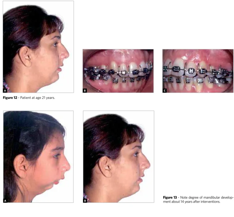
Figure 12 - Patient at age 21 years.
A B C
Figure 13 - Note degree of mandibular develop-ment about 14 years after interventions.
treatment achieved a better occlusion and satisfactory facial esthetics (Figs 10 to 13).
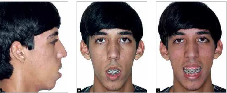
Shy and withdrawn, the second patient present-ed for treatment at 20 years of age. According to his mother, he had been bullied for many years because of his unsightly facial appearance. He underwent
orthodontic treatment and maxillomandibular os-teotomies, and was given psychological support postoperatively. About a year after surgery and orth-odontic treatment, he returned, now more talkative, lively and even responding to the jokes and remarks from members of our surgical team (Figs 14, 15, 16).
Figure 16 - 14 months postoperatively. Note improvement in interlabial relationship. Changes in behavior and attitude were remarkable.
A B C
Figure 15 - Preoperative occlusion. Due to difficulties in social adjustment, “Anticipated Benefit” was employed (A). Occlusion about 14 months after surgery (B).
Figure 14 - Hypoplastic mandible and greatly increased mandibular plane. Note long face and severe lip incompetence (A, B); patient was extremely shy and hardly ever smiled (C).
A
A
B
B
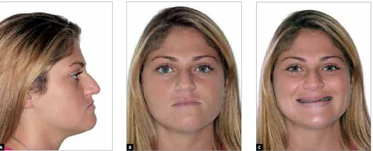
The third patient was a young woman aged 23, who exhibited several positive characteristics such as beautiful skin, eyes and hair which, however, were undermined by a complex dentofacial de-formity. She presented with maxillary retrusion,
maxillary vertical hypoplasia, mandibular progna-thism and lateral deviation of the mandible. As a result of the successful functional and esthetic out-come achieved, positive changes occurred in many areas of her life (Figs 17 to 20).
A
Figure 19 - Patient about 8 months after surgery.
A
Figure 17 - A) The maxilla was retruded, the mandible overly protruded and the nasal dorsum showed an unfavorable contour, B) patient had facial asym-metry with marked mandibular deviation; C) the maxilla was uneven and also hypoplastic in the vertical direction, which compromised the smile.
B C
Figure 18 - Preoperative occlusion (A) and approximately 8 months after surgery (B).
A B
Figure 20 - Changes in frontal view resulting from esthetic treatment (A, B). Profile before and after surgery (C, D). An improved facial symmetry and smile highlighted other patient features (E, F).
E
A B
C D
Antenor Araújo
» Post-Doc in Bucomaxillofacial Surgery, University of Texas.
Arno Locks
» PhD in Orthodontics, UNESP. Post-Doc, Royal School of Dentistry, University of Aarhus.
carlos Elias Ferreira de Freitas
» Professor of Surgery and Bucomaxillofacial Trauma, Hospital Geral Roberto Santos.
» Professor of Orthognatic Surgery, Specialization Courses in Orthodontics, UFBA and ABO-BA. » Head of the Bucomaxillofacial Surgery and Trauma
Division, SESAB.
» Head of the Bucomaxillofacial Surgery and Trauma Division, Hospital da Bahia.
carlos Estevanell tavares » PhD in Dentistry, UFRJ.
Marco Antonio Almeida
» Post-Doc in Orthodontics, University of North Carolina.
Weber Ursi
» PhD in Orthodontics, USP. 1. Gornic C, Nascimento PP, Melgaço CA, Ruellas ACO, Medeiros PJD, Sant’Anna EF.
Análise cefalométrica das vias aéreas superiores de pacientes Classe III submetidos a tratamento ortocirúrgico. Dental Press J Orthod. 2011 Sept-Oct;16(5):82-8.