Facial pattern of patients with post-foramen incisor cleft
Leopoldino Capelozza Filho1, Rodrigo Silva Caldas2, Rita de Cássia Moura Carvalho Lauris3, Arlete de Oliveira Cavassan4
Objective: The assessment and establishment of the facial growth pattern for patients with a cleft palate. Material: This cross-sectional retrospective study was based on front and profile photos of a sample of 71 patients at the HRAC-USP, 22 males and 49 females, Brazilians, young adults, with a mean age of 17 years 8 months, without previous orthodontic treat-ment and no associated syndromes. The method was the subjective facial diagnosis based on technical concepts, that is, the qualitative morphologic analysis of the face through clinical examination. Individuals were classified as Pattern I, II, III, Long Face or Short Face. Results: The distribution found with the frontal morphologic analysis was: Pattern I (69%), II (6%), III (7%), Long (18%) and Short (0%). As for the proile morphologic analysis, the distribution was: Pattern I (35%), II (38%), III (10%), Long (17%) and Short (0%).The distribution observed in the frontal analysis was very positive, since individuals Pattern I prevailed. For the proile evaluation, the anterior-posterior dysplasias were essentially shown, signiicantly increasing their participation. Long Face Pattern maintained a balance in both ratings and Short Face Pattern was not found in the sample used, probably related to the low prevalence in the general population. Conclusion: The prevalence of diferent Facial Patterns for patients with cleft palate was similar to that found in individuals without cleft.
Keywords: Cleft palate. Orthodontics. Growth.
How to cite this article: Capelozza Filho L, Caldas RS, Lauris RCMC, Cavassan AO. Facial pattern of patients with post-foramen incisor cleft. Dental Press J Orthod. 2012 Sept-Oct;17(5):35-42.
Submitted: April 09, 2009 - Revised and accepted: April 12, 2010
» Patients displayed in this article previously approved the use of their facial and in-traoral photographs.
Contact address: Leopoldino Capelozza Filho
Rua Dr. Sérvio Tulio Carrijo Coube, 2-70 – Infante Dom Henrique – Bauru/SP – Brazil ZIP CODE: 17012-632 – E-mail: [email protected]
1 Coordinator of the Specialization course, PROFIS and Sagrado Coração University
(USC).
2 Specialist in Orthodontics, Hospital of Rehabilitation of Craniofacial Anomalies,
São Paulo University (HRAC-USP).
3 PhD student in Rehabilitation Sciences, Hospital of Rehabilitation of Craniofacial
Anomalies, São Paulo University (HRAC-USP).
4 MSc in Orthodontics, Faculty of Dentistry of São Paulo University.
Objetivo: avaliar e determinar o padrão de crescimento facial de indivíduos com issura pós-forame incisivo. Métodos: esse estudo transversal retrospectivo usou fotografias frontais e de perfil de uma amostra de 71 pacientes matriculados no HRAC--USP (Bauru/SP), sendo 22 indivíduos do sexo masculino e 49 do feminino, jovens adultos brasileiros, com idade média de 17 anos e 8 meses, sem tratamento ortodôntico prévio ou síndromes associadas. O método utilizado foi o diagnóstico facial subjetivo, baseado em conceitos técnicos, constando da análise morfológica qualitativa da face. Os indivíduos foram classifi-cados, por dois ortodontistas do HRAC/USP, com base no conceito de padrão sugerido por Capelozza Filho: Padrão I, II, III, Face Longa e Face Curta. Resultados: a distribuição na análise morfológica frontal encontrada foi: Padrão I (69%), Padrão II (6%), Padrão III (7%), Padrão Face Longa (18%) e Padrão Face Curta (0%). Na análise morfológica de peril, a distribuição encontrada foi: Padrão I (35%), Padrão II (38%), Padrão III (10%), Padrão Face Longa (17%) e Padrão Face Curta (0%). A distribuição no aspecto frontal foi muito positiva, já que os indivíduos Padrão I predominaram. Na análise do peril, as displasias anteroposteriores foram expressas em essência, aumentando signiicativamente sua participação. Já o Padrão Face Longa manteve um equilíbrio em ambas as avaliações e o Padrão Face Curta não foi encontrado na amostra utilizada, provavelmente devido à baixa prevalência na popula-ção geral. Conclusão: a prevalência dos diversos padrões faciais para os pacientes com issura pós-forame incisivo foi semelhante à encontrada para indivíduos sem issura.
IntROduCtIOn
Cleft lip and/or palate are congenital malforma-tions that occur in the embryonic period and involve numerous consequences that can follow a person throughout life. The morphological change clinically manifested is variable and may involve the lip, palate or lip and palate.28
The cleft palate, which corresponds to about 23% of cleft patients,9 the object of study of this work, appears
in the prenatal life, the period between late embryonic and early fetal periods, speciically between the eighth and twelfth week of pregnancy, during which it is fused the secondary palate. The formation of the secondary palate comprises: 1) The growth of two individual pal-ate processes, one on each side, originpal-ated from the inner part of the maxillary processes, 2) the elevation of palatal processes obliquely positioned on each side of the tongue, and 3) inally, medial growth toward the midline to the junction of two palatine processes, which culminates in the disappearance of the epithe-lium that covers and individualizes it, characterized by a biological mechanism called “mesodermization”.
These events occur until the twelfth week of prenatal life. The fusion failure of the palatine processes, due to the absence of any of these events described above, determines the cleft palate, which morphological di-versity varies in length from a cleft uvula to the full impairment of the palate when it reaches the incisive foramen (Fig 2).3They can be classiied as complete or
incomplete and aggravating from posterior to anteri-or. It is believed in extra-genetic etiology for cleft pal-ate, although it has been mentioned that several genes are involved in the formation of palate.11,18
The Hospital for Rehabilitation of Craniofacial Anomalies (HRAC-USP) set a routine therapy that establishes the performance of palatoplasty at 12 months old. It seems obvious the logic to reconstruct the morphology and then seek an adjustment of the functions developed by the nasopharyngeal system.19
The anatomical restoration of cleft palate aims to develop normal speech, protection of the nasal respi-ratory mucosa and better functioning of the Eusta-chian tube. There is a consensus that the earlier the palatoplasty is performed, the better the functional responses.16 In patients with cleft palate, palatoplasty
may negatively inluence the sagittal maxillary per-formance, according to analysis of malar projection
on the face, although it does not compromise the fa-cial behavior pattern.29
The coniguration of the dentofacial characteristics of patients with cleft lip and / or palate has been based on cephalometry. In this context, the cephalometric pattern of patients with cleft palate displays diference in relation to cephalometric normative.27 Due to lack of
literature reports describing the facial pattern of pa-tients with this type of cleft, there is the necessity of a larger study that is not based solely on the cephalomet-ric pattern but also in the facial morphology.
OBJECtIVE
The purpose of this study was to diagnose the facial growth pattern in patients with cleft palate using fa-cial morphological analysis, through frontal and pro-ile assessments, deining the classiication based on the concept of pattern suggested by Capelozza Filho.5
MAtERIAL And MEtHOdS
For this retrospective cross-sectional study were selected frontal and profile photographs based on existing orthodontic records, 71 patients enrolled at HRAC-USP (Fig 1), 22 males and 49 fe-males, Brazilian, Caucasian, young adults in per-manent dentition stage, an average of 17 years and 8 months old, with no history of orthodontic treat-ment nor associated syndromes.
The sample distribution is in agreement with the literature, where there is a greater consensus on the frequency of cleft palate in women.26,27
The post-surgical morphological changes, so re-markable in cleft lip and palate,8 ultimately redesign
the maxilla during growth, they do not esthetic com-promise the cleft palate maxillas, proving that palato-plasty does not induce changes in the facial pattern of patients with cleft palate.1,2,26,29 Based on this concept,
we classiied the operated and non-operated patients disregarding the variable “surgery intervention”.
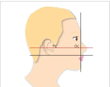
round black backgrounds (Fig 3). Thus the examiner deined the Frankfort’s horizontal plane (Fig 4) in the image and, in case of not being parallel to the ground, correctly oriented the photo in the album on the prop-er position for evaluation.
The two examiners, orthodontists at HRAC-USP, assigned scores based on technical concepts, con-sisting only of qualitative morphological analysis of
the face performed in two assessments and did not receive prior calibration about the photographs of this study. The purpose of the absence of calibration was to make sure that only the individual impres-sions were recorded when it comes to facial growth pattern of the patients. For the irst assessment, the album was delivered to the examiners and they were instructed to evaluate the photographs for 1 minute and diagnose patients according to the growth pat-tern for the frontal view and proile: a) Patpat-tern I, b) Pattern II, c) Pattern III d) Long Facial Pattern e) Short Facial Pattern. At this stage the examiners did not know that they would perform a second eval-uation intended to verify the intra-examiner match-up. A second assessment was done 10 days after the irst, and the same instructions were given.
Pattern I is identiied in a normal face. When there is a malocclusion, it is only dental, not associ-ated with any sagittal or vertical skeletal discrepancy (Fig 5). Patterns II and III (Figs 6 and 7) are charac-terized respectively by the positive and negative sag-ittal balance between maxilla and mandible. In the Long (Fig 8) and Short Facial Patterns (Fig 9) there is
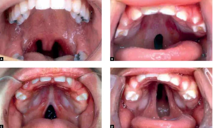
Figure 2 - A) Different extensions of cleft palate: In uvula (incomplete cleft palate); B) different extensions of cleft palate: In the soft palate (incomplete cleft palate); C)different extensions of cleft palate, partial hard palate (incomplete cleft palate); D) different extensions of cleft palate, total hard palate (complete cleft palate).
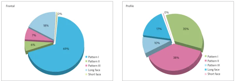
Figure 1 - Gender distribution for total sample.
A B
C D
Total Sample
female
male 49
22 50
40
30
20
10
a vertical discrepancy. In patients with skeletal dis-crepancies, malocclusions are usually consequences of these disharmonies.
On the proi le assessment, the Pattern I is charac-terized by a moderate degree of convexity. The max-illary expression on the face is identii ed by the pres-ence of the zygomatic projection and infraorbital de-pression, which can be verii ed also in the front view. The nasal base line, slightly inclined to the anterior, shows proper maxillary position. The rictus nasoge-niano with a slight posterior inclination, completes the assessment of the maxillary balance. The nasola-bial angle evaluates the nasal base in relation to the upper lip, which position is largely determined by the inclination of upper incisors. Therefore, this angle may be appropriated, open or closed in Pattern I pa-tients, as a consequence of the position of the upper front teeth, regardless the good maxillary position, al-ways observed in these patients.5,20,24
The maxillary balance (size, shape and position) may be verii ed on proi le assessment through the mentocervical angle. It should be expressive with-out being excessive and tend to parallelism with the Camper’s Plane. This parallelism contributes to a proper mentocervical angle. In addition, it is expected an esthetically pleasing mentolabial sulcus and built with equal participation of the lip and chin.5
Patients from patterns II and III present saggital discrepancy between the maxilla and identii ed man-dible, mainly on the lateral face assessment. Individu-als classii ed as Pattern I in front view and in proi le II
or III have a better prognosis when compared to pat-terns II or III in front and side view, in which the un-balance is severe enough to be identii ed in the frontal assessment due to its vertical consequences. Pattern II presents increased facial convexity as a result of maxillary excess, rarer, or by mandibular dei ciency. Generally, it is observed a maxilla with good expres-sion on the face, while the lower third is dei cient with a short mentocervical angle.22
In Pattern III, the facial convexity presents a de-crease,22 resulting in a straight proi le or rarely
con-cave, due to maxillary dei ciency, mandibular progna-thism or the combination of both. The middle facial height tends to look dei cient, even if it is normal, because the mandibular excess dislocate to anterior the soft tissue of the maxilla, masking the zygomatic projection reading.5 The lower face tends to increase,
especially in prognathism, and the mentocervical an-gle looks normal in maxillary dei ciency individuals or excessive in the prognathous.
The patients classii ed as Long and Short Facial Pat-terns have a visible vertical unconformity in the front and proi le evaluations.5 The long facial pattern is
char-acterized by excessive facial height, resulting in the absence of lip sealing, increased facial convexity, weak maxilla expression and short mentocervical angle.7 The
Short Facial Pattern patient is identii ed by disabilities in the vertical dimensions, compressed lips, maxilla with appropriate position and high mentocervical an-gle, due to counterclockwise rotation of the mandible.5
To perform the analysis of inter and intra-examiner
Po’ Or’
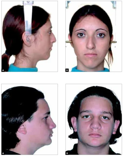
Figure 3 - Photography changed in the computer software, assembled and standardized as described by Bittner and Pancherz.4
Figure 5 - A) Frontal facial photograph of a Pattern I patient with cleft palate; B) facial profile photograph of a Pattern I patient with cleft palate.
A B
Figure 6 - A) Photograph of frontal facial Pat-tern II patients with cleft palate; B) facial pro-file photograph of a Pattern II patient with cleft palate.
A B
A B
A
A
B
B
Figure 8 - A) Frontal facial photograph of a Long Facial Pattern patient with cleft palate. B) Facial profile photograph of a Long Facial Pattern patient with cleft palate.
Figure 9 - A) - Frontal facial photograph of a Short Facial Pattern patient. B) Profile facial photograph of a Short Facial Pattern patient.
data, the Kappa statistic 15 was used, and the conidence
intervals were constructed by the methods proposed by Donner and Eliasziw.13
RESuLtS
The facial morphological analysis is the main diag-nosis tool to determine the Facial Pattern 21 that refers
to treatment protocols and speciic prognostic in dif-ferent age groups.5
This study, aiming to deine the prevalence of dif-ferent patterns for individuals with cleft palate, yielded the values shown in igures 10 and 11, with Kappa values ranging from 0.76 to 0.98 for the frontal analysis and 0.81 to 0.98 for the proile in the intra-examiner assess-ment, with a high level of agreement. Kappa statistics
in assessing inter examiner ranged from 0.45 to 0.79 for the frontal analysis and from 0.45 to 0.79 for the proile, with a rate of moderate to substantial agreement.
dISCuSSIOn
The distribution observed in the frontal aspect was potentially positive regarding the esthetic point of view, since Pattern I individuals were predominant in this analysis (69%), similar to results obtained in studies with patients without cleft, to the diferent patterns, not including Long Face and Short Face, where the pattern I obtained 85% of the sample.23
Figure 10 - Distribution of different patterns in frontal view. Figure 11 - Distribution of different patterns in the profile aspect.
incompatibility was 35% for the total sample, the same value found in patients without cleft.23 This change was
clearly expressed by the migration of individuals classi-ied as frontal Pattern I to the Patterns III and especial-ly II, by the proile classiication. As suggested by Reis,23
it is in this norm — the proile — that the malocclusions of Patterns II and III, anteroposterior dysplasia, are es-sentially expressed. The present work noticed the prev-alence of 38% and 10% respectively in the Patterns II and III in the proile. Thus, following the higher preva-lence of malocclusions in the Pattern II, many individu-als that presented it with a less signiicant magnitude, were not marked in the frontal analysis of the face, be-ing classiied as Pattern I (32%). The same happened, in lower proportions, with Standard III (3%).
For the prevalence of Pattern I individuals, we must consider them only when the classiication is repeated for the front and proile, so the prevalence would be 35%, similar to the projection for the general population of individuals without cleft, considering the characteristics of sample, in which patients with vertical discrepancies were not included.
From the cephalometric point of view, individuals with cleft palate present a retrognathic mature face, however, with an acceptable sagittal relation between the apical bases.2,25 This satisfactory sagittal relation is
followed by a facial growth with a predominant vertical component, due to mandibular structural morphol-ogy, facilitating clockwise rotation, regardless previous therapeutic approaches.1,12,26 The Long Facial Pattern,
vertical discrepancy visible in front and proile6
assess-ments, is an average prevalence deformity. In a recent
study,11 the prevalence was of 14.06% for Brazilians,
be-tween 12 and 15 years old, from this group, 0.68% have a deformity severe enough to justify the indication for an orthodontic surgical treatment. In this study, the fron-tal and proile evaluations had balanced results (18 and 17% respectively), similar to literature.10,14,17
The Short Facial Pattern, due to the low prevalence in the general population, has a negative vertical dis-crepancy, not being found in the sample used, prob-ably due to the predominance of vertical growth in individuals with cleft palate.
COnCLuSIOn
This study found the prevalence of diferent Pat-terns for individuals with cleft palate by qualitative morphological facial analysis, suggesting that:
• The prevalence of several Patterns for patients
with cleft palate was similar to the one of indi-viduals without cleft;
• Regarding the esthetic point of view, the distri
-bution in the frontal aspect was very positive, since Pattern I individuals prevailed;
• Analyzing the proile, the anteroposterior dys
-plasias were expressed in essence, increasing signiicantly their participation;
• The Long Facial Pattern, presenting a vertical
discrepancy visible in the frontal and profile assessments, maintained balanced in both evaluations.
• The Short Facial Pattern was not found in the
sample used, which is probably related to the low prevalence in the general population.
Pattern I Pattern I
0%
18%
7%
6%
69%
Frontal Profile
Pattern II Pattern II
Pattern III Pattern III
Long face Long face
Short face Short face
38% 10%
17% 0%
1. Bishara SE. Cephalometric evaluation of facial growth in operated and non-operated individuals with isolated clefts of the palate. Cleft Palate J. 1973 Jul;10:239-46..
2. Bishara SE. The influence of palatoplasty and cleft length on facial development. Cleft Palate J. 1973 Oct;10:390-8.
3. Bishara SE. Ortodontia. São Paulo: Ed. Santos; 2004.
4. Bittner C, Pancherz H. Facial morphology and malocclusions. Am J Orthod Dentofacial Orthop. 1990 Apr;97(4):308-15.
5. Capelozza Filho L. Diagnóstico em Ortodontia. Maringá (PR): Dental Press; 2004. 6. Capelozza Filho L, Cardoso MA, An TL, Bertoz FA. Características cefalométricas do padrão face longa: considerando o dimorfismo sexual. Rev Dent Press Ortodon Ortop Facial. 2007 Mar-Abr;12(2);49-60.
7. Capelozza Filho L, Cardoso MA, An TL, Lauris JRP. Proposta para classificação, segundo a severidade, dos indivíduos portadores de más oclusões do Padrão Face Longa. Rev Dent Press Ortodon Ortop Facial. 2007 Mar-Abr;12(4);124-58. 8. Capelozza Filho L, Cavassan AO, Silva Filho OG. Avaliação do crescimento craniofacial em portadores de fissura transforame incisivo unilateral: estudo transversal. Rev Bras Cirur. 1987 Mar-Abr;77(2):97-106.
9. Capelozza Filho L, Silva Filho OG. Abordagem interdisciplinar no tratamento das fissuras labiopalatinas. In: Mêlega JC. Cirurgia plástica fundamentos e arte: cirurgia reparadora da cabeça e pescoço. Rio de Janeiro: Medsi; 2002. p. 59-88. 10. Cardoso MA. Epidemiologia do Padrão Face Longa em escolares do ensino
fundamental do município de Bauru – SP [Tese de Doutorado]. Araçatuba (SP): Universidade Estadual Paulista, Faculdade de Odontologia; 2007. 112f. 11. Cardoso MA, Capelozza Filho L, An TL, Lauris JRP. Epidemiologia do Padrão Face
Longa em escolares do Ensino Fundamental do município de Bauru - SP. Dental Press J Orthod. [online]. 2011;16(2):108-19 .
12. Christensen K, Fogh-Andersen P. Isolated cleft palate in Danish multiple births, 1970-1990. Cleft Palate Craniofac J. 1993 Sep;30(5):469-74.
13. Dahl E. Craniofacial morphology in congenital clefts of the lip and palate: an x-ray cephalometric study of young adult males. Acta Odontol Scand. 1970;28:Suppl 57:11. 14. Donner A, Eliasziw M. A goodness-of-fit approach to inference procedures for the
Kappa statistic: confidence interval construction, significance-testing and sample size estimation. Stat Med. 1992 Aug;11(11):1511-9.
15. Fitzpatrick BN. The long face and V. M. E. Aust Orthod J. 1984 Mar;8(3):82-9. 16. Fleiss JL, Levin B, Paik MC. Statistical methods for rates and proportions. 3rd ed.
Hoboken (NJ): John Wiley; 2003.
REFERENCES
17. Jolleys A. A review of the results of operations on cleft palates with reference to maxillary growth and speech functions. Br J Plast Surg. 1954 Oct;7(3):229-41. 18. Linder-Aronson S, Woodside DG. Excess face height malocclusion: etiology,
diagnosis and treatment. Chicago (IL): Quintessence, 2000.
19. Miettinen PJ, Chin JR, Shum L, Slavkin HC, Shuler CF, Derynck R, Werb Z.. Epidermal growth factor receptor function is necessary for normal craniofacial development and palate closure. Nat Genet. 1999 May;22(1):69-73. 20. Piazentin SHA. A influência da palatoplastia primária nas alterações do ouvido
médio [Dissertação]. São Paulo: Pontifícia Universidade Católica de São Paulo; 1989.
21. Reis SAB, Abrão J, Capelozza Filho L, Claro CAA. Análise facial numérica do perfil de brasileiros Padrão I. Rev Dent Press Ortodon Ortop Facial. 2006 Nov-Dez;11(6):24-34.
22. Reis SAB, Abrão J, Capelozza Filho L, Claro CAA. Análise Facial Subjetiva. Rev Dent Press Ortodon Ortop Facial. 2006 Set-Out;11(5):159-72.
23. Reis SAB, Abrão J, Capelozza Filho L, Claro CAA. Estudo comparativo do perfil facial de indivíduos Padrões I, II e III portadores de selamento labial passivo. Rev Dent Press Ortodon Ortop Facial. 2006 Jul-Ago;11(4):36-45.
24. Reis SAB. Análise facial numérica e subjetiva do perfil e análise da relação oclusal sagital de brasileiros, adultos, leucodermas, não tratados ortodonticamente [Dissertação]. São Paulo: UMESP; 2001.
25. Reis SAB, Capelozza Filho L, Cardoso MA, Scanavini MA. Características cefalométricas dos indivíduos padrão I. Rev Dent Press Ortodon Ortop Facial. 2005;10(1):67-78.
26. Shibasaki Y, Ross RB. Facial growth in children with isolated cleft palate. Cleft Palate J. 1969 Jul;6:290-302.
27. da Silva Filho OG, Cavassan AO, Normando ADC. Influência da palatoplastia no padrão facial de pacientes portadores de fissura pós-forame incisivo. Rev Bras Cirur. 1989 Nov-Dez;79(6):315-22.
28. da Silva Filho OG, Cavassan AO, Sampaio LL. Avaliação do padrão cefalométrico em pacientes portadores de fissura pós-forame incisivo, não operado. Rev Bras Cirur. 1989 Maio-Jun;79(3):137-47.
29. da Silva Filho OG, Ferrari Junior, FM, Rocha DL, Souza Freitas JAS. Classificação das fissuras lábio-palatais: breve histórico, considerações clinicas e sugestão de modificação. Rev Bras Cirur. 1992 Mar-Abr;82(2):59-65.