© 2013 Dental Press Journal of Orthodontics 2 Dental Press J Orthod. 2013 Mar-Apr;18(2):2-3 Matheus Melo Pithon1
RAPID MAXILLARY EXPANSION DOES NOT INDUCE SIDE EFFECTS
With computerization of the contemporary world, access to information has become rapid and practical. Following this trend, this has beneited the biomedical ield, because nowadays clinicians do not need to wait for a scientiic meeting to keep up-to-date. However, with the great amount of scientiic information available, what can really be taken into consideration and brought to the daily clinic? With the intention of solving these questions and attempting to separate “the wheat from the chaf”, systematic reviews appear with the goal of “ishing” in an “ocean” of information for the most reliable answer to a certain question. In line with this trend and in seeking answers there is an old, but ever up to date topic: Rapid maxillary expansion. Italian researchers assessed whether this procedure would induce side efects in growing indi-viduals.1 Ater an extensive search of the literature, they arrived to the conclusion that rapid maxillary expansion does not cause injuries in growing individuals. However, the authors open a parenthesis and emphasize that as a result of the low quality of studies found and available, no scientiically based conclusion could be drawn, leav-ing the suggestion of conductleav-ing future researches with greater methodological strictness.
DO NOT EXAGGERATE WHEN ASKING YOUR PA-TIENTS FOR RADIOGRAPHS AND TOMOGRAPHS Röentgen undoubtedly was one of the greatest ge-niuses of humanity, with his brilliant discovery (the “X-ray”), everything that could not be seen with the naked eye became visible. Since it is not all sweetness and light, in order to achieve this feat, it is necessary for the individual to be exposed to radiation and its known harmful efects. However, the orthodontist who follows-up scientiic de-velopment and stays alert to new technologies, could think,
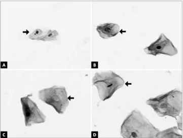
“I use cone beam computerized tomography which, in a single shot, acquires all the images”. Therefore, we ask: With the advent of tomographs, has there been an end to problems with exposure to radiation? What would be the real risks from the point of view of cellular changes in patients submitted to this new technology and to conven-tional radiographic exams? To elucidate these questions, Brazilian researchers proposed to evaluate the behavior of oral cavity cells submitted to protocols of conventional and tomographic orthodontic documentation.2 The results found (Fig 1) demonstrated cytotoxicity in cells of the oral cavity submitted to conventional radiographs and cone beam computerized tomography. A larger number of cell deaths were found in the group in which computerized tomography was used. The authors concluded the article brilliantly, pointing out that orthodontic documentation must be done only when necessary, thereby avoiding un-necessary radiographic and tomographic exposure.
Figure 1 - Evaluation of the nuclear alterations in the cone beam com-puterized tomography sequence (CBCT) or radiographic exposure (400x magnification, Feulgen/Fast Green stain): cells (A) micronucleated (ar-row) and normal, (B) karyorrhexis (arrow), karyolysis (C) (arrow), and (D) pyknosis (arrow). Source: Lorenzoni et al,2 2013.
How to cite this article: Pithon MM. Orthodontics highlights. Dental Press J Orthod. 2013 Mar-Apr;18(2):2-3.
Submitted: February 20, 2013 - Revised and accepted: February 20, 2013
Contact address: Matheus Melo Pithon E-mail: [email protected]
1 Professor of Orthodontics, State University of Southwest Bahia. MSc and PhD
in Orthodontics, UERJ. Diplomate by the Brazilian Board of Orthodontics and Facial Orthopedics.
orthodontics
highlights
A
C
B
© 2013 Dental Press Journal of Orthodontics 3 Dental Press J Orthod. 2013 Mar-Apr;18(2):2-3 orthodontics highlights
Pithon MM
© 2013 Dental Press Journal of Orthodontics 3
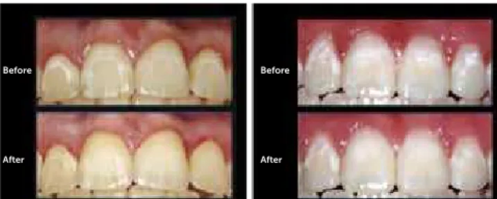
Figure 2 - Examples of photos before and after. Source: Huang et al,3 2013.
GOOD TOOTHBRUSHING! NOTHING BETTER FOR PREVENTING WHITE SPOT LESIONS IN PATIENTS USING FIXED ORTHODONTIC APPLIANCE
Much has been developed and commercialized with the aim at improving and even promising to put an end to white spot lesions in patients wearing fixed orthodontic appliances. Mouth wash solutions, rem-ineralizing toothpastes, chewing gums, fluoridated varnishes, among others are used for these purpos-es. In this context, a pertinent question arispurpos-es. Are these products really effective in the prevention and improvement of white spot lesions? In seeking an answer to this question, American researchers at the University of Washington conducted a randomized controlled clinical study3 that evaluated the effica-cy of a remineralizing toothpaste and a fluoridated varnish, in comparison with normal brushing with regard to improving white spot lesions. The results attained (Fig 2) refuted the manufacturer’s claims, since the authors reached the conclusion that good toothbrushing has the same benefit as the one ob-tained with the use of these substances.
IT IS POSSIBLE TO OPTIMIZE BONDING OF FIXED RETAINERS
We all know about the importance of orthodon-tic retention after the removal of fixed orthodonorthodon-tic appliances. There are various methods described in the literature for retention, such as movable and fixed retainers, bonded directly to the teeth with orth-odontic composites. As their use does not need pa-tient’s cooperation, fixed retainers have been shown to be more effective. But how does one optimize the adaptation procedure of this device, in view of the technical difficulty of direct adaptation because of its lingual location? One of the methods clinicians use to optimize bonding is to perform it without the ad-dition of adhesive resin after acid etching. But what would be the clinical repercussion of this procedure? Swedish, Chinese and Danish researchers conducted a clinical study in which part of the patients had their fixed retainers bonded conventionally with the addi-tion of fluid resin, and another group without
per-forming this stage.4 The results found demonstrated
that clinically, the addition of fluid resin did not in-fluence the longevity of retainer bars. So here is this clinical hint for speeding up your appointments.
1. Lione R, Franchi L, Cozza P. Does rapid maxillary expansion induce adverse efects in growing subjects? Angle Orthod. 2013;83(1):172-82. 2. Lorenzoni DC, Fracalossi AC, Carlin V, Ribeiro DA, Sant’Anna EF.
Mutagenicity and cytotoxicity in patients submitted to ionizing radiation. Angle Orthod. 2013;83(1):104-9.
3. Huang GJ, Rolof-Chiang B, Mills BE, Shalchi S, Spiekerman C, Korpak
AM, et al. Efectiveness of MI Paste Plus and PreviDent luoride varnish for treatment of white spot lesions: a randomized controlled trial. Am J Orthod Dentofacial Orthop. 2013;143(1):31-41.
4. Tang AT, Forsberg CM, Andlin-Sobocki A, Ekstrand J, Hagg U. Lingual
retainers bonded without liquid resin: a 5-year follow-up study. Am J Orthod Dentofacial Orthop. 2013;143(1):101-4.
5. Long H, Pyakurel U, Wang Y, Liao L, Zhou Y, Lai W. Interventions for accelerating orthodontic tooth movement. Angle Orthod. 2013;83(1):164-71.
REFERENCES
ACCELERATION OF TOOTH MOVEMENT, IS IT POSSIBLE?
Without a shadow of doubt, reduction in treatment time is the orthodontists’ dream and patients’ wish. But in the light of present knowledge, how can we accelerate tooth movement and consequently diminish treatment time? There are various mechanisms described in the lit-erature, said to be accelerators of tooth movement. Among these: Application of laser, electromagnetic ields, pulsed currents, invasive procedures such as dentoalveolar and periodontal distraction and corticotomies. In an endeavor to elucidate which of the methods would be efective and safe for accelerating orthodontic movement, Chinese re-searchers developed a systematic review.5 The authors con-cluded that low level laser therapy is safe, but incapable of accelerating orthodontic movement, whereas corticotomy is safe and capable of accelerating tooth movement. The other methods need further well designed studies in order to conirm their results. Therefore, in the light of scientiic evidence, when a patient requests an accelerated treatment, corticotomy appears to be the option available.
Before Before