Occlusal assessment in surgically assisted
unilateral cleft lip and palate patients
Leanne Matias Portela Leal1, Marcus Vinicius Neiva Nunes do Rego2, Cosme José Albergaria da Silva Filho3, Leopoldino Capelozza Filho4, Mauricio de Almeida Cardoso5
Objective:The aim of this study was to assess the magnitude of occlusal changes in individuals with unilateral clet lip and palate (CLP). The study was conducted on study casts of 25 subjects, 14 men and 11 women aged from 7 to 20 years, without previous orthodontic treatment and with surgical repair carried out at São Marcos Hospital, Teresina, Piauí State, Brazil. Methods: The casts were assessed by three orthodontists based on the occlusal scores established by Atack et al., whose scores range from 1 to 5, according to the magnitude of transverse and sagittal changes. Re-sults: Intra and inter-observer reproducibility of occlusal scores was satisfactory and statistically signiicant according to the Spearman Correlation test with signiicance level set at 5%. With regard to the distribution of occlusal scores, 30.67% of the subjects achieved scores 1 and 2, 22% score 3 and 47.53% achieved scores 4 and 5. Conclusions: Four was the score most frequently assigned by the observers, disclosing a high degree of transverse and sagittal disorders in the occlusion of patients.
Keywords:Growth. Malocclusion. Clet lip.
How to cite this article: Leal LMP, Rego MVNN, Silva Filho CJA, Capelozza Filho L, Cardoso MA. Occlusal assessment in surgically assisted unilateral clet lip and palate patients. Dental Press J Orthod. 2013 July-Aug;18(4):120-5.
Submitted: May 18, 2011 - Revised and accepted: February 15, 2012 » The authors report no commercial, proprietary or inancial interest in the prod-ucts or companies described in this article.
Contact address: Mauricio de Almeida Cardoso
Rua Arnaldo de Jesus Carvalho Munhoz, 6-100 – Bauru/SP, Brazil CEP: 17018-520 – E-mail: [email protected]
1 Masters student in Orthodontics, Sacred Heart University (USC).
2 Professor of Orthodontics at the Graduate and Postgraduate programs,
UNINOVAFAPI. Professor of Orthodontics at the Postgraduate program, Federal University of Piauí (UFPI).
3 Graduated in Dentistry, UNINOVAFAPI.
4 PhD in Orthodontics, College of Dentistry — Bauru/USP. Professor of
Orthodontics at the Graduate and Postgraduate (specialization and Masters courses) programs, USC.
5 PhD in Orthodontics, State University of São Paulo (UNESP). Professor of
Orthodontics at the Graduate and Postgraduate (specialization and Masters courses) programs, USC.
Objetivo:o propósito do presente estudo foi avaliar a magnitude das alterações oclusais em 25 indivíduos com issura transforame incisivo unilateral, sendo 14 do sexo masculino e 11 do sexo feminino, com idades entre 7 e 20 anos, não tratados ortodonticamente e operados no Hospital São Marcos, na cidade de Teresina/PI. Métodos: os modelos de estudo desses pacientes foram avaliados por três ortodontistas, utilizando-se como referência os índices oclusais de Atack et al., cujos escores variam de 1 a 5, de acordo com a magnitude das alterações transversais e anteroposteriores. Resultados: os níveis de coniabilidade e reprodutibilidade do índice oclusal intraexaminador e interexaminado-res, respectivamente, mostraram-se bastante satisfatórios e estatisticamente signiicativos quando aplicado o teste de correlação de Spearman, com intervalo de coniança de 5%. Quanto à distribuição dos índices oclusais, 30,67% dos indivíduos apresentaram os índices 1 e 2; 22% o índice 3; e 47,53% os índices 4 e 5. Conclusão: o índice oclusal 4 foi o mais referido, de acordo com todos os escores atribuídos pelos examinadores, evidenciando um acentuado grau de alterações transversais e anteroposteriores na oclusão dos pacientes.
INTRODUCTION
Individuals with cleft lip and palate exhibit func-tional and morphological changes and may, in some cases, manifest psychosocial changes given that the affected region is highly visible, causing a negative esthetic impact. As for their morphological diversi-ty, lip and palate clefts yield different levels of sever-ity and consequences, requiring not only changes in treatment protocol, but also an interdisciplinary team composed of an orthodontist, pediatrician, plastic surgeon, speech pathologist, psychologist, geneticist and social worker, to promote adequate esthetic and functional rehabilitation as well as seamless social and psychological integration to patients.6
In Brazil, clefts are currently classified on the ba-sis of a system proposed by Spina,18 which uses the
incisive foramen, the only single structure connect-ing the primary and secondary palates durconnect-ing intra-uterine life, as anatomical reference. These palates ultimately formed the midface. Thus, this classifica-tion defines clefts based on length, in line with their embryonic origin.17 According to this classification,
clefts are divided into four different groups: Group I = Pre-foramen clefts; Group II = Trans-foramen inci-sor clefts; Group III = Post-foramen inciinci-sor clefts; and Group IV = Rare facial clefts.14
Some treatments should be provided as soon as patients with this anomaly are born in order to im-prove the quality of life of these individuals. Soon after the third month of life, the lip should be re-constructed (cheiloplasty) and in the twelfth month, the palate should be reconstructed (palatoplasty). These surgeries are called primary surgeries as they are aimed at restoring the anatomic integrity left un-finished in intrauterine life. If necessary, secondary surgeries are performed to close the fistulas, as well as pharyngoplasty while finishing touches are ap-plied to the primary surgeries.
The craniofacial growth of individuals with uni-lateral cleft lip and palate (CLP) should be constantly monitored due to the adverse effects of primary sur-geries on the anteroposterior and transverse growth of the maxilla. Some changes in the occlusion are fre-quently found, especially anterior crossbite and pos-terior crossbite.12
The impact of primary plastic surgery on the mid-face growth makes it important to study the
magni-tude of occlusal changes undergone by patients with unilateral cleft lip and palate (CLP) subjected to sur-gery at São Marcos Hospital in Teresina, Piauí State (PI), Brazil, an institution recognized by the Ministry of Health as a reference center in the Northeastern region of Brazil.
MATERIAL AND METHODS
Sample characterization
The NOVAFAPI Institutional Review Board (CEP/NOVAFAPI) reviewed this research project and found that it met the provisions of resolution No. 196/96, issued by the National Council of Health (CNS/MS). Therefore, the project was authorized and filed under number 0430-08.
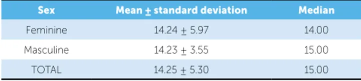
The sample comprised 25 individuals aged from 7 to 20 years old, with unilateral CLP, subjected to primary plastic surgery at the São Marcos Hospital in the city of Teresina, Piauí State, Brazil. Out of 25 patients who participated in this study, 14 (56%) were men while 11 (44%) women. Patients’ age range was 14 years and 3 months (Table 1). According to the number of primary plastic surgeries and the time when they were performed, the first lip repair was carried out by means of the Millard’s technique, on average, when the patient was 7 months old; whereas
Sex Mean ± standard deviation Median
Feminine 14.24 ± 5.97 14.00
Masculine 14.23 ± 3.55 15.00
TOTAL 14.25 ± 5.30 15.00
Table 1 - Mean, median and standard deviation of ages (in years) of patients with unilateral CLP who underwent surgery at São Marcos Hospital. Data displayed according to sex.
Table 2 - Distribution of the 25 patients with unilateral CLP who underwent surgery at São Marcos Hospital. Data displayed according to the number of surgeries and the time when the irst plastic surgeries were performed (in months).
Surgery Mean ± standard deviation Median
Number of procedures
cheiloplasty 1.29 ± 0.62 1.00
palatoplasty 1.29 ± 0.62 1.00
Age (months)
1st cheiloplasty 7.63 ± 8.24 3.00
palatoplasty was performed at 30 months of age, us-ing a modified version of Von Langenback’s tech-nique (Table 2).
The following inclusion criteria were applied: Plastic surgeries performed at São Marcos Hospital; cheiloplasty (lip surgery) before 2 years of age; pala-toplasty (surgery of the palate) before 3 years of age; no prior orthopedic treatment performed to achieve expansion and/or reverse traction of the maxilla; no syndrome in the craniofacial region; no alveolar bone graft in the cleft area.
Assessment of occlusal characteristics in the study casts
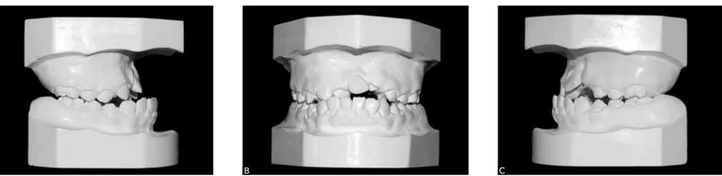
The occlusal characteristics of the 25 patients with unilateral CLP were evaluated on study casts (Fig 1) by 3 examiners (orthodontists and professors at the
NOVA-FARI Specialization Course in Orthodontics) using the occlusal index proposed by Atack et al.2 (Table 3).
The casts were carefully arranged in ascending order on a bench concealing the patients’ personal identiica-tion data. This evaluaidentiica-tion was performed individually with no communication between examiners, who were previously calibrated for application of the occlusal in-dex, while changing the numbering on the casts in both assessments to prevent memorization.
In order to verify method reliability, all casts were reassessed 15 days after the first evaluation.8
Statistical analysis
Method reliability
Evaluation results were exported to a database cre-ated with SPSS software, version 13.0. Inter-exam-iner correlation was determined in both assessments
Table 3 - Evaluation index of the occlusal characteristics using casts (inter-arch relationship) in individuals with unilateral CLP.3
Figure 1 - Study casts of an individual from the sample used in this study, showing unilateral CLP. A) Right lateral view. B) Frontal view. C) Left lateral view.
Group Occlusal characteristics Long-term outcome
prognosis
1
– Positive overjet with normal inclination or lingual inclination of the incisors; – Absence of crossbite and open bite;
– Satisfactory morphology of the upper dental arch
Excellent
2
– Positive overjet with normal inclination or labial inclination of the incisors; – Unilateral crossbite / tending towards crossbite;
– Tendency towards open bite adjacent to the cleft
Good
3
– End-on relationship with normal inclination or labial inclination of the incisors, or negative overjet with incisors inclined lingually;
– Tendency towards open bite adjacent to the cleft
Regular
4
– Negative overjet with normal inclination or labial inclination of the incisors; – Tendency towards unilateral/bilateral crossbite;
– Tendency towards open bite adjacent to the cleft
Bad
5
– Negative overjet with labial incisor inclination
– Bilateral crossbite;
– Substantially changed upper jaw morphology
Very bad
RESULTS
Intra and inter-observer agreement in determining the occlusal index
Table 5 depicts a correlation between the first and second scores assigned to each observer (p), and shows whether the degree of agreement is acceptable. A statistically significant correlation (R) was found for all examiners.
As regards inter-observer correlation, the degree of agreement ranged between “good” and “very good” (Table 6). The good repeatability accom-plished in the test was expected because all examiners were orthodontists and the index designed by Atack et al.3 proved to be easy to apply.
Magnitude of occlusal changes exhibited by the patients with CLP
The results showed that 30.67% of patients were included in indices 1 and 2, 22% in index 3, and 47.33% were included in indices 4 and 5 (Fig 2). (initial and after 15 days), as well as intraexaminer
correlation with the purpose of verifying method re-liability using Spearman’s correlation test with a sig-nificance level set at 5% (p < 0.05).
To define the degree of agreement, the scale pro-posed by Altman2 was applied to the values shown in
Table 4. Only “good” and “very good” scores were considered satisfactory.
Spearman correlation. N = 25, *signiicant at p < 0.05.
Captions for Level of Agreement: P - Poor, M - Mild, R - Regular, G - Good, VG - Very good.
Table 5 - Intraobserver agreement in determining the occlusal index in the 25 patients with unilateral CLP who underwent surgery at São Marcos Hospital. Teresina, Piauí State, Brazil.
Examiner Correlation
P R LA
Orthodontist 1 0.000* 0.896 VG
Orthodontist 2 0.000* 0.627 G
Orthodontist 3 0.000* 0.917 VG
Spearman correlation. N = 25, *signiicant at p < 0.05. LA (Level of Agreement): P - Poor, M - Mild, R - Regular, BG- Good, VG - Very good. P column= correlation between the irst and second score assigned to each observer. R column = correlation for all examiners.
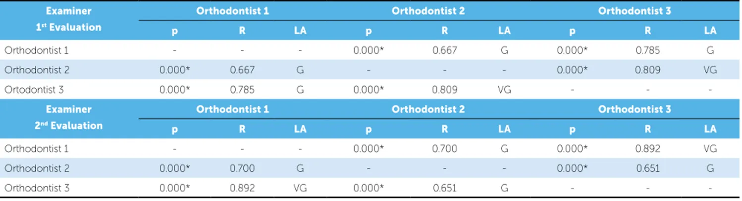
Table 6 - Inter-observer agreement in the irst and second evaluations in determining the occlusal index in the 25 patients with unilateral CLP who underwent surgery at São Marcos Hospital.
Examiner
1st Evaluation
Orthodontist 1 Orthodontist 2 Orthodontist 3
p R LA p R LA p R LA
Orthodontist 1 - - - 0.000* 0.667 G 0.000* 0.785 G
Orthodontist 2 0.000* 0.667 G - - - 0.000* 0.809 VG
Ortodontist 3 0.000* 0.785 G 0.000* 0.809 VG - -
-Examiner 2nd Evaluation
Orthodontist 1 Orthodontist 2 Orthodontist 3
p R LA p R LA p R LA
Orthodontist 1 - - - 0.000* 0.700 G 0.000* 0.892 VG
Orthodontist 2 0.000* 0.700 G - - - 0.000* 0.651 G
Orthodontist 3 0.000* 0.892 VG 0.000* 0.651 G - -
-Table 4 - Relevance of the level of agreement as described by Altman.2
Value Level of agreement (LA)
≤ 0.2 Poor
0.21 to 0.40 Regular
0.41 to 0.60 Mild
0.61 to 0.80 Good
0.81 to 1.00 Very good
Figure 2 - Distribution of occlusal indices in the 25 patients with unilateral CLP who underwent surgery at São Marcos Hospital.
Indices 1 - 2
22% 47.33%
30.67%
Index 3
Indices 4 - 5
Choosing a straightforward occlusal index is of par-amount importance since it interferes in method reli-ability and in determining the number of examiners. The results of this study agree with those of Okada,11
who also found the occlusal index of Atack et al.3 to be
straightforward and easy to apply, since there was no statistically signiicant intraobserver diferences, even when diferent groups of examiners were used, such as oral maxillofacial surgeons, plastic surgeons and or-thodontists. Therefore, the ease with which the occlu-sal assessment method was applied precluded the need for a greater number of examiners.
The quality of a given rehabilitation protocol used by a reference center in treating patients with cleft lip and palate can be measured by the magnitude of oc-clusal changes that occur, especially in patients with CLP, since sequelae inherent to the morphology of the clefts as a result of the surgical repair procedure, involve significant changes in the transverse and an-teroposterior growth of the maxilla. Results showed that cases considered “very bad” (indices 4 and 5) accounted for almost half of the sample, confirming the negative effect of primary plastic surgery in the growth of midface structures, for which there seems to be consensus in the literature.1,5,7,9-12
On assessing the magnitude of occlusal alterations observed in patients rehabilitated at São Marcos Hos-pital (Teresina, Piauí State), this magnitude was found to be close to results achieved by reference centers both in Brazil and abroad - such as the Hospital for Reha-bilitation of Craniofacial Anomalies, Bauru, São Paulo State, and the Rehabilitation Center of Oslo, Norway. The indices found in this study were compared to those obtained by Okada,11 which were assessed by the Bauru
and Oslo centers. For indices 1 and 2, the data showed a percentage of 30.67% in Teresina, 34% in Bauru and 60% in Oslo. As for index 3, the percentage was 22% in Teresina, 27.72% in Bauru and 22% in Oslo. For indi-ces 4 and 5 (which represent the major occlusal sequelae and comprise the cases requiring orthognathic surgery) the percentages were 47.33% in Teresina, 38.20% in Bauru and 18% in Oslo. Thus, it can be asserted that the Oslo Center results were higher than those of the Brazilian centers, especially when comparing the per-centage of cases with greater occlusal sequelae.
With regard to the average occlusal index based on all scores assigned by examiners in both assessments,
DISCUSSION
Lip and palate clefts are a congenital defect with diverse clinical manifestations. Its involvement can range from a small notch in the lip vermilion or mu-cosa of the uvula to a complete breakdown of the maxilla. In summary, from an embryonic point of view, clefts can affect the primary and secondary palate with different levels of severity. Given their higher prevalence, this study aimed at assessing the behavior of post-surgical complete unilateral clefts involving the primary and secondary palate, also known as unilateral cleft lip and palate (CLPs). Cor-roborating the literature,3 this study also showed a
higher prevalence of unilateral CLP in males. According to surveys conducted at the Hospital for Rehabilitation of Craniofacial Anomalies (HRAC-USP), these clets account for approximately 33% of occurrences.16 Among unilateral CLPs, there are
nu-merous variables that inluence their anatomical con-iguration and diferentiate one patient from another, such as clet width, the presence of Simonart’s band and degree of nose asymmetry. Simonart’s band is formed by a bridge of sot tissue that joins the mesi-al and distmesi-al edges of a complete lip and pmesi-alate clet, which can positively afect the morphology of postop-erative upper dental arches when the band is bulky.11
Primary plastic surgery reconstructs lesions in the lip and palate and in so doing influences the behavior of the nasomaxillary complex, of which expression of genetic growth potential is restricted. The more numerous the morphological variables inherent in clefts are, the greater will be the number of therapeu-tic variables that may influence the patient’s face and occlusion in the long-term. Among these variables, one could mention the amount of time required to complete surgery, surgical techniques, number of repetitive surgeries as well as the extra-surgical care provided pre- and postoperatively. Undoubtedly, the surgeon’s skill in manipulating young tissue plays a key role in ultimately shaping the face.11,13,14
there was an index of 3.28 in Teresina, 3.07 in Bauru and 2.52 in the Oslo Center. In examining the medi-an scores assigned by examiners in the present study, index 4 proved to be the most frequent, indicating a high degree of transverse and anteroposterior changes in the occlusion of the patients.
How can one explain Oslo’s higher results in terms of occlusion conditions? Many variables certainly play a role in the diversity of the outcomes, but according to Shaw et al.13 one major factor is the standardization
of the protocol and the surgical techniques adopted, along with a small number of surgeons, with surgeries being performed by virtually one single surgeon.
When the results obtained in Teresina were com-pared to those of Bauru, a higher percentage of cases with greater occlusal sequelae were found in Tere-sina. However, age is yet another key factor to be taken into account when interpreting these results. The vast majority of patients in this study were at the permanent dentition stage, whereas those assessed by Okada11 in Bauru were at the early mixed
den-tition stage. Given that plastic surgery cumulatively affects the growth of the midface, patients at the stage of occlusal maturity tend to produce more sequelae in transverse and anteroposterior maxillary growth
compared to younger patients. It is worth noting that variables such as the anatomical extent of the cleft, the presence of Simonart’s band, surgical procedure variables such as the surgical technique, surgeon skills and pre and postoperative care were not assessed.
The indings of this study conirmed the assump-tion that the maxillomandibular relaassump-tionship of a patient with clet lip and palate undergoes — starting from the stage of embryo formation — the strains imposed by the clet and the resulting functional deviations. Fur-thermore, maxillomandibular relationship is potentially undermined by reconstructive surgeries (lip repair and palatoplasty), which hinder maxillary growth to varying degrees, resulting in changes in craniofacial growth and deiciencies in the intra and inter-arch relationships.
CONCLUSIONS
» With regard to the magnitude of occlusal changes, it was found that 30.67% of the subjects exhibited indices 1 and 2, 22% index 3 and 47.53% exhib-ited indices 4 and 5.
» On examining the scores assigned by examiners, occlusal index 4 proved to be the most frequent, indicating a high degree of transverse and antero-posterior changes in the occlusion of the patients.
1. Aiello CA, Silva Filho OG, Freitas JAS. Fissuras labiopalatais: uma visão contemporânea do processo reabilitador. In: Mugayar LRF, colaboradores. Pacientes portadores de necessidades especiais. Manual de odontologia e saúde bucal. São Paulo: Pancast, 2000. p. 111-39.
2. Altman DG. Practical statistics for medical research. p. 403-409. London: Chapman & Hall; 1991.
3. Atack N, Hathorn IS, Semb G, Dowell T, Sandy JR. A new index for assessing surgical outcome in unilateral cleft lip and palate subjects aged ive: reproducibility and validity. Cleft Palate Craniofac J. 1997;34(3):242-6. 4. Capelozza Filho L, Cavassan AO, Silva Filho OG. Avaliação do crescimento
craniofacial em portadores de issuras transforame incisivo unilateral. Estudo transversal. Rev Bras Cirur. 1987;77(2):97-106.
5. Cavassan AO, Silva Filho OG. Abordagem ortodôntica. In: Trindade IEK, Silva Filho OG. Fissuras labiopalatinas: uma abordagem interdisciplinar. São Paulo: Ed. Santos; 2007. cap. 4, p. 213-38.
6. Correia JP, Carvalho LRR, Rego MVNN. Fissuras labiais. In: Carreirão S. Cirurgia plástica. São Paulo: Atheneu; 2005. p. 220-30.
7. Faraj JORA, André M. Alterações dimensionais transversas do arco dentário com issuras labiopalatinas, no estágio de dentadura decídua. Rev Dental Press Ortod Ortop Facial. 2007;12(5):100-8.
8. Gravely JF, Benzies PM. The clinical signiicance of tracing error in cephalometry. Br J Orthod. 1974;1(3):95-101.
9. Houston WJ. The analysis of errors in orthodontic measurements. Am J Orthod. 1983;83(5):382-90.
10. Mazzottini R. Variações nas dimensões do arco dentário superior em issurados unilaterais, em função da época do tratamento cirúrgico [tese]. Bauru (SP): Universidade de São Paulo; 1985.
REFERENCES
11. Okada TO. Avaliação dos efeitos da queiloplastia e palatoplastia primária sobre o crescimento dos arcos dentários de crianças com issura transforame incisivo unilateral dos 5-6 anos de idade [tese]. Araraquara (SP): Universidade Estadual Paulista; 2001; 205 p.
12. Ribeiro AA, Leal L, Thuin R. Análise morfológica dos issurados de lábio e palato do centro de tratamento de anomalias craniofaciais do Estado do Rio de Janeiro. Rev Dental Press Ortod Ortop Facial. 2007;12(5):109-18. 13. Mars M, Asher-McDade C, Battström V, Dahl E, McWilliam J, Molsted K, et
al. A six-centre international study of treatment outcome in patients with clefts of the lip and palate: Part 3. Dental arch relationships. Cleft Palate Craniofac J. 1992;29:405-408.
14. Silva Filho OG. Crescimento facial. In: Trindade IEK, Silva Filho OG. Fissuras labiopalatinas: uma abordagem interdisciplinar. São Paulo: Ed. Santos; 2007. p. 173-98.
15. Silva Filho OG, Freitas JAS. Caracterização morfológica e origem embrionária. In: Trindade IEK, Silva Filho OG. Fissuras labopalatinas: uma abordagem interdisciplinar. São Paulo: Ed. Santos; 2007. p. 17-49. 16. Silva Filho OG, Freitas JAS, Okada T. Fissuras labiopalatais: diagnóstico
e uma ilosoia interdisciplinar de tratamento. In: Pinto VG. Saúde bucal coletiva. 4a ed. São Paulo: Ed. Santos; 2000. p. 481-527.
17. Silva Filho OG, Ferrari Júnior FM, Rocha DL, Freitas JAS. Classiicação das issuras labiopalatais: breve histórico, considerações clínicas e sugestão de modiicação. Rev Bras Cir. 1992;82(2):59-65.