83
IMAGES IN NEUROLOGY
DOI:
10.1590/0004-282X20130206
Brainstem reversible leukoencephalopathy
syndrome
Síndrome da leucoencefalopatia reversível de tronco cerebral
Vera L. Braatz, Paulo José Lorenzoni, Cláudia S. K. Kay, Domingos C. Chula, Rosana H. Scola,
Lineu C. Werneck
A 39-year-old man presented with progressive
hea-dache, nauseas, blurriness and cortical blindness. Blood
pressure: 230x140 mmHg. Glasgow score: 15.
Fundos-copy revealed hypertensive retinopathy and papille
-
de ma. Neurological examination was normal. Brain ma
g-netic resonance imaging revealed area of swelling and high
signal on FLAIR-weighted images on the brainstem and
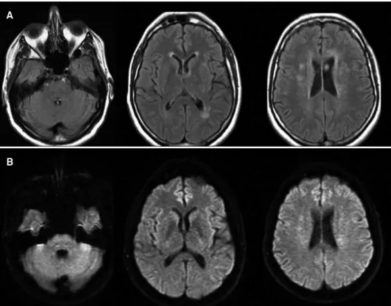
cerebral whitematter abnormalities (Figure 1). Hyperten
-sion management improved his clinical and radiological
indings (Figure 2).
Posterior reversible leukoencephalopathy syndrome (PRES),
with classical reversible cerebral vasogenic edema, occurred
predominantly in the posterior distribution (occipital and
pa-rietal lobes) on brain imaging
1-3. he brainstem involvement
is an atypical feature which may be encountered in midbrain
(13%), pons (20%) and medulla oblongata (5%)
1-3. his PRES
variant should be diferentiated from brainstem infarction,
pontine glioma, infective encephalitis, central pontine
mye-linolysis and others demyelinating disorders because PRES is
potentially full reversible after treatment
1-3.
Divisão de Neurologia, Departamento de Clínica Médica,, Hospital de Clínicas, Universidade Federal do Paraná, Curitiba PR, Brazil.
Correspondence: Rosana Herminia Scola; Serviço de Doenças Neuromusculares; Hospital de Clínicas, Universidade Federal do Paraná; Rua General Carneiro 181 / 3o andar;
80060-900 Curitiba PR - Brasil; E-mail: [email protected] Conflict of interest: There is no conflict of interest to declare.
Received 05 February 2013; Received in final form 11 July 2013; Accepted 18 July 2013.
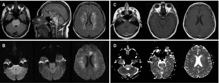
Figure 1.
Initial brain magnetic resonance imaging revealed area of swelling and high signal on the brainstem (mainly in pons
and medulla oblongata) and cerebral white-matter abnormalities, which are compatible with the brainstem variant of posterior
reversible leukoencephalopathy syndrome (A, FLAIR-weighted images; B, diffusion-weighted images; C, T1-weighted images after
contrast infusion; D, apparent diffusion coefficient).
A
B
C
84
Arq Neuropsiquiatr 2014;72(1):83-841. Kitaguchi H, Tomimoto H, Miki Y, et al. A brainstem variant of reversible posterior leukoencephalopathy syndrome. Neuroradiology 2005;47:652-656.
2. Liman TG, Bohner G, Heuschmann PU, Endres M, Siebert E. The clinical and radiological spectrum of posterior reversible
References
encephalopathy syndrome: the retrospective Berlin PRES study. J Neurol 2012;259:155-164.
3. Stevens CJ, Heran MK. The many faces of posterior reversible encephalopathy syndrome. Br J Radiol 2012;85:1566-1575.