971
Arq Neuropsiquiatr 2010;68(6):971-973
Letter
Glial microtumor with 17-year
of postoperative follow up
José Alberto Gonçalves da Silva1, Maria Desterro Leiros da Costa2 , Luiz Ricardo Santiago Melo1, Antônio Fernandes de Araújo1, Jurandy Lins de Araújo3, Vamberto Augusto Costa Filho4, Adailton Arcanjo dos Santos Junior5
Correspondence
José Alberto Gonçalves da Silva Av. Minas Gerais 1150 58030-092 João Pessoa PB - Brasil E-mail: [email protected]
Received 30 August 2009
Received in final form 13 November 2009 Accepted 23 November 2009
PEQUENO GLIOMA DE BAIXO GRAU COM 17 ANOS DE SEGUIMENTO PÓS-OPERATÓRIO
1Neurosurgical Unit of the Santa Isabel Hospital, João Pessoa PB, Brazil; 2Neurologist, Head of the Department of Movement
Disorders of the Federal University of Paraíba; 3Neuroanesthesiologist; 4Radiologist, Head of the CEDRUL Radiological Clinic
of Paraíba; 5Associated Physician.
Reports about biological behavior of gliomas most commonly describe the lat-est stages of their natural course1-3.
How-ever, studies about the earliest stages of human astrocytomas are rare. Nishio et al.1 stated that 1% to 2% of necropsies may
contain some type of undiagnosed glioma. Identiication of microscopic foci of astro-cytic cells at the primary phase of a glioma is extremely rare. Tamura et al.3 reported
that whereas the majority of small gliomas tends to be benign and malignant tumors invade neighboring tissues early on their
development. Rubinstein4 reported that
the most primitive cells of the reticular sys-tem found in the meninges and perivascu-lar sheaths of cerebral vessels, along with the microglia, originate the reticular cell sarcoma (microglioma group). However,
Amacher et al.5 believe that the
congen-ital cerebellar medulloblastomas which occur in adults’ stem are originated from the remnants of primitive cells located in the fetal external granular layer. hese au-thors observed the presence of microscop-ic medulloblastoma on necropsy exam in a neonate with 8 hours of life, in which neo-plastic cells were found within the exter-nal granular layer, invading the perivascu-lar spaces and internal granuperivascu-lar layers.
he present report is based on the rar-ity of benign small low grade glioma hav-ing 17 years’ postoperative follow up, no further focal convulsive crisis since sur-gical removal, and no use of antiepilep-tic drugs.
CASE
An 18-year old male patient report-ed a history of epileptic crisis on January 14th 1991, when started tonic-clonic
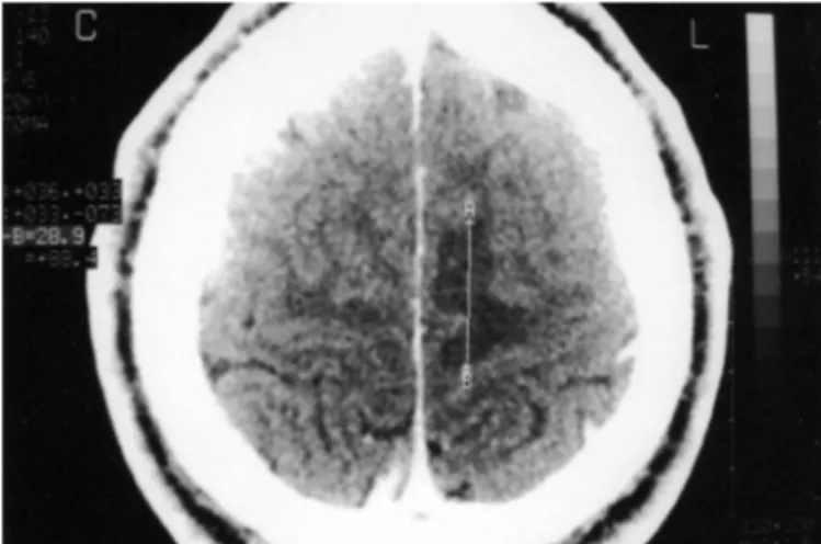
move-ments in the right foot which rose to the homolateral side including the hemiface, and was followed by transient paralysis of this side. On the following day, the patient presented another crisis having the same clinical characteristics as the irst. Brain computed tomography scan (CT) revealed a parasagital tumoral lesion in the superi-or frontal gyrus measuring 8 mm × 5 mm (Figs 1 and 2). Cerebrospinal luid (CSF) exam was negative for both neurocystic-ercosis and neuroschistosomiasis. It was prescribed to the patient Carbamazepine 200 mg twice a day and instructed to re-turn for follow up in 6 months. On Au-gust 30th 1991 the patient had 4 successive
epileptic crisis with the same features as previously outlined. A repeat CT was
per-formed on September 10th 1991 which
re-vealed the same features as the first CT scan. Left frontoparietal craniotomy was
performed on September 12th 1991 with
Arq Neuropsiquiatr 2010;68(6)
972
Glial microtumor Silva et al.
was performed on June 10th 2008 and revealed normal
neurological exam and no convulsive crisis and with no antiepileptic drugs were used throughout the follow up
period. he latest cranial CT was performed on June 12th
2008 and it did not show any evidence of tumor recur-rence (Fig 3). he patient signed an inform consent for this publication.
DISCUSSION
he present study reports the results obtained of 17 years, after removal of a small low grade glioma mea-suring 8 mm × 5 mm located in the left superior fron-tal gyrus.
here are few reports of glial small tumors without signs of intracranial expansion in the literature1,2. Cranial
CT can appear normal in inspite of early glioma has grown in patients presenting transient neurological symptoms.
Some small glioma casuistics have been described. For instance, Nishio et al.1,2 reported 4 cases of
astro-cytic tumorette with the largest measuring 1,2 cm, com-prising 3 cases of low-grade astrocytoma and one of
ma-lignant astrocytoma. Nishio et al.2 described 8 cases of
small tumors, the largest measuring 15 mm. he anato-mopathological exam revealed the presence of 6 cases of low grade gliomas (ibrillar astrocytoma), and two ana-plastic astrocytomas. From these 8 patients, 5 presented epileptic crisis only. Tamura et al.3 described also 8
cas-es of small gliomas, 6 of them were benign and two ma-lignant. Tumors were located in separate gyrus and mea-sured less than 20 mm.
Glioblastomas which develop in the cerebral cortex or in the subcortical area, trigger more precocious epilep-tic crisis than gliomas found in deeper layers of the brain. Focal epileptic crisis can often outpoint the beginnings of a glioma1-3,6, as demonstrated in this study.
he early stages of development of gliomas in the cen-tral nervous system have already been clearly described.
Graeber et al.7 believe that potential proliferation of
mi-croglia in cerebral gliomas may be induced by tumor growth, or by the microglia gave rise to a glioma. Korn-blith8 stated that several growth factor peptides have
in-luence in self-perpetuation of gliomas.
In regard to small human gliomas, Engel et al.6
de-scribed small neoplastic lesions in the temporal lobe, composed of well-diferentiated oligodendrongliomas and Schwann cells. In all four cases reported by Nishio et al.1,
all glioma foci observed in surgical specimens were com-posed of neoplastic astrocytic cells.
In terms of surgical treatment, the majority of the au-thors propose early surgery, thereby achieving a high cure rate in benign gliomas2,5,6,9,10.
his case presented had early diagnosis of
microtu-Fig 1. Brain CT scan showing the tumor measuring 8 mm × 5 mm. Fig 2. Area of cortical edema caused by the lesion.
Arq Neuropsiquiatr 2010;68(6)
973
Glial microtumor Silva et al.
mor and subsequent surgical treatment which resulted in cure of both the tumor lesion and the epilepsy caused by this tumor.
REFERENCES
1. Nishio S, Takai Y, Baaky RAE. Astrocystic tumorette: microscopic to minute foci of glioma unexpectedly found in autopsy or surgical specimens. J. Neu-rosurg Sci 1987;31:201-206.
2. Nishio S, Morioka T, Takeshita I, Fukui M. Glial tumourettes (glial microtu-mours): their clinical and histopathological manifestations. Acta Neurochir (Wien) 1996;138:818-823.
3. Tamura M, Shibasaki T, Horikoshi S, Ono N, et al. Small gliomas: metabolism and blood low. Neurol Med Chir (Tokyo) 1994;34:91-94.
4. Rubinstein LJ, Sarcomas. Deinition, cytogenesis and classiication. In
Ru-binstein LJ (Ed). Tumors of the central nervous system. Washington, Armed Forces Institute of Pathology 1972:190-191.
5. Amacher AL, Torres QU, Rittenhouse S. Congenital medulloblastoma: an in-quiry into origins. Case report. Child’s Nerv Syst 1986;2:262-265. 6. Engel Jr. J, Brown WJ, Kuhl DE, Phelps ME, Mazziorra JC, Crandall PH.
Patho-logical indings underlying focal temporal lobe hypometabolism in partial epilepsy. Ann Neurol 1982;12:518-528.
7. Graeber MB, Scheithauer BW, Kreutzberg GW. Microglia in brain tumors. Glia 2002;40:252-259.
8. Kornblith PL. Perpetual motion and glioma growth. Surg Neurol 1997;47: 282-283.
9. Laws Jr. ER, Taylor WF, Clifton MB, Okazaki H. Neurosurgical management of low-grade astrocytoma of the cerebral hemispheres. J Neurosurg 1984; 61:665-673.