Article
J. Braz. Chem. Soc., Vol. 22, No. 6, 1177-1182, 2011. Printed in Brazil - ©2011 Sociedade Brasileira de Química
0103 - 5053 $6.00+0.00
A
*e-mail: [email protected]
Biotransformation of Sclareolide by Filamentous Fungi:
Cytotoxic Evaluations of the Derivatives
Arturo Cano,a María Teresa Ramírez-Apanb and Guillermo Delgado*,b
aFacultad de Estudios Superiores Zaragoza, Universidad Nacional Autónoma de México,
Av. Guelatao no. 66 (Eje 7 Oriente), Col Ejército de Oriente, Iztapalapa 09230, Mexico, D.F.
bInstituto de Química, Universidad Nacional Autónoma de México, Ciudad Universitaria,
Circuito Exterior, Coyoacán 04510, Mexico, D.F.
O esclareolido (1) foi incubado com oitodiferentesespécies defungos ilamentosos usados convencionalmente para bio-oxidações. O composto 1 metabolizadopelo fungo Aspergilus niger
em um meio de cultura A forneceu o 3-cetoesclareolido (2) e 3β-hidroxiesclareolido (4). Quando em um meio de cultura B (mais rico em nutrientes em relação ao meio de cultura A), foram obtidos os compostos 2, 4, e ainda 3α,6β-diidroxiesclareolido (16), 1-cetoesclareolido (17), 3-ceto-15-hidroxiesclareolido (18) e 3β,15-diidroxiesclareolido (19). Os produtos 16-19
resultantes da biotransformação de 1 são relatados como substâncias inéditas. A fermentação de 1 com Cunninghamella blackesleeana usando o meio de cultura A forneceu os compostos
2 e 4, enquanto que empregando o meio de cultura B, forneceu os compostos 2, 4, 16 e 17.Os compostos 2, 4 e 17 foram obtidos também com Curvularia lunata. A biotransformação de 1
com Beauveria bassiana forneceu o composto 4 com rendimento satisfatório; com Rhizopus oligosporus e com Mucor miehei forneceu os compostos 2 e 4, enquanto que comR. nigricans and
Fusarium moliniforme os compostos2, 4 e 16 foram obtidos. Aavaliação dos efeitos citotóxicos do composto 1 e dos produtos obtidos frente as linhagens de células cancerosas humanas selecionadas (U251, PC-3, K562, HCT-15, MCF-7 e SKUL-1) indicaram que o composto
16 (3α,6β- diidroxiesclareolido) apresenta um efeito citotóxico moderado (IC50 < 100 µM)
contra a U251, a PC-3, a HCT-15 e a MCF-7.
Sclareolide (1) was incubated with eightdifferentspecies ofilamentous fungi conventionally used for bio-oxidations. Compound 1 was metabolizedwith Aspergillus niger in medium A to yield 3-ketosclareolide (2) and 3β-hydroxysclareolide (4), while in medium B (containing major number of nutrients with respect to medium A), compounds 2, 4, 3α,6β-dihydroxysclareolide (16), 1-ketosclareolide (17), 3-keto-15-hydroxysclareolide (18) and 3β,15-dihydroxysclareolide (19) were obtained. The biotransformationproducts 16-19 were found to be new substances. Fermentation of 1 with Cunninghamella blackesleeana using medium A afforded 2 and 4, while using medium B yielded 2, 4, 16 and 17.Compounds 2, 4 and 17 were also obtained with Curvularia lunata. Biotransformation of 1 with Beauveria bassiana yielded 4 in satisfactory yield, with
Rhizopusoligosporus and Mucor miehei afforded 2 and 4, while withR. nigricans and Fusarium moliniforme yielded 2, 4 and 16. Cytotoxic evaluation of 1 and the obtained products against selected human cancer cell lines (U251, PC-3, K562, HCT-15, MCF-7 and SKUL-1) indicated that 16 (3α,6β-dihydroxysclareolide) displayed moderate cytotoxic (IC50 < 100 µM) against U251,
PC-3, HCT-15 and MCF-7.
Keywords: biotransformation, sclareolide, ilamentous fungi, microbiological oxidation
Introduction
The use of natural catalysts is now considered one of the most valuable routes for the synthesis of fine
chemicals employing ecologically competitive procedures, particularly at industrial scale.1-3 The selectivity and
mildness of the biotransformations can be considered as advantages to similar, chemical-based methods4-6 and this is
Biotransformation of Sclareolide by Filamentous Fungi: Cytotoxic Evaluations of the Derivatives J. Braz. Chem. Soc. 1178
chemistry. Biotransformations are typically carried out using either whole cells or isolated enzymes. Although in recent years some enzymes responsible for fungal hydroxylation have been isolated, whole-cell fermentation is the technique most often employed.7-9 A major challenge
for the transformations using biocatalytic methods is to determine the appropriate microorganism and conditions, so, we proceeded with the screening using different fungal strains and media, with sclareolide (1, Figure 1) as substrate and then the processes were scaled up. In addition and in the search of new bioactive agents from natural products,10,11
the cytotoxic evaluation against several human tumor cell lines of 1 and the obtained products (2, 4, 16-19) were carried up. Here we report our results.
Sclareolide (1) is a natural product isolated from several plant species which displays antifungal,12 phytotoxic13 and
cytotoxic14 activities against several human tumor cell lines.
This compound has also been used as starting material for the synthesis of various bioactive natural products.15,16
Regarding the biotransformation of sclareolide (1) it has been previously reported that the incubation of 1
with Mucor plumbeus afforded 3-ketosclareolide (2), 1β-hydroxysclareolide (3) and 3β-hydroxysclareolide (4).17
The bioconversion of 1 with Cephalosporium aphidicola
gave 2, 4 and 3β,6β-dihydroxysclareolide (5).18
The microbial transformation of 1 by Curvularia lunata yielded 2-4, 1α,3β-hydroxysclareolide (6) and 1β,3β-dihydroxysclareolide (7).19 The incubation of
sclareolide with Cunninghamella elegans afforded 2, 4, 6
and 8-10.20Cunninghamella blackesleeana metabolized
compound 1 to afford 2, 5, 7 and 11-13. Biotransformation of 1 with C. echinulata yielded 5-hydroxysclareolide (14) and 7β-hydroxysclareolide (15).21
Results and Discussion
A preliminary screening and preparative bioconversion of 1 with eight fungal species showed a widespread capacity to transform the starting material into more polar products in different yields (Table 1), which were separated by vacuum column chromatography.22,23
The crude organic extract obtained from incubation of sclareolide (1) with A. niger in culture medium A was separated by silica gel chromatographed to yield the known metabolites 2 and 4. However, when the same microorganism was grown in culture medium B (containing major number of nutrients with respect medium A), compounds 2, 4 and four new metabolites:
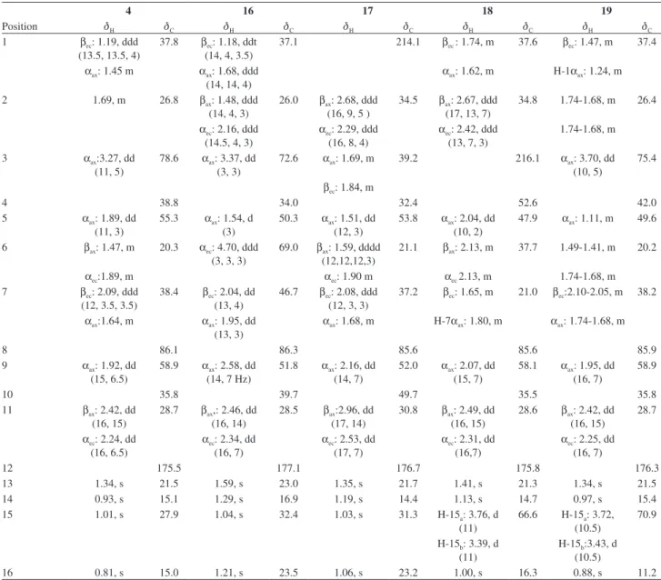
16-19 were obtained. Compound 2 was identiied as the 3-ketosclareolide by comparison of its spectral data with those reported in the literature.17,19 The 1H NMR spectrum
of 4 (Table 2)revealed a signal at dH 3.27 (dd, 3J 11, 5 Hz,
1H,) indicative of an axial proton at C-3 (dC 78.6). The
location of the hydroxyl group at C-3 was conirmed on the basis of HMBC experiments and this compound was identiied as 3β-hydroxysclareolide (4).17,19,20 Unambiguous
and detailed 1H NMR assignments (in comparison with
previous reports17) are shown in Table 2.
Table 1. Yields (%) of the isolated products of transformation of 1 with eight ilamentous fungia
Products (%)
Fungi 1b 2 4 16 17 18 19
A. nigerc 6.5 22.7 37.4 - - -
-A. niger d 2.8 11.7 27.7 5.9 10.1 12.4 9.4
C. blackesleeanac 6.5 18.8 57.3 - - -
-C. blackesleeanad 5.3 7.2 20.6 28.3 8.7 -
-C. lunata d 5.5 20.9 38 - 18.8 -
-B. bassianac 14.9 - 51.3 - - -
-R. oligosporusc 17.3 12.1 32.1 - - -
-R. nigricansc 18.9 12.9 28.5 4.5 - -
--M. miehei c 17.9 12.1 25.5 - - -
-F. moliniformec 10.3 16.1 28.9 32.3 - -
-aYields calculated after puriication. bRecovered starting material. cUsing
culture medium A. d Using culture medium B (see Experimental).
Figure 1. Chemicals structures ofsclareolide (1) and their derivatives compounds 2, 4, 16-19.
O O R1
R2
1 R1 = H, R2 = H
3 R1 =βOH, R2 = H
4 R1 = H, R2 = βOH
6 R1 = αOH, R2 = βOH
7 R1 = βOH, R2 = βOH
O O O 2 O O R1 R2
5 R1 = βOH, R2 = βOH
12 R1 = αOH, R2 = αOH
16 R1 = αOH, R2 = βOH
O O
R1
R2
8 R1 = H, R2 = αOH, R3 = H
9 R1 =βOH, R2 = αOH, R3 = H
14 R1 = H, R2 = H, R3 = αOH
O O
R3
R1
R2
13 R1 = OH, R2 = H
15 R1 =H, R2 = βOH
O O 10 O O O 11 O O O O O O OH O O HO OH
17 18 19
Cano et al. 1179 Vol. 22, No. 6, 2011
For compound 16, the HRFABMS established a molecular formula of C16H26O4 [M+H]+, at m/z 283.1915,
calculated for (C16H26O4 + H) 283.1909. The
1H NMR
spectrumrevealed two signals at dH 3.37 (dd, 3J 3, 3 Hz,
1H) and dH 4.70 (ddd,
3J 3, 3, 3 Hz, 1H), indicative of two
equatorial hydrogens at C-3 (dC 72.6) and C-6 (dC 69.0),
respectively. The locations of the hydroxyl groups at C-3 and C-6 in 16 were determined on the basis of HMBC experiments, in particular by the 16CH3– (dH 1.21) and the
15CH3– (dH 1.04) crosspeaks with C(3) (dC 72.6) and the
crosspeaks between H-6 (dH 4.70) and C-4(dC 34.0), C-5
(dC 50.3), C-10 (dC 39.7), C-7 (dC 46.7), and C-8 (dC 86.3).
COSY, HMQC and NOESY experiments allowed complete assignments for all protons and carbons (Table 2) and the structure was conirmed as 3α,6β-dihydroxysclareolide (16). The epimer at C(3) has been reported previously.18
The new metabolite 17 was obtained as a white crystalline solid. Its HRFABMS exhibited [M+H]+ at m/z 265.1805,
corresponding to the molecular formula C16H25O3 (calculated
for C16H24O3 + H: 265.1804). The IR spectral data displayed
absorptions for γ-lactone (1774 cm−1) and ketone (1708 cm−1).
These data were similar to those for compound 2.17,18 The 13C NMR data (Table 2) showed resonances for sixteen carbons
and the DEPT experiments established the multiplicity for each carbon signal and they revelead the presence of four methyls, ive methylenes, two methines and ive quaternary carbons. The 1H NMR (500 MHz) data of 17 (Table 2) showed
singlets at dH 1.35, 1.19, 1.03 and 1.06 which were assigned to
13CH3–, 14CH3–, 15CH3– and 16CH3–, respectively, by direct
comparison with similar compounds. In particular, 13CH3– is
located downield (dH 1.35) due to it is linked to the carbon
closing the γ-lactone (C-8) and the signal at dH 1.19, which Table 2.1H and 13C NMR data (d in ppm, multiplicite) of compounds 4, 16-19 (500 and 125 MHz, CDCl
3). Coupling constants (J in Hz) in parenthesis
4 16 17 18 19
Position dH dC dH dC dH dC dH dC dH dC
1 βec: 1.19, ddd
(13.5, 13.5, 4)
37.8 βec: 1.18, ddt
(14, 4, 3.5)
37.1 214.1 βec : 1.74, m 37.6 βec: 1.47, m 37.4
αax: 1.45 m αax: 1.68, ddd
(14, 14, 4)
αax: 1.62, m H-1αax: 1.24, m
2 1.69, m 26.8 βax: 1.48, ddd
(14, 4, 3)
26.0 βax: 2.68, ddd
(16, 9, 5 )
34.5 βax: 2.67, ddd
(17, 13, 7)
34.8 1.74-1.68, m 26.4
αec: 2.16, ddd
(14.5, 4, 3)
αec: 2.29, ddd
(16, 8, 4)
αec: 2.42, ddd
(13, 7, 3)
1.74-1.68, m
3 αax:3.27, dd
(11, 5)
78.6 αax: 3.37, dd
(3, 3)
72.6 αax: 1.69, m 39.2 216.1 αax: 3.70, dd
(10, 5)
75.4
βec: 1.84, m
4 38.8 34.0 32.4 52.6 42.0
5 αax: 1.89, dd
(11, 3)
55.3 αax: 1.54, d
(3)
50.3 αax: 1.51, dd
(12, 3)
53.8 αax: 2.04, dd
(10, 2)
47.9 αax: 1.11, m 49.6
6 βax: 1.47, m 20.3 αec: 4.70, ddd
(3, 3, 3)
69.0 βax: 1.59, dddd
(12,12,12,3)
21.1 βax: 2.13, m 37.7 1.49-1.41, m 20.2
αec:1.89, m αec: 1.90 m αec 2.13, m 1.74-1.68, m
7 βec: 2.09, ddd
(12, 3.5, 3.5)
38.4 βec: 2.04, dd
(13, 4)
46.7 βec: 2.08, ddd
(12, 3, 3)
37.2 βec: 1.65, m 21.0 βec:2.10-2.05, m 38.2
αax:1.64, m αax: 1.95, dd
(13, 3)
αax: 1.68, m H-7αax: 1.80, m αax: 1.74-1.68, m
8 86.1 86.3 85.6 85.6 85.9
9 αax: 1.92, dd
(15, 6.5)
58.9 αax: 2.58, dd
(14, 7 Hz)
51.8 αax: 2.16, dd
(14, 7)
52.0 αax: 2.07, dd
(15, 7)
58.1 αax: 1.95, dd
(16, 7)
58.9
10 35.8 39.7 49.7 35.5 35.8
11 βax: 2.42, dd
(16, 15)
28.7 βax,: 2.46, dd
(16, 14)
28.5 βax:2.96, dd
(17, 14)
30.8 βax: 2.49, dd
(16, 15)
28.6 βax: 2.42, dd
(16, 15)
28.7
αec: 2.24,dd
(16, 6.5)
αec: 2.34, dd
(16, 7)
αec: 2.53, dd
(17, 7)
αec: 2.31, dd
(16,7)
αec: 2.25, dd
(16, 7)
12 175.5 177.1 176.7 175.8 176.3
13 1.34, s 21.5 1.59, s 23.0 1.35, s 21.7 1.41, s 21.3 1.34, s 21.5
14 0.93, s 15.1 1.29, s 16.9 1.19, s 14.4 1.13, s 14.7 0.97, s 15.4
15 1.01, s 27.9 1.04, s 32.4 1.03, s 31.3 H-15a: 3.76, d
(11)
66.6 H-15a: 3.72,
(10.5)
70.9
H-15b: 3.39, d
(11)
H-15b:3.43, d
(10.5)
Biotransformation of Sclareolide by Filamentous Fungi: Cytotoxic Evaluations of the Derivatives J. Braz. Chem. Soc. 1180
showed NOESY crosspeak with H-11β (dH 2.96) and HMBC
crosspeak with the signal at dC 52.00 (C-9) was assigned to
14CH3–. The location of the ketone at C-1 was determined by
the HMBC crosspeak of the signal at dH 1.19 (14CH3–) with
the resonance at dC 214.1. Assignments for all hydrogens and
carbons were done by COSY, HMQC, HMBC and NOESY experiments.
Compound 18 was obtained as white crystalline solid and its IR spectral data displayed absorptions at 3459 (O-H), 1773 (C=O) and 1174 (C-O) cm−1. The HREIMS exhibited
[M+H]+ at m/z 281.1751 establishing the molecular formula
C16H24O4 (calculated for C16H24O4 + H: 281.1753). The 13C NMR spectrum of 18 (DEPT experiment) displayed
resonances for sixteen carbon atoms, including three methyls, six methylenes, two methines and ive quaternary carbons. Its 1H NMR spectrum (CDCl
3, 500 MHz) showed three
singlets at dH 1.41, 1.13 and 1.00 due the H-13, H-14 and
H-16 methyls, respectively, due to the observed NOESY crosspeaks between C-13 and C-14 and between C-14 with C-16. A notable difference with the 4,4’-dimethyl products was the absence of one methyl group and the presence of an AB system (dH 3.76 and 3.39,
2J 11 Hz), which was assigned
to the methylene protons at C-15 (dC 66.6). The signal at dC 216.1 corresponds to a ketone carbonyl located at C-3 due to
HMBC correlations of this carbon with the AB system (H-15a
and H-15b), H-2βax (dH 2.67), H-2αec (dH 2.42), H-1βec (dH 1.74)
and with 16CH3– (dH 1.00). The 1H and 13C NMR chemical
shifts assigments for compound 18 (Table 2), conirmed this compoundas 3-keto-15-hydroxysclareolide.
The other novel biotransformation product, 19, was a crystalline solid. The IR spectral data displayed absorptions at 3489 (O-H) and 1767 (C=O) cm−1. The HREIMS of 19
exhibited a molecular ion m/z 283.1908 corresponding to the molecular formula C16H26O4 (calculated for
C16H26O4 + H: 283.1909). The 13C NMR spectrum of 19
showed resonances for sixteen carbons including three methyls, six methylenes, three methines and four quaternary carbons. The 1H NMR data of 19 (Table 2) showed singlets
at dH 1.34, 0.97 and 0.88, assigned to 13CH3–, 14CH3–,
16CH3–, respectively, by comparison with the above
described substances. Additionally, the signals at dH 3.72
and 3.43 (2J 10, 5 Hz) established the presence of an AB
system for the hydroxymethylene protons located at C-15 (dC 70.9) which overlapped with a doublet of doublet signal
corresponding to H-3αax (dH 3.70, 3J 10, 5 Hz). The signal
at dC 75.4, assigned to C-3, showed HMBC correlations
with the AB system for H-15a and H-15b, and with 16CH3–
(dH 0.88). Assignments for all hydrogens and carbons for
compound 19 were done by COSY, HMQC, HMBC and NOESY experiments and veriied its structure as 3β,15 -dihydroxysclareolide (Table 2).
An interesting variety of products and yields were obtained by the biotransformation of sclereolide (1) with different fungi. The transformation of 1 with A. niger in medium A (containing minor number of nutrients) produced 2 and 4, while using medium B yielded six compounds (2, 4, 16-19) in variable yields (from 5.9% to 27.1%), affording a keto group at C-1 (17) for the irst time. Using medium B with A. niger,
C. blackesleeana and C. lunata yielded various products in relatively variable yields, while medium A produced fewer products, with better yields though (see Table 1). The transformation of 1 with B. bassiana afforded exclusively product 4 in acceptable yield (51.3%), in comparison with other biotransformations.24 These results gave additional
evidences of the importance of the species, strains and media employed in the structural diversity of the products and the yields of the biotransformations.
Sclareolide (1) and derivatives 2, 4, 16-19 were tested for in vitro cytotoxic activity against human cancer cell lines U251 (central nervous system), PC-3 (prostate cancer), K562 (leukemia), HCT-15 (colon), MCF-7 (breast) and SKUL-1 (lung) following standard procedures.25 Compound 1 displayed
activity against PC-3 tumor cell line (IC50 71.12 ± 4.7 µM) and
16 displayed activity against U251 (IC50 87.40 ± 5.4 µM), PC-3
(IC50 34.47 ± 7.4 µM), HCT-15 (IC50 92.23 ± 4.2 µM), MCF-7
(IC50 89.11 ± 2.4 µM). All the other IC50 values were above a
100 µM and were considered not active.25 It is interesting that
the presence of the hydroxyl groups at C-3α and C-6β increased the cytotoxicity with respect to the starting material (1).
Experimental
General experimental procedures
Melting points were determined on a Fischer-Jones apparatus. Optical rotation were measured on a Perkin-Elmer 341 polarimeter. Infrared spectra are registred Nicolet Magna FT-IR 750 spectrometer. 1H and 13C NMR spectra were taken
on a Varian Unity-plus 500 (at 500 and 125 MHz) instrument. EI-MS: Jeol JMS-AX505HA mass spectrometer and Jeol JMS-SX 102 A) for HREIMS. TLC spots were revealed by spraying with ceric ammonium sulfate, followed by heating. Vacuum column chromatographies were done following the reported procedures.22,23 Column chromatography (CC) were
performed using silica gel 70-230, TLC using silica gel 60F254
(Merck) plates and preparative TLC using silica gel 60F254
(Merck) Plates. Sclareolide was purchased from Sigma-Aldrich.
Microorganisms
A. niger (ATCC 16404), B. bassiana (ATCC 13144),
Cano et al. 1181 Vol. 22, No. 6, 2011
F. moliniforme (ATCC 10209), M. miehei (ATCC 16457),
R. oligosporus (ATCC 22959), R. nigricans (ATCC 6227b), were obtained from the Instituto de Biotecnología, Universidad Nacional Autónoma de México, maintained on potato dextrose agar (PDA) and stored at 4 °C.
Media and culture conditions
The media were prepared by mixing the following ingredients in 1 L of distilled water. Medium A (YEPGA): (10 g) peptone, (10 g) yeast extract, (10 g) beef extract and (50 g) glucose. The pH was adjusted to 7 (NaOH 1 mol L−1)
before autoclaving. Medium B was prepared by mixing the following ingredients: glucose (10 g), glycerol (10 g), peptone (5 g), yeast extract (5 g), KH2PO4 (5 g) and NaCl (5 g) at pH 7
(adjusted with NaOH 1 mol L−1) for A. niger, C. blackesleeana,
C. lunata.
Incubation experiments
Firstly,the eight fungi were tested for their ability to metabolize 1 onthe analytical scale (10 to 15 mg of substrate, 25 mL medium culture), and then the same biotransformation experiments were carried out on preparative scale (150 to 310 mg). Both analytical and preparative biotransformations were conducted following similar procedures. Erlenmeyer lasks (250 mL) containing 125 mL of medium A or the medium B were inoculated with a dense suspension (2 mL) of the corresponding fungi. Incubations were maintained at 25 °C with gyratory shaking (125 rpm) for 24 h (A. niger, R. nigricans, R. oligosporus, M. miehei), 48 h (F. moniliforme) or 72 h (C. blackesleeana, C. lunata, B. bassiana). Then, the substrates in acetone (5-10 mL) were added and the process continued for 14 days. These biotransformation experiments were monitored by TLC, including two controls, a “culture control” and a “substrate control” to eliminate the possibility that the isolated products were microbial secondary metabolites and/or that the culture media did not perform any chemical transformation on the substrate.
Recovery and puriication of metabolites
Cultures were iltered and fungal cells were washed thoroughly with water and the iltrate and washings were combined, saturated with NaCl and extracted with CH2Cl2
(three times). The organic extracts were combined, dried with Na2SO4 and evaporated under reduced pressure. The
organic residues were subjected to chromatography by VCC using gradient elution system with hexane/EtOAc. CC and preparative TLC allowed the inal puriication of the compounds.
Biotransformation of 1 by A. niger in medium A
After 24 h of inoculation with a dense suspension of the spores of A. niger, 20 erlenmeyer lask cultures (YEPGA) received 204.3 mg of sclareolide (1) in 10 mL of acetone. After incubation for 14 days the cultures were processed as indicated above to yield a crude dark oily residue (194.3 mg). Column chromatography of the organic extract yielded 3-ketosclareolide (2, 44.2 mg, 22.7%), 3β-hydroxysclareolide (4, 72.7 mg, 37.4%) and sclareolide (1, 12.6 mg, 6.5%).
Biotransformation of 1 by A. niger in medium B
The substrate 1 (210 mg) was dissolved in acetone (10 mL), distributed among 20 erlenmeyer lask cultures (medium B), previously (24 h) inoculated with a dense suspension of spores of A. niger. The biotransformation was allowed to proceed for 14 days and the cultures were processed as indicated above to obtain starting material 1 (6 mg, 2.8%), 2 (24.5 mg, 11.7%), 4
(58.2 mg, 27.7%), 16 (22.4 mg, 5.9%), 17 (21.2 mg, 10.1%)
18 (26 mg, 12.4%) and 19 (19.7 mg, 9.4%).
3α,6b-Dihydroxysclareolide (16): mp.188-190°C; [α]25
D+ 30.3 (c 0.30, CHCl3); IR νmax/ cm−1 (CHCl3): 3618,
2933, 1758; 1H NMR (CDCl
3, 500 MHz) and
13C NMR (CDCl 3,
125 MHz): see Table 2; HRFABMS [M+H]+ Found: 283.1915.
Calc. for C16H26O4+H: 283.1909; EIMS m/z 267 (24%), 249
(56), 169 (83), 43 (100).
1-Ketosclareolide (17): mp. 152-153 °C; [α]25
D + 83.8
(c 0.11, CHCl3); IR νmax/cm−
1 (CHCl
3): 2958, 1774,
1708, 1232, 1199, 925. 1H NMR (CDCl
3, 500 MHz) and 13C NMR (CDCl
3, 125 MHz): see Table 2; HRFABMS
[M+H]+ Found: 265.1805. Calc. for C
16H24O3+H: 265.1804;
EIMS m/z 264 (17%), 205 (86), 55(69), 43 (100).
3-Keto-15-hydroxysclareolide (18): mp. 169-171 °C; [α]25
D+ 29.3 (c 0.20, MeOH); IR νmax/cm−1 (CHCl3): 3459,
2985, 2952, 1773, 1698, 1431, 1234, 1174, 920; 1H NMR
(CDCl3, 500 MHz) and 13C NMR (CDCl3, 125 MHz): see
Table 2; HRFABMS [M+H]+ Found: 281.1751. Calc. for
C16H24O4+H: 281.1753); EIMS m/z 280 (12%), 278 (36),
277 (11), 250 (67), 235 (48), 81 (41), 43 (100).
3b-15-Dihydroxysclareolide (19): mp. 157-158 °C; [α]25
D+ 49.2 (c 0.13, MeOH); IR νmax/cm−1 (CHCl3): 3489,
2944, 1767, 1603,1457, 1349, 1243, 920. 1H NMR (CDCl 3,
500 MHz) and 13C NMR (CDCl
3, 125 MHz): see Table 2.
HRFABMS [M+H]+ Found: 283.1908. Calc. for C
16H26O4+H:
283.1909; EIMS m/z 282 [M+, 20%], 264 [M+–H
Biotransformation of Sclareolide by Filamentous Fungi: Cytotoxic Evaluations of the Derivatives J. Braz. Chem. Soc. 1182
(57), 233 (56), 181 (12), 173 (30) 147 (69), 121 (64), 93 (52), 81 (41), 43 (100), 18 (36).
Biotransformation of 1 by C. blackesleeana in medium B
The substrate 1 (307.5 mg) was dissolved in acetone (15 mL), distributed among 20 erlenmeyer lask cultures (medium B), previously inoculated (72 h) with a dense suspension of spores of C. blackesleeana. The fermentation was allowed to proceed for 14 days, the cultures were processed as indicated above to obtain starting material
1 (16.3 mg, 5.3%), 2 (22.3, 7.2%), 4 (63.4 mg, 20.6%), 3α,6β-dihydroxysclareolide (16,86.9 mg, 28.3%) and 1-ketosclareolide (17, 26.7 mg, 8.7%).
Biotransformation of 1 by C. lunata, B. bassiana, R. oligosporus, R. nigricans, M. miehei and F. moliniforme
Compound 1 (150 mg) was reacted with the microorganisms following the procedure described above, obtaining the results shown in Table 1.
Cytotoxic assays
Human tumor cell lines of central nervous system (U251), prostate cancer (PC-3), leukemia (K562), colon (HCT-15), breast (MCF-7), lung (SKLU-1) were supplied by the National Cancer Institute (NCI). The cytotoxic activities of 1, 2, 4, 16-19
were determined using the protein-binding dye sulforhodamine B in a microculture assay to measure cell growth, following the protocols described in the literature.18 Results were expressed
as concentration giving 50% inhibition (IC50). The IC50 values
(mean ± standard error) were 100 µM, and those with minor values are reported in the text. The positive control was adriamycin (IC50 = 0.32 ± 0.02 µM against U251).
Supplementary Information
Supplementary information data are available free of charge at http://jbcs.org.br as a PDF ile.
Acknowledgments
The autors thank Rocío Patiño, Beatriz Quiroz, Angeles Peña, María Isabel Chávez, Luis Velasco, Javier Pérez and Antonio Nieto from the Instituto de Química, UNAM for technical assistence. Financial supports from Consejo Nacional de Ciencia y Tecnología (Project 102158), Dirección General de Asuntos del Personal Académico, UNAM and Carrera de Biología, Facultad de Estudios Superiores Zaragoza, UNAM, are gratefully acknowledged.
References
1. Panke, S.; Wubbolts, M.; Curr. Opin. Chem. Biol. 2005, 9, 188. 2. Huisman, G. W.; Gray, D.; Curr. Opin. Biotechnol. 2002, 13,
352.
3. Straathof, A. J. J.; Panke, S.; Schmid, A.; Curr. Opin. Biotechnol.
2002, 13, 548.
4. Davis, B. G.; Boyer, V.; Nat. Prod. Rep.2001, 18, 1669. 5. De Raadt, A.; Herfried, G.; Curr. Opin. Biotechnol. 2002, 13,
537.
6. Zaks, A.; Curr. Opin. Chem. Biol. 2001, 5, 130.
7. Lehman, L. R.; Stewart, J. D.; Curr. Org. Chem. 2001, 5, 439. 8. Urlacher, V.; Schmid, R. D.; Curr. Opin. Biotechnol. 2002, 13,
557.
9. Van Beilen, J. B.; Duetz, W. A.; Schmid, A.; Whitholt, B.; Trends Biotechnol. 2003, 4, 170.
10. Lee, K. H.; J. Nat. Prod. 2010, 73, 500.
11. León, A.; Reyes, B. M.; Chávez, M. I.; Toscano, R. A.; Delgado, G.; J. Mex. Chem. Soc.2009, 53, 193.
12. Upar, K. B.; Mishra, S. J.; Nalawade, S. P.; Singh, S. A.; Khandare, R. P.; Bhat, S. V.; Tetrahedron: Asymmetry 2009, 20, 1637.
13. Atta-ur-Rahman; Farooq, S.; Choudhary, M. I.; Curr. Org. Chem. 1999,3, 309.
14. Rodríguez, E.; Towers, G. H. N.; Mitchell, J. C.; Phytochemistry 1976,15, 1573.
15. Oh, S.; Jeong, I. H.; Shim, W. S.; Wang, Q. S.; Lee, S.; Bioorg. Med. Chem. Lett.2006, 16, 1656.
16. Zoretic, P. A.; Fang, H.; Ribeiro, A. A.; Dubay, G.; J. Org. Chem. 1998, 63, 1156.
17. Aranda, G.; El-Korbi, M. S.; Lallermand, J. V.; Neuman, A.; Tetrahedron1981, 47, 8339.
18. Hanson, J. R.; Truneh, A.; Phytochemistry1996, 42, 1021. 19. Atta-ur-Rahman; Farooq, A.; Choudhary, M. I.; J. Nat. Prod.
1997, 60, 1038.
20. Choudhary, M. I.; Musharraf, S. G.; Sami, A.; Atta-ur-Rahman; Helv. Chem. Acta2004, 87, 2685.
21. Ata A.; Conci, L. J.; Betteridge, J.; Orhan, I.; Sener, B.; Chem. Pharm. Bull. 2007, 55, 118.
22. Pelletier, S. W.; Chokshi, H. P.; Desai, H. K.; J. Nat. Prod. 1986, 49, 892.
23. Coll, J. C.; Bowden, B. F.; J. Nat. Prod. 1986, 49, 934. 24. García Granados, A.; Martínez, A.; Parra, A.; Rivas, F.; Curr.
Org. Chem. 2007, 11, 679.
25. Monks, A.; Seudiero, D.; Skehan, P.; Shoemaker, R.; Paull, K.; Vistica, D.; Hose, C.; Langley, J.; Cronise, P.; Vaigro-Wolff, A.; Gray-Goodrich, M.; Campbell, H.; Mayo, J.; Boyd, M.; J. Nat. Cancer Inst. 1991, 83, 753.
Submitted: March 30, 2010
Supplementary Information
J. Braz. Chem. Soc., Vol. 22, No. 6, S1-S7, 2011. Printed in Brazil - ©2011 Sociedade Brasileira de Química
0103 - 5053 $6.00+0.00
S
I
*e-mail: [email protected]
Biotransformation of Sclareolide by Filamentous Fungi:
Cytotoxic Evaluations of the Derivatives
Arturo Cano,a María Teresa Ramírez-Apanb and Guillermo Delgado*,b
aFacultad de Estudios Superiores Zaragoza, Universidad Nacional Autónoma de México,
Av. Guelatao no. 66 (Eje 7 Oriente), Col Ejército de Oriente, Iztapalapa 09230, Mexico, D.F.
bInstituto de Química, Universidad Nacional Autónoma de México, Ciudad Universitaria,
Circuito Exterior, Coyoacán 04510, Mexico, D.F.
Figure S1. 1H NMR (500 MHz, CDCl
Biotransformation of Sclareolide by Filamentous Fungi: Cytotoxic Evaluations of the Derivatives J. Braz. Chem. Soc.
S2
Figure S2. 13C NMR (125 MHz, CDCl
3) of 3α,6β-dihydroxysclareolide (16).
Cano et al. S3 Vol. 22, No. 6, 2011
Figure S4. 1H NMR (500 MHz, CDCl
3) of 1-ketosclareolide (17).
Figure S5. 13C NMR (125MHz, CDCl
Biotransformation of Sclareolide by Filamentous Fungi: Cytotoxic Evaluations of the Derivatives J. Braz. Chem. Soc.
S4
Figure S6. HMBC (500 MHz, CDCl3) of 1-ketosclareolide (17).
Figure S7. 1H NMR (500MHz, CDCl
Cano et al. S5 Vol. 22, No. 6, 2011
Figure S8. 13C NMR (125 MHz, CDCl
3) of 3-keto-15-hydroxysclareolide (18).
Figure S9. 1H NMR (500 MHz, CDCl
Biotransformation of Sclareolide by Filamentous Fungi: Cytotoxic Evaluations of the Derivatives J. Braz. Chem. Soc.
S6
Figure S10. 13C NMR (125 MHz, CDCl
3) of 3β,15-dihydroxysclareolide (19).
Cano et al. S7 Vol. 22, No. 6, 2011