376
Rev. Col. Bras. Cir. 2010; 37(5): 376-378
Kamamoto KamamotoKamamoto KamamotoKamamoto New technique for dynamic closure of the abdominal wall
Technical Note
Technical Note
Technical Note
Technical Note
Technical Note
New technique for dynamic closure of the abdominal wall
New technique for dynamic closure of the abdominal wall
New technique for dynamic closure of the abdominal wall
New technique for dynamic closure of the abdominal wall
New technique for dynamic closure of the abdominal wall
Nova técnica para o fechamento dinâmico da parede abdominal
Nova técnica para o fechamento dinâmico da parede abdominal
Nova técnica para o fechamento dinâmico da parede abdominal
Nova técnica para o fechamento dinâmico da parede abdominal
Nova técnica para o fechamento dinâmico da parede abdominal
F
ABIOK
AMAMOTO1; B
ERNARDON
OGUEIRAB
ATISTA2; F
LAVIOT
OKESHI3A B S T R A C T
A B S T R A C T
A B S T R A C T
A B S T R A C T
A B S T R A C T
Advances in care of trauma patients and severe abdominal infections are responsible for an increasing number of laparostomies. The management of this entity is complex and several techniques have been described for its treatment. Recently the concept of dynamic closure of the abdominal wall was introduced in the literature with high success rates. The objective of this report is to serve as a foreword for a new approach for the treatment of laparostomy developed at the University Hospital of the University of São Paulo. This is a simple and low cost method, easily performed by a general surgeon. The procedure was also used prophylactically as reinforcement in tight abdominal closures. It is described in detail as well as the results in the first patients. Although promising, refinements and further studies are needed to validate the technique.
Key words Key words Key words Key words
Key words: Laparostomy. Abdominal hernia. Abdominal compartment syndrome.
Work done at the University Hospital of the University of São Paulo – São Paulo – Brazil.
1. M.D., Surgery Department, University Hospital, University of São Paulo, São Paulo, Brazil; 2. Resident, Plastic Surgery, Hospital das Clínicas, University of São Paulo Mecial School, São Paulo, Brazil; 3. M.D., Surgery Department, University Hospital, University of São Paulo, São Paulo, Brazil.
INTRODUCTION
INTRODUCTION
INTRODUCTION
INTRODUCTION
INTRODUCTION
A
dvances in the treatment of abdominal compartment
syndrome as well as the techniques of damage control
have contributed to significant gains in survival of patients
victims of trauma and severe abdominal infections
1-3. They
created, however, a difficult management problem: by
reducing the mortality of these patients, laparostomy
appears as increasingly frequent, and its appropriate
closure is a challenge for surgeons. We present a new
proposal for the management of this entity developed at
the University Hospital of the University of São Paulo, São
Paulo, Brazil.
METHODS
METHODS
METHODS
METHODS
METHODS
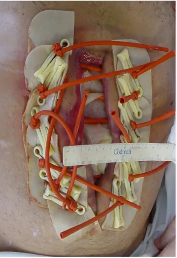
The technique is aimed at primary closure of the
abdominal cavity, covering all layers of the abdominal
wall. For that it begins by isolating the abdominal contents
through the suture of a protective film (open bag of saline)
to the healthy parietal peritoneum. Following this, punctures
are made bilaterally, along the wound’s longitudinal axis,
transfixing all layers of the abdominal wall. We adopt the
distances of 2.5 cm between the holes and approximately
4 cm from the edge of the wound. We then pass a 14F
NELATON catheter through each pair of holes (to the right
and to the left of the wound), connecting its two edges, in
a “U” fashion, so that each catheter stays perpendicular to
the wound axis and its extremities come out of the skin. The
catheters’ ends are fixed with plastic clamps, applying some
tension to the wound (Figure 1).
Six patients underwent abdominal wall closure
using the described technique. In four the primary closure
of the abdominal cavity was possible, with big tension
on the suture line. We then decided to apply the
technique to prevent complications by distributing the
tension across the wall thickness and around the wound’s
craniocaudal axis. The other two patients had already
been submitted to laparostomy resulting from a
complication of previous laparotomies. In these cases,
every two or three days the catheters were tensioned
and the clamps repositioned until proper tension was
reached or the patient referred pain (whichever occurred
first). The procedure was repeated until proper
rapprochement of the wound was achieved, this way
aiming for the dynamic closure of the defect. After about
three weeks we carried out the final tension-free closing
of the wall and retrieved the catheters.
RESULTS
RESULTS
RESULTS
RESULTS
RESULTS
Kamamoto Kamamoto Kamamoto Kamamoto Kamamoto
New technique for dynamic closure of the abdominal wall 377
Rev. Col. Bras. Cir. 2010; 37(5): 376-378
wound. In both cases in which we sought closure of an
open abdomen, it was successfully achieved in one
patient. The other patient presented with rupture of
the wall at the insertion points of three of the six
catheters installed due to excessive tension applied
postoperatively.
DISCUSSION
DISCUSSION
DISCUSSION
DISCUSSION
DISCUSSION
The technique of dynamic closure appears to be
a promising tool in closing the abdominal wall after
laparostomy, as well as effective prophylaxis in sutures that
have apply tension in the abdominal wall.
Usually, when faced with a tense suture, the
general surgeon makes use of stiches with an
unabsorbable, thick, suture in total plan. Although data
have not been measured objectively, it was observed
that patients undergoing the described procedure
reported less pain after surgery, with more effective
drug therapy.
With regard to the definitive treatment of
laparostomy, this technique allows complete closure of
the abdominal wall, covering all of its plans, restoring its
normal anatomy and all the functions of containment
and resistance of the muscle wall, without generating
new morbidity factors for the patient, facts that make it
superior to the techniques of closure with autologous,
xenologous or synthetic substitutes (meshes and
flaps)
4. Other studies in the literature reinforce the idea
that the progressive (dynamic) closure techniques of the
abdominal wall are effective in obtaining a continent
abdominal wall
1,5.
Figure 1 Figure 1 Figure 1 Figure 1
-Figure 1 - Photo showing the appearance of the final procedure in which 14F NELATON catheters are passed through punctures in the abdominal wall, allowing the distribution of tensile strength throughout the wound.
R E S U M O
R E S U M O
R E S U M O
R E S U M O
R E S U M O
Os avanços nos cuidados com o paciente traumatizado e com infecções abdominais graves são responsáveis por um número crescente de peritoneostomias. O manejo desta entidade é complexo e várias técnicas foram descritas para seu tratamento. Recentemente foi introduzido na literatura o conceito de fechamento dinâmico da parede abdominal, com elevadas taxas de sucesso. O objetivo deste trabalho é de servir como nota prévia de uma nova abordagem para o tratamento das peritoneostomias, desenvolvida no Hospital Universitário da Universidade de São Paulo. Trata-se de um procedimento simples e de baixo custo, facilmente realizado por cirurgião geral. O procedimento também foi utilizado como reforço em fechamentos abdominais tensos, de maneira profilática. O procedimento é descrito em detalhes, assim como os resultados nos primeiros pacientes. Apesar de promissora, refinamentos técnicos e estudos complementares são necessários para a validação da técnica.
Descritores DescritoresDescritores
DescritoresDescritores: Peritoneostomia. Hernia abdominal. Sindrome compartimental abdominal.
REFERENCES
REFERENCES
REFERENCES
REFERENCES
REFERENCES
1. Cothren CC, Moore EE, Johnson JL, et al. One hundred percent fascial approximation with sequential abdominal closure of the open abdomen. Am J Surg 2006; 192: 238-242.
2. Costa A. Making a virtue of necessity: managing the open abdomen. Anz J Surg 2006; 76: 356-363.
3. Hultman CS, Pratt B, Cairns BA, et al. Multidisciplinary Approach to Abdominal Wall Reconstruction After Decompressive Laparotomy for Abdominal Compartment Syndrome. Ann Plast Surg 2005; 54 (3): 269-275.
378
Rev. Col. Bras. Cir. 2010; 37(5): 376-378
Kamamoto KamamotoKamamoto KamamotoKamamoto New technique for dynamic closure of the abdominal wall
5. Jernigan TW, Fabian TC, Croce MA, et al. Staged Management of Giant Abdominal Wall Defects Acute and Long-Term Results. Ann Surg 2003; 238: 349-357.
Received in 20/05/2010
Accepted for publication in 20/07/2010 Conflict of interest: none
Source of funding: none
How to cite this article: How to cite this article:How to cite this article: How to cite this article: How to cite this article:
Kamamoto F, Batista BN, Tokeshi F. New technique for dynamic closure of the abdominal wall. Rev Col Bras Cir. [periódico na Internet] 2010; 37(5). Disponível em URL: http://www.scielo.br/rcbc