*e-mail: [email protected] Recebido: 08/05/2012 / Aceito: 01/08/2012
The inluence of cell dimensions on the vulnerability of ventricular
myocytes to lethal injury by high-intensity electrical ields
Jair Trapé Goulart, Pedro Xavier de Oliveira, José Wilson Magalhães Bassani, Rosana Almada Bassani*
Abstract Application of high intensity electric ields (HIEF) to the myocardium is commonly used for cardiac deibrillation/cardioversion. Although effective at reversing life-threatening arrhythmias, HIEF may cause myocyte damage due to membrane electropermeabilization. In this study, the inluence of cell length and width on HIEF-induced lethal injury was analyzed in isolated rat cardiomyocytes in parallel alignment with the ield. The ield-induced maximum variation of membrane potential (∆Vmax) was estimated with the Klee-Plonsey model. The studied myocyte population was arranged in two group pairs for comparison: the longest vs. the shortest cells, and the widest vs. narrowest cells. Threshold ield intensity was signiicantly lower in the longest vs. shortest myocytes, whereas cell width inluence was not signiicant. The threshold ∆Vmax was comparable in all groups. Likewise, a signiicant leftward shift of the lethality curve (i.e., relationship of the probability of lethality vs. ield intensity) of the longest cells was observed, evidencing greater sensitivity to HIEF-induced damage. However, the lethality curve as a function of ∆Vmax was similar in all groups, conirming a prediction of the Klee-Plonsey model. The similar results for excitation and injury at threshold and HIEF stimulation, respectively, indicate that: a) the effect of cell length on the sensitivity to the ield would be attributable to differences in ield-induced membrane polarization that lead to excitation or lethal electroporation; b) the Klee-Plonsey model seems to be reliable for analysis of cell interaction with HIEF; c) it is possible that increased cell length in hypertrophied hearts enhances myocyte fragility upon deibrillation/cardioversion.
Keywords Electric ield, Cardiac myocytes, Lethal cell injury, Modeling, Deibrillation.
Inluência das dimensões celulares sobre a vulnerabilidade de miócitos
ventriculares ao efeito letal de campos elétricos de alta intensidade
Resumo Campos elétricos de alta intensidade (HIEF) são aplicados ao miocárdio durante desibrilação e cardioversão. Embora eicazes na reversão de arritmias potencialmente letais, HIEF podem lesar cardiomiócitos por eletropermeabilização da membrana. Neste estudo, a inluência das dimensões celulares sobre o efeito letal de HIEF foi estudada em cardiomiócitos isolados de rato alinhados paralelamente ao campo. A máxima variação do potencial de membrana induzida pelo campo (∆V
max) foi calculada com o modelo de Klee‑Plonsey.
As células estudadas foram distribuídas em dois pares de grupos de acordo com seu comprimento e largura. A intensidade limiar do campo não dependeu da largura celular, mas sim do comprimento (menor nas células mais longas, p < 0.001), enquanto ∆Vmax no limiar foi comparável entre os grupos. Nas células mais
longas, observou‑se desvio à esquerda (p < 0.01) da curva que descreve a relação entre probabilidade de letalidade e a intensidade do campo, evidenciando maior sensibilidade à ação deletéria de HIEF. Porém, a curva de letalidade em função de ∆V
max foi semelhante em todos os grupos, o que conirma a predição pelo
Introduction
Several pathophysiological conditions may be associated with disturbances in heart rhythm, which may result in impairment of cardiac pumping function.
Ventricular ibrillation is considered the most severe of
the arrhythmia types, as it frequently leads to sudden cardiac death. The latter, with estimated incidence of 0.1-0.2% of the adult population per year in Western industrialized countries, represents ~50% of the deaths
related to coronary disease (Zipes et al., 2006). Electrical deibrillation, namely the application of high intensity electric ields (HIEF) to the heart, is the
most commonly used and most effective procedure
to reverse ventricular ibrillation in the emergency setting (Zipes et al., 2006). As a rule, it is considered that the likelihood of success of deibrillation keeps a
positive relationship with shock strength, but only up to a certain point: increasing shock intensity above the
optimal range diminishes success rate (Dosdall et al., 2010; Fotuhi et al., 1999). While poor underlying
myocardial conditions may be partially accountable
for the therapeutic failure of very strong shocks, HIEF itself may exert deleterious effects on the heart. Such
effects include sustained membrane depolarization, cell damage and conduction block that may facilitate
post-shock arrhythmia reinitiation (e.g., Dosdall et al., 2010; Fedorov et al., 2008; Fotuhi et al., 1999; Knisley
and Grant, 1985; Oliveira et al., 2008; Sowell and Fast, 2012; Yabe et al., 1990).
The deleterious effects of HIEF are attributable to membrane electropermeabilization (electroporation),
i.e., the opening of hydrophilic pores, due to transition of the membrane phospholipid organization brought
about by the energy delivered by shock (Weaver and Chizmadzhev, 1996). The electroporated
membrane loses its selective permeability, so that
large transmembrane lux of ions (including cytotoxic calcium ions) and water, as well as loss of intracellular components, may ensue. When exposed to very strong
shocks, the cardiac myocyte develops irreversible hypercontracture, calcium overload and electrical
refractoriness, and loses its physical integrity (lethal injury; Knisley and Grant, 1985; Oliveira et al., 2005, 2008).
External field application generates a
spatially-varying electrical potential gradient in the
extracellular medium that drives the rearrangement of
charges on the membrane surfaces, which results in
change in the transmembrane electrical potential (Vm). Experimental studies indicate that the occurrence of
electroporation is highly dependent on the variation
of membrane potential (∆Vm) that results from the
ield application, and will take place when Vm exceeds a critical value (Fedorov et al., 2008; O’Neill and Tung, 1991).
Mathematical models have been valuable tools to simulate and predict membrane polarization and
electroporation in response to external ields (e.g., Krassowska and Filev, 2007; Valič et al., 2003). However, most of them cannot give much information
on Vm. In our laboratory, we have employed the model
described by Klee and Plonsey (1976) (K-P model), which allows the estimation of the maximal ∆Vm
(∆Vmax) upon imposition of an external ield. While this model produces consistent results for threshold
ield stimulation (Bassani et al., 2006; Gomes et al.,
2001; Oliveira et al., 2008), it has not been ascertained yet if it also applies for HIEF.
According to the K-P model, the only factors that directly determine ∆Vmax, in addition to the ield
intensity, are the cell dimensions and ield orientation with respect to the cell. In our experimental setting, ield-induced extensive electroporation can be indicated by cell lethal injury (Oliveira et al., 2008). Assuming
that the critical ∆Vmax for this effect is similar for a
particular cell type (i.e., rat ventricular myocytes) under a given set of experimental conditions, a simple way to test whether the K-P model is applicable to HIEF
is to investigate whether ∆Vmax behaves as predicted
when varying a ield-independent parameter. The objective of the present study was to evaluate the inluence of cell dimensions (length and width)
on the sensitivity of isolated rat ventricular myocytes
to the lethal effect of external HIEF, as well as to the
estimated ∆Vmax developed in response to the latter.
In addition to providing a test of the usefulness of the K-P model for ield intensities close to the range of those attained during deibrillation, this study also
addresses a point of potential clinical importance, as cardiomyocyte hypertrophy is commonly associated with conditions that increases the probability of
cardiac ibrillation and sudden death occurrence (e.g., Bender et al., 2012; Brouwer et al., 2011;
Reinier et al., 2011).
Materials and Methods
Rat cardiomyocyte preparation
Myocytes were isolated from the left ventricle of
adult (4-6 month-old) male Wistar rats, not previously submitted to any type of experimental manipulation.
Myocyte isolation was carried out by coronary
perfusion with collagenase I, as described by Penna and Bassani (2010). Cells were used within 12 hours
after isolation. The protocols for animal care and
The cell suspension was plated on a perfusion chamber, of which the bottom was a glass coverslip treated with collagen to enhance cell adhesion. A pair of wire platinum electrodes was inserted into slits along the chamber lateral inner walls, spanning most of the length of the chamber. The chamber was
placed on a microscopy system (Ricardo et al., 2006), and cells were perfused (~3 mL/min) with modiied Tyrode´s solution (mM composition: 140 NaCl; 6 KCl; 1.5 MgCl2.6 H2O; 1 CaCl2.2 H2O; 10 HEPES; 11.1 glucose; pH 7.4) at 23 °C. Vacuum suction of the
solution at the chamber outlet ensured that the height
of the solution column in the chamber (thus, solution volume and conductivity) could be kept constant.
Perfusion was interrupted during measurement of stimulus amplitude.
By means of a CCD camera, the cell image was projected on a video monitor, and captured (mod. Dazzle Digital VideoCreator 150, Pinnacle Systems, North Canton, USA) for data storage and ofline analysis. Cell length and width, i.e., major and minor axes (2c and 2a, respectively), were measured on the screen using a graduated scale (<1 µm resolution) that was calibrated with aid of a graticule (10 µm resolution, Carl Zeiss, Göttingen, Germany) projected on the screen (estimated error <3%).
Only cells that met the following requirements
were selected for this study: a) presence of clear
striations, as well as preserved structure and contractile
function; b) parallel orientation of the cell major axis to the ield direction, which was necessary to isolate cell dimension and ield intensity as the only factors
determining ∆Vmax; and c) location at least at 2 mm from the electrodes, which is required for low error
(<2%) in ield estimation (Oliveira et al., 2008).
Field and ∆V
max estimation
The ield intensity (V/cm) was calculated as in the case of a parallel plate capacitor (Gomes et al., 2001),
as the ratio of the stimulus voltage and the distance
between the electrodes (0.75 cm).
For both threshold and HIEF stimulation, ∆Vmax
was estimated as in Equation 1 (Klee and Plonsey, 1976), assuming for the myocyte a prolate spheroidal
geometry, and that the membrane could be represented
by dielectric shell (Bassani et al., 2006; Gomes et al.,
2001; Oliveira et al., 2008):
2 2 2 2 2 2 1/2
max
V (E, ,a,c) E (a A sin c C cos )
∆ θ = θ + θ (1)
where E is the ield intensity; a and c are half the length of cell minor and major axes, respectively; A and C are geometrical parameters (described below);
and θ is the angle between the ield lines and the cell
major axis.
{
}
12 2 3
1 0.5 − 0.25(1 ) ln (1 ) (1 ) − − = − ε − − ε ⋅ − ε + ε ε
A (2)
{
}
12 0.5(1 2) ln (1 ) (1 ) 3 −
− −
= ε − − ε ⋅ − ε + ε ε
C (3)
2 2 1/ 2
(1 )
ε = −a c (4)
As θ was null due to the parallel cell orientation,
the equation could be simpliied to:
max
∆V =EcC (5)
Experimental protocol
Only one cell was studied for each cell suspension
sample plated on the chamber. Initially, the stimulation
threshold for each cell was determined as follows.
Cells were subjected to suprathreshold stimulation at 0.5 Hz with biphasic voltage pulses (10 ms total duration), and then stimulus amplitude was gradually
decreased until stimulation failed to elicit a twitch.
The threshold stimulus amplitude, deined as the least
stimulus voltage that evoked contractile response
(Gomes et al., 2001), was used for estimation of the threshold stimulation ield.
Cyclic stimulation (0.5 Hz, 1.2 times the threshold amplitude) continued for 30 seconds. Two seconds
after the last near-threshold stimulus, a high power
stimulator (mod S48K, Grass, West Warwick, USA) delivered a single test stimulus (HIEF stimulation; 10 ms-long, monophasic pulse). The test stimulus amplitude was a factor (from 8 to 30) of the threshold. The irst test amplitude used was the lowest, i.e.,
8-fold the threshold voltage. The cell then was rested for a variable period, until it was fully recovered
from the shock (i.e., it recovered quiescence and full responsiveness to near-threshold stimulation). The
protocol was repeated, increasing the amplitude of
the test pulse until the production of lethal injury. The latter was identiied by sustained hypercontracture
accompanied by irreversible loss of responsiveness to electrical stimulation and of discernible cellular
structure (Oliveira et al., 2008).
Experimental groups and statistical analysis Twenty three cells were used in this study, which were arranged in 2 pairs of groups: the cells with the greatest and lowest 2c values formed the long and
short groups, respectively (10 cells in each group),
and wide). The threshold values of electric ield and ∆Vmax, as well as cell dimensions (2c and 2a), were compared within each group pair with Student´s t test for unpaired samples.
For each group, the maximum non-lethal and the minimum lethal ield intensities (or the corresponding ∆Vmax values) determined in each cell were used as
the primary data for the survival analysis (Kleinbaum, 1996), which generates a table of the probability of lethality as a function of ield intensity or ∆Vmax. These lethality curves were compared within each group
pair with the Mantel-Cox test (Mantel, 1966). For all statistical comparisons, p < 0.05 was considered as indicative of statistically signiicant difference.
To describe the lethality curves and to provide a
mean parameter of sensitivity to the ield or ∆Vmax,
the data from the probability tables were itted with a monoexponential function:
1
1 ( 50 )
=
+ n
P
L X (6)
where P is the probability of lethality, X is the ield
intensity or ∆Vmax; L50 is the X value for P= 0.5; and
n is the Hill coeficient. In all non-linear regressions,
R2 was greater than 0.95.
Values are presented as means ± standard error. All analyses were performed with Prism 5.03 (GraphPad
Software, Inc, San Diego, USA).
Results
Threshold stimulation
As shown in Table 1, it was possible to arrange the original cell population in group pairs with different
dimensions. Signiicant difference between the long
and short groups was detected for 2c (p < 0.001), but
not for 2a (p > 0.63), whereas the opposite occurred
for the comparison between wide and narrow groups (p < 0.001 for 2a; p > 0.78 for 2c).
The threshold ield was ~25% lower in the long
group, compared to the short group (p = 0.001).
However, the threshold ∆Vmax values were comparable
in these groups (p > 0.74). On the other hand, neither the ield nor ∆Vmax at threshold was signiicantly
different between wide and narrow groups (p > 0.08).
HIEF stimulation
Cell death occurred in the range of ield values typically found during cardiac deibrillation (up to 190 V/cm; Yabe et al., 1990).
As it happened for threshold stimulation,
statistically signiicant difference was observed in
the response of the long and short groups to HIEF
(p < 0.003; Mantel-Cox test). This difference was
characterized by a marked leftward shift in the lethality curve of the former group, which indicated
that lower ield amplitude was required to produce lethal injury in longer cells (Figure 1). For instance, the mean ield intensity estimated to cause death in
50% of the cells was 20% lower in the long group than that in the short group (Table 2). However, a comparable relationship between the probability of
lethal damage and ield intensity was observed in the wide and narrow groups (p > 0.35; Figure 1; Table 2).
Regarding the inluence of cell dimensions on
the probability of lethality as a function of ∆Vmax,
no signiicant differences were observed for the long × short and the wide × narrow comparison pairs (p > 0.14; Figure 2), which indicates that the sensitivity
to membrane polarization was not affected by cell
dimensions, as expected. The variation of the mean L50 values among groups (Table 2) did not reach 10%.
Discussion
The present study shows that myocyte length is an important determinant of the cell sensitivity to
externally applied electric ield, not only for threshold excitation, but also for lethal injury brought about by HIEF stimulation. This is to our knowledge the irst time that the inluence of cardiomyocyte dimension has been demonstrated for the ield range that may be reached during electrical deibrillation of the heart.
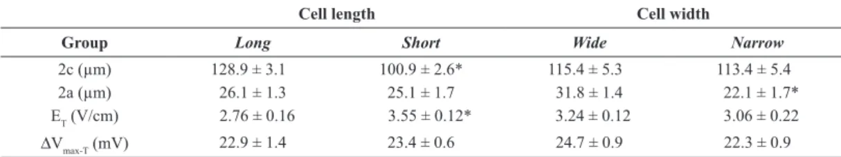
Table 1. Cell dimensions and threshold variables in isolated rat ventricular myocytes. Means ± standard error values of cell length (2c), cell width (2a), threshold electric ield (ET) and estimated maximum variation in transmembrane electrical potential at threshold (∆Vmax-T) are
presented for two pairs of groups: one pair included cells with the greatest and lowest 2c values (long and short), and the other, with cells
showing the greatest and lowest 2a values (wide and narrow), in a total population of 23 myocytes. N = 10 for each group. *p < 0.05 for
comparison of the groups within the pair (Student’s t test).
Cell length Cell width
Group Long Short Wide Narrow
2c (µm) 128.9 ± 3.1 100.9 ± 2.6* 115.4 ± 5.3 113.4 ± 5.4
2a (µm) 26.1 ± 1.3 25.1 ± 1.7 31.8 ± 1.4 22.1 ± 1.7*
ET (V/cm) 2.76 ± 0.16 3.55 ± 0.12* 3.24 ± 0.12 3.06 ± 0.22
Nevertheless, our results indicate that, in both types
of stimulation, the ield effects are exerted within a
narrow range of membrane polarization, which was
not signiicantly affected by the cell dimensions. It is well accepted that the application of an external ield results in the production of electrical potential
gradient in the medium that surrounds the outer surface of the membrane. The electrical potential, which is null at the point equidistant to the electrodes and
increases with the proximity of the electrodes, leads
to space-dependent variation of Vm, which attains its
maximum modulus (∆Vmax) at the membrane regions
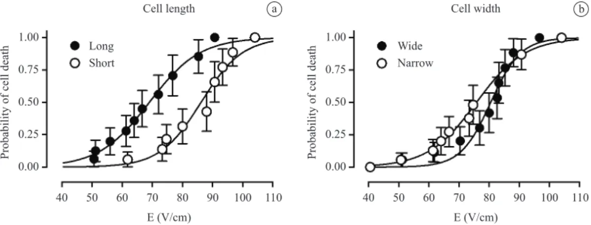
a b
Figure 1. Curves that describe the probability of ield-induced lethal injury to isolated rat ventricular myocytes as a function of the ield intensity (E), determined in the cell groups formed according to the values of cell length (panel a) and width (panel b). Data are presented as means and respective standard error. The mean parameters of the itted curves are presented in Table 2. Statistical difference was observed only for the curves shown in panel a (p < 0.001; Mantel-Cox test).
Table 2. Mean values of the parameters of the lethality curves in isolated rat ventricular myocytes. The values of ield intensity and maximum change in membrane potential (∆Vmax) associated with the probability of death in 50% of the cell population (L50), as well as the Hill
coeficients (n) of the curves, are presented for the pairs of cell groups arranged according to cell length (long and short) and cell width (wide and narrow). The lethality curves for ield intensity and ∆Vmax are presented in Figures 1 and 2, respectively.
Cell length Cell width
Group Long Short Wide Narrow
Field: L50 (V/cm) 69.0 86.2 80.5 75.8
n 0.052 0.064 0.087 0.054
∆Vmax: L50 (mV) 585 579 605 550
n 0.005 0.010 0.010 0.010
Figure 2. Curves that describe the probability of ield-induced lethal injury to isolated rat ventricular myocytes as a function of the estimated maximum variation of membrane potential (∆Vmax), determined in the cell groups formed according to the values of cell length (panel a) and
width (panel b). Data are presented as means and respective standard error. The mean parameters of the itted curves are presented in Table 2. No statistical difference was observed for the curves shown in either panel a or b (p > 0.14; Mantel-Cox test).
closest to the electrodes (negative at the anodal end and positive at the cathodal end) (Knisley et al., 1993; Neunlist and Tung, 1997; Sharma and Tung, 2002). If the myocyte major dimension (length) is oriented parallel to the ield (i.e., to the imaginary axis between the electrodes), the ∆Vmax developed at the cell
extremities in response to a certain ield intensity should
be considerably greater than that if the cell orientation is transversal: in the latter case the membrane sites where ∆Vm reaches its maximum are much farther
from the electrodes. This has been conirmed by both modeling and experimental observations (e.g.,
Knisley et al., 1993; Oliveira et al., 2008; Sharma and Tung, 2002; Valič et al., 2003), and should result in the need of greater ield intensity for Vm to reach the excitation threshold when orientation is transversal. This was the proposed mechanism to explain why lower ield intensity is required for excitation when the orientation of the cell major axis with respect to the external ield is parallel (Bassani et al., 2006;
Oliveira et al., 2008; Tung et al., 1991).
By the same token, when a ield of a given intensity
is applied to cells with parallel orientation, however with different lengths, ∆Vmax should be the greater as the longer is the cell. Our present results are in agreement with this prediction, in that, although ∆Vmax at threshold was similar in the long and short cell
groups, the ield intensity required for its attainment was lower for longer cells. Cell width, on the other hand, did not show signiicant inluence on myocyte sensitivity to the ield. This was expected, because, for parallel cell orientation, the minor axis of the cell is transversal to the axis between the electrodes. Thus,
cell width should not be an important determinant
of maximal cell polarization under our experimental conditions (see Equations 1-5).
The presently obtained threshold ∆Vm values were close to those determined theoretically
and experimentally in rat ventricular myocytes (Bassani et al., 2006; Oliveira et al., 2008;
Oshiyama et al., 2012). While the results at threshold
level were fully compatible with the K-P model
predictions and experimental data, the main question raised was regarding stimulation with HIEF. Although
the K-P model was used previously in this condition
to examine polarization by HIEF applied at different directions (Oliveira et al., 2008), apparent
space-dependent differences in electroporation severity
made it dificult to ascertain whether the model was applicable for ield-induced cell death.
Differently from threshold excitation, which is
a clear-cut phenomenon, electropermeabilization
is graded with regard to both extension and pore
lifetime, depending on shock parameters, such as
intensity and duration (Fedorov et al., 2008; Neunlist and Tung, 1997; Oliveira et al., 2005; Tovar and Tung, 1992). Possibly for this reason, the range of the
critical Vm for permeabilization described for cardiac myocytes and millisecond shock duration is quite wide
(250-1000 mV) (e.g., Cheek and Fast, 2004; Neunlist and Tung, 1997; O’Neill and Tung, 1991; Tovar and Tung, 1992). However, in the case of HIEF-induced cell
death, it is plausible to conclude that permeabilization
should be suficiently extensive and long-lasting to
allow sustained calcium overload and disruption of cell structure. Moreover, it can also be assumed that,
under identical experimental conditions, the ∆Vmax
necessary to cause such severe injury should be similar
for any rat ventricular cardiomyocyte, no matter its
dimensions. Thus, conirmation of the hypothesis that the K-P model was applicable to HIEF-induced cell death required that the inluence of cell dimensions on ield sensitivity reproduced what had been seen
at threshold stimulation, and that ∆Vmax values were insensitive to cell dimensions. Our results show that
these requirements were met: a) signiicantly lower ield intensities (by ~20%) were necessary to produce lethal injury in long vs. short cells; b) cell width did not exert signiicant inluence on ield sensitivity; and c) the estimated ∆Vmax was fairly similar in all groups
(as seen by the absence of signiicant difference in
the lethality curves as a function of ∆Vmax, and the variation below 10% in the ∆Vmax values associated
with 50% probability of cell death).
According to our ∆Vmax values, lethal injury should ensue when Vm exceeds –480 mV at the cell
anodal extremity (assuming a resting Vm of – 80 mV) (Bassani et al., 2004; Oshiyama et al., 2012). Although within the experimentally determined
Vm critical range for electropermeabilization in
cardiac myocytes (O’Neill and Tung, 1991), this value is likely to represent an overestimate. It should be stressed that lethal injury must require
considerably severe electroporation. Although transient and mild electroporation may occur for ∆Vmax values below 200 mV (Cheek and Fast, 2004), it does not necessarily lead to cell death, but only
to temporary hypoexcitability and spontaneous activity (Oliveira et al., 2005, 2008). Thus, higher
membrane polarization should be necessary to induce
permeabilization that leads to cell death. In addition,
electroporation is a self-limiting phenomenon: the ion
luxes that develop through the electropores curtail membrane polarization due to the ield (Cheek and Fast, 2004; Neunlist and Tung, 1997). Because the K-P
model does not take into account possible dielectric breakdown of the membrane, the estimated ∆Vmax
should be regarded as the maximum polarization
charging, and thus is probably higher that the actual values. Nevertheless, the present observation that
it was not signiicantly affected by cell dimensions argues in favor of the usefulness of this model for HIEF
application, as long as one is aware of its limitations regarding the absolute values of ∆Vmax.
Finally, the greatest sensitivity to the HIEF
deleterious effects in longer cells raises the possibility that hypertrophied myocytes would be more susceptible
to lethal injury during deibrillation. Although in the
whole heart cells are electrically connected and thus the myocardium should behave as a functional syncytium, a remarkable similarity of the electrical interaction with
external ield has been observed in isolated myocytes and whole hearts of neonatal rats (Gomes et al., 2001, 2002). Increase in cell length is known to occur in some physiological (pregnancy; Virgen-Ortiz et al., 2009) and pathophysiological conditions, such as mitral insuficiency (Dillon et al., 2012), spontaneous arterial hypertension (R.A. Bassani, unpublished results), dilated cardiomyopathy (Kaistura et al., 1995), and familial hypertrophic cardiomyopathy (Brouwer et al., 2011). Cardiac hypertrophy has been considered
an independent factor associated with increased
risk of arrhythmia and sudden death (Bender et al.,
2012; Reinier et al., 2011), particularly in the case of hypertrophic cardiomyopathy (Brouwer et al., 2011). Although during deibrillation/cardioversion only a fraction of the cardiac myocytes are expected to be in parallel orientation with the ield, our results
are suggestive that these cells might be at risk of
severe injury, which might compromise the success of deibrillation (due to possible conduction block)
and recovery of adequate cardiac pumping function.
Accordingly, increased deibrillation threshold has been reported in hypertrophic hearts (Almquist et al.,
2005; Kalighu et al., 1997; Ott and Reiter, 1997).
Conclusion
From the present results, it is possible to conclude that the cell major dimension exerts signiicant inluence
on the polarization of ventricular myocytes induced
by parallel external electrical ields, not only at the excitation threshold, but also at high, deibrillator-type ield intensities. As in both cases the results are in
agreement with the predictions from the K-P model, it appears that this model is also applicable to the
interaction of cells with HIEF. Additionally, the inding of higher sensitivity to HIEF lethal injury associated
with greater myocyte length raises the possibility of
greater susceptibility to HIEF-induced damage in
hypertrophied myocardial cells.
Acknowledgements
The authors are grateful to the team of the Área de
Pesquisa e Desenvolvimento at CEB/UNICAMP
for the valuable technical support. This study was
funded by CNPq (Proc. 302996/2011-7) and CAPES (scholarship to J.T.G.).
References
Almquist AK, Montgomery JV, Haas TS, Maron BJ. Cardioverter-defibrillator implantation in high-risk patients with hypertrophic cardiomyopathy. Heart Rhythm. 2005; 2:814-9. PMid:16051115. http://dx.doi. org/10.1016/j.hrthm.2005.05.008
Bassani RA, Altamirano J, Puglisi JL, Bers DM. Action potential duration determines sarcoplasmic reticulum Ca2+ reloading in mammalian ventricular myocytes. Journal of Physiology. 2004; 559:591-607. PMid:15243136 PMCid:1665117. http://dx.doi.org/10.1113/ jphysiol.2004.067959
Bassani RA, Lima KA, Gomes PAP, Oliveira PX, Bassani JWM. Combining stimulus direction and waveform for optimization of threshold stimulation of isolated ventricular myocytes. Physiological Measurement. 2006; 27:851-63. PMid:16868351. http:// dx.doi.org/10.1088/0967-3334/27/9/008
Bender SR, Friedman DJ, Markowitz SM, Lerman BB, Okin PM. Electrocardiographic left ventricular hypertrophy predicts arrhythmia and mortality in patients with ischemic cardiomyopathy. Journal of Interventional Cardiac Electrophysiology. 2012; 34:237-45.PMid:22354775. http://dx.doi.org/10.1007/s10840-011-9661-2
Brouwer WP, Van Dijk SJ, Stienen GMJ, Van Rossum AC, Van der Velden J, Germans T. The development of familial hypertrophic cardiomyopathy: from mutation to bedside. European Journal of Clinical Investigation. 2011; 41:567-78. PMid:21158848. http:// dx.doi.org/10.1111/j.1365-2362.2010.02439.x
Cheek ER, Fast VG. Nonlinear changes of transmembrane potential during electrical shocks: role of membrane electroporation. Circulation Research. 2004; 94:208-14. PMid:14670844. http://dx.doi.org/10.1161/01. RES.0000111526.69133.DE
Dillon AR, Dell’Italia LJ, Tilson M, Killingsworth C, Denney T, Hathcock J, Botzman L. Left ventricular remodeling in preclinical experimental mitral regurgitation of dogs. Journal of Veterinary Cardiology. 2012; 14:73-82. PMid:22386719. http://dx.doi.org/10.1016/j.jvc.2012.01.012
Fotuhi PC, Epstein AE, Ideker RE. Energy levels for deibrillation: what is of real clinical importance: American Journal of Cardiology. 1999; 83:24D-33D. http://dx.doi. org/10.1016/S0002-9149(98)00966-7
Gomes PAP, Bassani RA, Bassani JWM. Electric ield stimulation of cardiac myocytes during postnatal development. IEEE Transactions on Biomedical Engineering. 2001; 48:630-6. PMid:11396593. http:// dx.doi.org/10.1109/10.923781
Gomes PA, De Galvão KM, Mateus EF. Excitability of isolated hearts from rats during postnatal development. Journal of Cardiovascular Electrophysiology. 2002; 13:355-60. P M i d : 1 2 0 3 3 3 5 2 . http://dx.doi.org/10.1046/ j.1540-8167.2002.00355.x
Kaistura J, Zhang X, Liu Y, Szoke E, Chen W, Olivetti G, Hintze TH, Anversa P. The cellular basis of pacing-induced dilated cardiomyopathy: myocyte cell loss and myocyte cellular reactive hypertrophy. Circulation. 1995; 92:2306-17. PMid:7554216. http://dx.doi.org/10.1161/01.CIR.92.8.2306 Kalighi K, Daly B, Leino EV, Shorofsky SR, Kavesh NG, Peters RW, Gold MR. Clinical predictors of transvenous deibrillator energy requirement. American Journal of Cardiology. 1997; 79:150-3. http://dx.doi.org/10.1016/ S0002-9149(96)00702-3
Klee M, Plonsey R. Stimulation of spheroidal cells: the role of cell shape. IEEE Transactions on Biomedical Engineering. 1976; 23:347-54. PMid:1278928. http://dx.doi. org/10.1109/TBME.1976.324597
Kleinbaum DG. Survival Analysis: a Self-learning Text. New York: Springer-Verlag; 1996.
Knisley SB, Grant AO. Asymmetrical electrically induced injury of rabbit ventricular myocytes. Journal of Molecular and Cellular Cardiology. 1985; 27:1111-22. http://dx.doi. org/10.1016/0022-2828(95)90047-0
Knisley SB, Blitchington TF, Hill BC, Grant AO, Smith WM, Pilkington TC, Ideker R. Optical measurements of transmembrane potential changes during electrical field stimulation of ventricular cells. Circulation Research. 1993; 72:255-70. PMid:8418982. http://dx.doi. org/10.1161/01.RES.72.2.255
Krassowska W, Filev PD. Modeling electroporation in a single cell. Biophysical Journal. 2007; 92:404-17. PMid:17056739 PMCid:1751390. http://dx.doi.org/10.1529/ biophysj.106.094235
Mantel N. Evaluation of survival data and two new rank order statistics arising in its consideration. Cancer Chemotherapy Reports. 1966; 50:163-70. PMid:5910392.
Neunlist M, Tung L. Dose-dependent reduction of cardiac transmembrane potential by high intensity electrical shocks. American Journal of Physiology. 1997; 273:H2817-25. PMid:9435619.
Oliveira PX, Bassani RA, Bassani JWM. Cytosolic Ca2+ accumulation in ventricular myocytes after stimulation with high-intensity electric ields. Biophysical Journal. 2005; 88 (suppl. 1):1 (abstr. 1514).
Oliveira PX, Bassani RA, Bassani JWM. Lethal effect of electric ields on isolated ventricular myocytes. IEEE Transactions on Biomedical Engineering. 2008; 55:2635-42. PMid:18990634. http://dx.doi.org/10.1109/ TBME.2008.2001135
O’Neill RJ, Tung L. Cell-attached patch clamp study of the electropermeabilization of amphibian cardiac cells. Biophysical Journal. 1991; 59:1028-39. http://dx.doi. org/10.1016/S0006-3495(91)82318-9
Oshiyama NF, Bassani JWM, Bassani RA. Coniguração do potencial de ação em miócitos ventriculares isolados de ratos neonatos e adultos. In: Proceedings of the I Simposio Brasileiro de Eletroisiologia Celular; 2012; Belo Horizonte, MG. Belo Horizonte; 2012. p. 40 (abstr.).
Ott P, Reiter MJ. Effect of ventricular dilatation on deibrillation threshold in the isolated perfused rabbit heart. Journal of Cardiovascular Electrophysiology. 1997; 8:1013-9. PMid:9300299. http://dx.doi.org/10.1111/j.1540-8167.1997. tb00625.x
Penna LB, Bassani RA. Increased spontaneous activity and reduced inotropic response to catecholamines in ventricular myocytes from footshock-stressed rats. Stress. 2010; 13:73-82. PMid:19697264. http://dx.doi. org/10.3109/10253890902951778
Reinier K, Dervan C, Singh T, Uy-Evanado A, Lai S, Gunson K, Jui J, Chugh SS. Increased left ventricular mass and decreased left ventricular systolic function have independent pathways to ventricular arrhythmogenesis in coronary artery disease. Heart Rhythm. 2011; 8:1177-82. PMid:21376836 PMCid:3123721. http://dx.doi.org/10.1016/j. hrthm.2011.02.037
Ricardo RA, Oliveira PX, Bassani RA, Bassani JWM. Compact cell image projector: application to study the relationship between stimulus interval and contraction amplitude in isolated rat cardiomyocytes. Revista Brasileira de Engenharia Biomédica. 2006; 22:151-60.
Sharma V, Tung L. Spatial heterogeneity of transmembrane potential responses of single guinea-pig cardiac cells during electric ield stimulation. Journal of Physiology. 2002; 542:477-92. PMid:12122146 PMCid:2290429. http://dx.doi.org/10.1113/ jphysiol.2001.013197
Sowell B, Fast VG. Ionic mechanism of shock-induced arrhythmias: role of intracellular calcium. Heart Rhythm. 2012; 9:96-104. PMid:21878203. http://dx.doi. org/10.1016/j.hrthm.2011.08.024
Tovar O, Tung L. Electroporation and recovery of cardiac cell membrane with rectangular voltage pulses. American Journal of Physiology. 1992; 263:H1128-36. PMid:1415761. Tung L, Sliz N, Mulligan MR. Inluence of electrical axis of stimulation on excitation of cardiac muscle cells. Circulation Research. 1991; 69:722-30. PMid:1873867. http://dx.doi. org/10.1161/01.RES.69.3.722
Authors
Jair Trapé Goulart
Department of Biomedical Engineering, School of Electrical and Computer Engineering – FEEC, University of Campinas – UNICAMP, Rua Alexander Fleming 163,
Cidade Universitária Zeferino Vaz, CEP 13083-881, Campinas, SP, Brasil.
Pedro Xavier de Oliveira, José Wilson Magalhães Bassani, Rosana Almada Bassani* Department of Biomedical Engineering, School of Electrical and Computer Engineering – FEEC, University of Campinas – UNICAMP, Rua Alexander Fleming 163,
Cidade Universitária Zeferino Vaz, CEP 13083-881, Campinas, SP, Brasil. Center for Biomedical Engineering – CEB, University of Campinas – UNICAMP,
Rua Alexander Fleming 163, Cidade Universitária Zeferino Vaz, CEP 13083-881, Campinas, SP, Brasil. Virgen-Ortiz A, Marin JL, Elizalde A, Castro E, Stefani E,
Toro L, Muñiz J. Passive mechanical properties of cardiac tissue in heart hypertrophy during pregnancy. Journal of Physiological Science. 2009; 59:391-6. PMid:19565322. http://dx.doi.org/10.1007/s12576-009-0047-5
Weaver JC, Chizmadzhev YA. Theory of electroporation: a review. Bioelectrochemistry and Bioenergetics. 1996; 41:135-60. http://dx.doi.org/10.1016/ S0302-4598(96)05062-3
Yabe S, Smith W, Daubert J, Wolf P, Rollins D, Ideker R. Conduction disturbances caused by high current density electric ields. Circulation Research. 1990; 66:1190-203. PMid:2335021. http://dx.doi.org/10.1161/01.RES.66.5.1190