The inluence of hydroxyurea on oxidative stress in sickle cell anemia
Lidiane de Souza Torres1
Danilo Grünig Humberto da Silva1 Edis Belini Junior1
Eduardo Alves de Almeida1 Clarisse Lopes de Castro Lobo2 Rodolfo Delini Cançado3 Milton Artur Ruiz4
Claudia Regina Bonini-Domingos1
1Universidade Estadual Paulista – UNESP, São José do Rio Preto, SP, Brazil
2Instituto de Hematologia “Arthur de Siqueira Cavalcanti” – HEMORIO, Rio de Janeiro, RJ, Brazil 3Faculdade de Ciências Médicas da Santa Casa de São Paulo – FCMSCSP, São Paulo, SP, Brazil 4Universidade de São Paulo – USP, São Paulo, SP, Brazil
Conlict-of-interest disclosure:
The authors declare no competing inancial interest
Submitted: 12/16/2011 Accepted: 8/8/2012
Corresponding author:
Lidiane de Souza Torres Departamento de Biologia,
Laboratório de Hemoglobinas e Genética das Doenças Hematológicas. Universidade Estadual Paulista, UNESP
Rua Cristóvão Colombo, 2265, Jardim Nazareth 15054-000 São José do Rio Preto, SP, Brazil [email protected]
www.rbhh.org or www.scielo.br/rbhh DOI: 10.5581/1516-8484.20120106
Introduction
Sickle cell anemia (SCA) is one of the most common genetic disorders in the world. It is characterized by homozygous hemoglobin (Hb) S and represents the most severe form of sickle cell disease (SCD)(1). Hb S is caused by a mutation in the β-globin gene in which the sixth amino acid is changed from glutamic acid to valine due to a substitution of adenine for thymine(2). SCA presents a
series of clinical manifestations which are inluenced by genetic and environmental factors. These
factors result in many phenotypes, mainly mediated by the polymerization of Hb S, hemolysis and cell adhesion to endothelium which leads to vascular occlusion(3,4). Blood transfusions, folic acid
supplementation and hydroxyurea (HU) are the most common treatments in SCA. HU is an oral
drug whose main effect in SCA is the increased synthesis of fetal Hb (Hb F) which reduces the
frequency of vaso-occlusive episodes, pain crises, hospitalizations and blood transfusions(5).
Oxidative stress is one of the factors that modulates the phenotypic expression of SCA. This stress inluences the vaso-occlusive process by increasing the adhesive properties
of erythrocytes, leukocytes and platelets to the endothelium(6). Normal erythrocytes suffer
oxidative stress resulting from the production of reactive oxygen species (ROS) due to the oxygen metabolism. This metabolic ROS production is increased in patients with hemoglobinopathies, causing oxidative damage such as lipid peroxidation. The release of Hb in plasma and ischemia-reperfusion cycles are characteristic of SCD, increasing oxidative
stress and requiring a more effective antioxidant system(7,8).
The literature suggests that excess ROS has implications in the pathophysiology of SCA. Thus an evaluation of the oxidative stress in these patients may provide important information regarding the current use of medications, such as HU, and may lead to new therapeutic strategies. Therefore, considering the intense generation of ROS with the presence and hemolysis of Hb S in SCA, the inluence of HU on oxidative stress was evaluated using cell damage markers and antioxidant capacity by comparing patients taking HU with those who
were not taking the medication and a control group.
Methods
Subjects
Peripheral blood samples of 33 SCA patients (21 females and 12 males; mean age: 28 ± 15
years) from blood banks in São Paulo and Rio de Janeiro, southeastern Brazil were evaluated.
Objective: The oxidative stress in 20 sickle cell anemia patients taking hydroxyurea and 13 sickle cell anemia patients who did not take hydroxyurea was compared with a control group of 96 individuals without any hemoglobinopathy.
Methods: Oxidative stress was assessed by thiobarbituric acid reactive species production, the Trolox-equivalent antioxidant capacity and plasma glutathione levels.
Results: Thiobarbituric acid reactive species values were higher in patients without speciic medication,
followed by patients taking hydroxyurea and the Control Group (p < 0.0001). The antioxidant capacity was higher in patients taking hydroxyurea and lower in the Control Group (p = 0.0002 for Trolox-equivalent antioxidant capacity and p < 0.0292 for plasma glutathione). Thiobarbituric acid reactive species levels were correlated with higher hemoglobin S levels (r = 0.55; p = 0.0040) and lower hemoglobin F concentrations (r = -0.52; p = 0.0067). On the other hand, plasma glutathione levels were negatively correlated with hemoglobin S levels (r = -0.49; p = 0.0111) and positively associated with hemoglobin F values (r = 0.56; p = 0.0031).
Conclusion: Sickle cell anemia patients have high oxidative stress and, conversely, increased antioxidant activity. The increase in hemoglobin F levels provided by hydroxyurea and its antioxidant action may explain the reduction in lipid peroxidation and increased antioxidant defenses in these individuals.
All patients were treated with folic acid supplementation however
only 20 of the patients were taking HU. A Control Group was
formed of 96 individuals without hemoglobinopathies and not
taking HU (57 females and 39 males; mean age: 23 ± 6 years)
from southeastern Brazil.
All SCA patients were screened using a questionnaire.
Pregnant women, smokers and individuals who drank signiicant quantities of alcohol were excluded from the study as were patients
who had had strokes, pain or hemolytic crisis or had received blood transfusions within the two months prior to the start of the study. Patients who had taken medications known to affect the analyzed parameters (such as acetylsalicylic acid, antibiotics or vitamins) within the 24 hours prior to sample collection were
also excluded from the study. All subjects gave their informed
consent and the study was approved by the Data Safety Monitoring Board (DSMB) according to Brazilian regulations.
Genotype investigation
All samples were submitted to classical hemoglobin diagnosis techniques including electrophoresis at alkaline
and acid pH, to evaluate the Hb migration proile and high
performance liquid chromatography (HPLC) to measure Hb fractions(9-11). Genomic DNA was extracted employing the
phenol-chloroform method for molecular analysis(12). The
ampliication of the segment that encodes Hb S was performed using speciic primers (sense: 5’-GGCAGAGCCATCTATTGCTTA-3’; antisense: 5’-ACCTTAGGGTTGCCCATAAC-3’) and cleavage
was achieved by the action of restriction endonuclease FastDigest
Ddel (Fermentas, USA).(13)
Biochemical analysis
Oxidative stress analysis was evaluated based on the detection of lipid peroxidation and antioxidant capacity markers in plasma. The lipid peroxidation levels were calculated by the thiobarbituric acid reactive species (TBARS) technique. This method is based on the reaction of malondialdehyde and other aldehydes, which are by-products of membrane damage caused by ROS, with thiobarbituric acid (TBA) at low pH and high temperature forming a complex with
maximum light absorption at 535 nm(14).
The antioxidant capacity was evaluated using Trolox (6-hydroxy-2,5,7,8-tetramethylchroman-2-carboxylic acid), a potent antioxidant similar to vitamin E. The Trolox-equivalent antioxidant capacity (TEAC) method is based on the reaction
between 2,2′-azino-bis (3-ethylbenzothiazoline-6-sulphonic acid)
(ABTS) (SIGMA, A1888), 2,2’-azino-bis (3-ethylbenzothiazoline-6-sulfonate) and potassium persulfate (K2S2O8). This reaction
produces the radical cation ABTS•+, a green/blue chromophore.
The addiction of one antioxidant to this radical cation reduces ABTS, resulting in a solution discoloration that is evaluated at 734 nm to determine the total antioxidant capacity(15).
Plasma glutathione (GSH) concentrations were determined in EDTA-treated plasma samples by the quantiication in HPLC
coupled to a coulometric electrochemical detector (Coulochem III
ESA, Bedford, MA) set with a potential of 650 mV(16). Under these
conditions, GSH is clearly eluted in ~ 6 min. GSH was extracted
Table 1 - Differences between hemoglobin proiles of the SCA patients taking HU or not
Sickle cell anemia patients
p-value
(-HU) (+HU)
Hb S 86.63 ± 4.42 80.44 ± 7.47 0.015
Hb A2 3.63 ± 1.28 4.11 ± 0.50 0.250
Hb F 6.47 ± 4.23 11.89 ± 6.92 0.018
-HU: patients not taking hydroxyurea; +HU: patients taking hydroxyurea.
from the plasma samples by adding perchloric acid to the plasma
sample (10% inal concentration). After vigorous stirring and 10 min on ice, the mixture was centrifuged at 825 g for 10 min at 4°C. The extract was then iltered through Millex syringe ilter units (0.22 μm) and directly injected into the HPLC. Calculations were based on a calibration curve previously constructed by injecting known GSH standards into the HPLC system.
Statistical analysis
Data were tested for normality and homoscedasticity using
the Shapiro-Wilk and Levene tests, respectively. Analysis of variance (ANOVA) was employed complemented by Tukey’s test for data with normal distribution. The Kruskal-Wallis test was employed followed by Dunn’s test for non-parametric data. The correlations between Hb concentrations and lipid peroxidation and plasma GSH levels were achieved by the Pearson linear correlation test. The level of signiicance was assumed as 0.05
and analysis was made using the Statistica 8.0 software.
Results
Hemoglobin fraction concentration in SCA patients
The Hb proiles of SCA individuals were compared depending on whether patients took HU or not. Results showed
higher Hb F levels (p = 0.018) and lower Hb S concentrations
(p = 0.015) among individuals taking HU compared to those who were not taking this medication (Table 1).
Lipid peroxidation – thiobarbituric acid reactive species dosage
The generation of ROS was indirectly measured though the analysis of lipid peroxidation. The values were higher for SCA patients (1450 ± 549 ng/mL) compared to the Control Group (239 ± 159 ng/mL)(p < 0.0001). Patients receiving HU had lower lipid peroxidation than those without speciic medication however they still had higher levels than the Control Group (p < 0.0001).
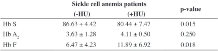
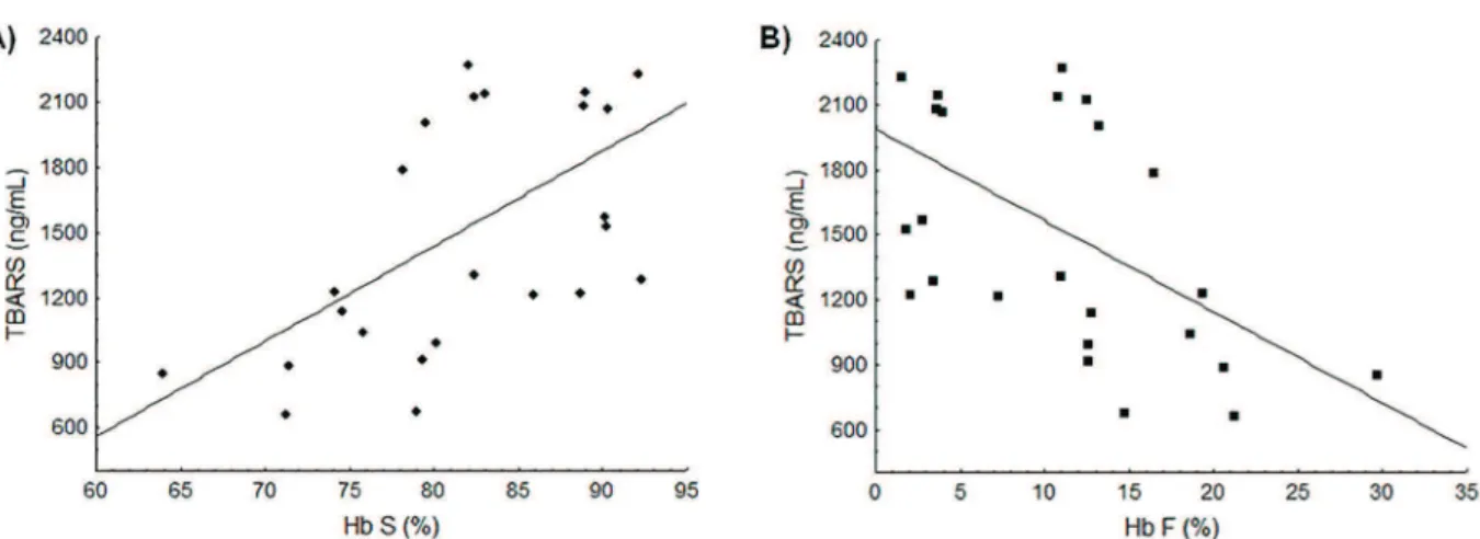
Correlation analysis was carried out comparing Hb S and Hb F
levels and the TBARS dosage. Results showed that TBARS values
in SCA patients were positively correlated with Hb S concentrations
(r = 0.55; p = 0.0040) relecting increased lipid peroxidation in the
presence of Hb S (Figure 1A). Negative correlation was observed
between TBARS and Hb F levels (r = -0.52; p = 0.0067), showing
Figure 1 - Association between the Hb S and Hb F concentrations and lipid peroxidation levels. (A) Positive linear correlation between Hb S and TBARS levels (r = 0.55; p-value = 0.0040). (B) Negative linear correlation between Hb F and TBARS levels (r = -0.52; p-value = 0.0067).
Figure 2 - Association between the Hb S and Hb F concentrations and the GSH levels. (A) Negative linear correlation between Hb S and GSH levels (r = -0.49; p-value = 0.0111). (B) Positive linear correlation between Hb F and GSH levels (r = 0.56; p-value = 0.0031).
Antioxidant Capacity – Trolox-equivalent antioxidant capacity assay and plasma glutathione levels
The overall antioxidant capacity analyzed by TEAC showed
higher values for SCA patients (2.04 ± 0.15 mM) than for the
Control Group (1.93 ± 0.15 mM; p = 0.0028). Patients taking HU had higher TEAC values when compared to the group of patients that did not take HU (p = 0.0002). No correlation was observed between Hb concentrations and TEAC levels.
The GSH concentration in the Control Group was 0.41 ± 0.38 μM with this value being about two times higher in SCA patients (0.88 ± 0.69 μM; p = 0.0292). Patients taking HU had higher GSH levels that were signiicantly different to the Control Group (p < 0.0001). Higher Hb S concentrations were correlated with decreased GSH levels (r = -0.49; p = 0.0111: Figure 2A) while Hb F concentrations were positively correlated with GSH
values (r = 0.56; p = 0.0031: Figure 2B).
Discussion
The oxidative stress and antioxidant markers were evaluated in SCA patients who were either taking HU or
not and the results were compared to the results of a control
group. The correlation between Hb S levels and TBARS values, as well as lipid peroxidation in SCA patients suggest that oxidative stress may result from high levels of meta Hb S
which is less stable than meta Hb A, leading to the formation of hemichromes and hemolysis with the release of heme iron.
Oxidative stress may be even higher during vaso-occlusive
crises and painful episodes(17).
Hb F levels were higher in SCA patients. It is known that in SCA patients, the concentration of Hb F ranges from 1% to 30% and it is inherited as a quantitative trait. A trend of increased Hb
F levels in SCA patients taking HU was observed in this study
Conclusion
The results of this study show that the inluence of Hb S on the oxidative status is relected by increased lipid peroxidation and antioxidant status in SCA patients. High Hb F concentrations are associated with less oxidative stress. Treatment using HU decreased lipid peroxidation and contributed to the body’s antioxidant defenses.
Acknowledgements
The authors would like to thank the following Brazilian foundations: Fundação de Amparo à Pesquisa do Estado de São Paulo (FAPESP), Conselho Nacional de Desenvolvimento Cientíico e Tecnológico (CNPq), Coordenação de Aperfeiçoamento de Pessoal de Nível Superior (CAPES) and the Ministry of Health, for the inancial support.
References
1. Frenette PS, Atweh GF. Sickle cell disease: old discoveries, new concepts, and future promise. J Clin Invest. 2007;117(4): 850-8.
2. Higgs DR, Wood WG. Genetic complexity in sickle cell disease. Proc Natl Acad Sci USA. 2008;105(33):11595-6. Comment on: Proc Natl Acad Sci USA. 2008;105(33):11869-74.
3. Figueiredo MS. Fatores moduladores da gravidade da evolução clínica da
anemia falciforme. Rev Bras Hematol Hemoter. 2007;29(3):215-7. 4. Rees DC, Williams TN, Gladwin MT. Sickle-cell disease. Lancet.
2010;376(9757):2018-31.
5. Kovacic P. Hydroxyurea (therapeutics and mechanism): metabolism, carbamoyl nitroso, nitroxyl, radicals, cell signaling and clinical applications. Med Hypotheses. 2011;76(1):24-31.
6. Chaves MA, Leonart MS, do Nascimento AJ. Oxidative process
in erythrocytes of individuals with hemoglobin S. Hematology.
2008;13(3):187-92.
7. Kuypers FA. Red cell membrane lipids in hemoglobinopathies. Curr Mol Med. 2008;8(7):633-8.
8. Kato GJ, Hebbel RP, Steinberg MH, Gladwin MT .Vasculopathy in sickle
cell disease: biology, pathophysiology, genetics, translational medicine,
and new research directions. Am J Hematol. 2009;84(9):618–25. 9. Marengo-Rowe AJ. Rapid electrophoresis and quantitation of
haemoglobin on cellulose acetate. J Clin Pathol. 1965;18(6):790-2. 10. Vella F. Acid agar gel electrophoresis oh human hemoglobins. Am J Clin
Pathol. 1968;49(3):440-2.
11. Bio-Rad Laboratories. Instruction Manual: Variant β-thalassemia short program. Hercules, California: Bio-Rad Laboratories; 1999.
12. Sambrook J, Fritcsh EF, Manatis T. Molecular cloning: a laboratory
manual. 2nd ed. New York: Cold Spring Harbor Laboratory Press; 1989.
13. Saiki RK, Scharf S, Faloona F, Mullis KB, Horn GT, Erlich HA, et al. Enzymatic ampliication of beta-globin genomic sequences and
restriction site analysis for diagnosis of sickle cell anemia. Science.
1985;230(4732):1350-4.
14. Percário S. Dosagem do dialdeído malônico. Newslab. 2004;6:46-50. 15. Re R, Pellegrini N, Proteggente A, Pannala A, Yang M, Rice-Evans
C. Antioxidant activity applying an improved ABTS radical cation decolorization assay. Free Radic Biol Med. 1999;26(9-10):1231-7. 16. Rodríguez-Ariza A, Toribio F, López-Barea J. Rapid determination
of glutathione status in ish liver using high-performance liquid SCA pathophysiology can be viewed as a cycle driven
by hemolysis, oxidative stress, inlammation, cell adhesion to endothelium and vaso-occlusion. During hemolysis, iron is
transformed from ferric to ferrous iron, thus producing meta
Hb, with heme iron release. The hydrophobic character of free
heme iron allows it to merge with cell membranes, increasing
the susceptibility to oxidant-mediated destruction as well as the generation of ROS, characterizing lipid peroxidation. Thus, the level of TBARS is a good indicator of pro-oxidant stimuli,
since it measures malondialdehyde and other aldehydes which are subproducts of cell membrane destruction(19).
Patients who are not taking HU but are taking folic acid, showed increased lipid peroxidation levels compared to patients taking HU. Folate acid is important in the formation of red
blood cells because it participates in the purine and pyrimidine
metabolism for DNA and RNA synthesis(20). In turn, HU is a
deoxyribonucleotide reductase which is able to increase Hb F synthesis due to its myelotoxicity. Studies have shown that this drug leads to increased Hb F levels in approximately 50% of
patients with SCD. High Hb F concentrations may decrease the severity of illness because of inhibition in the polymerization of Hb S. Hb F alter contact sites between Hb molecules, impairing polymer formation with consequent reduction in the sickling process(21-23). This is a potential reduction factor of hemolysis and the consequent cell damage caused by substances released
during this process, for example, iron. Kaul et al. demonstrated that increased Hb F expression reduces oxidative stress in SCD
transgenic mice(24). In the current work, this antioxidant property
of HU is observed by lower TBARS values obtained in patients
in the group taking this drug compared to patients who were not.
The generation of free radicals is counteracted by enzymatic and non-enzymatic antioxidants, including antioxidants from the
diet such as vitamins C and E and GSH(25). Some studies have
found decreased GSH concentrations in erythrocytes from SCD
patients(26,27). However, Reid et al. and Kiessling et al. showed that
GSH synthesis is not impaired in SCA patients, thus suggesting
that lower GSH levels may be explained by the high demand(28,29).
Reid et al. showed that GSH synthesis was increased in 57% of SCA patients compared to control subjects, indicating that the consumption
of GSH may exceed its synthesis(28). On the other hand, GSH levels
may also be inluenced by diet, for example, due to deiciencies in
the amino acid precursors of GSH synthesis, especially cysteine(30).
Maintaining adequate levels of GSH is important for
many critical cell functions with disruptions in these processes
being observed in several human diseases. GSH deiciency is manifested primarily as an increased susceptibility to oxidative
stress and cell damage, the results of which may be an important factor in the progression of the disease. On the other hand, high
GSH levels may increase antioxidant capacity and resistance to oxidative stress(31).
Regarding treatment, HU was associated with increased antioxidant capacity. Liu et al. tested the activity of HU against free radicals such as DPPH (2,2-diphenyl-1-picril-hydrazyl) and
hydroxyl radicals; they found that HU has antioxidant activity(32).
xxx chromatography and electrochemical detection. J Chromatogr B Biomed Appl. 1994;656(2):311-8.
17. Fibach E, Rachmilewitz E. The role of oxidative stress in hemolytic anemia. Curr Mol Med. 2008;8(7):609-19.
18. Green NS, Barral S. Genetic modiiers of HbF and response to hydroxyurea in sickle cell disease. Pediatric Blood Cancer. 2011;56(2):177-81. 19. Belcher JD, Beckman JD, Balla G, Balla J, Vercellotti G. Heme degradation
and vascular injury. Antioxid Redox Signal. 2010;12(2):233-48.
20. Brasil. Ministério da Saúde. Agencia de Vigilância Sanitária – ANVISA. Manual de diagnóstico e tratamento de doenças falciformes [Internet]. Brasília: Ministério da Saúde; 2001.[cited 2011 Jan 12]. Available from: http://www.ebah.com.br/content/ABAAAA2R4AB/manual-diagnostico-tratamento-doencas-falciformes
21. Steinberg MH. Management of sickle cell disease. N Engl J Med. 1999;340(13):1021-30.
22. Charache S, Terrin ML, Moore RD, Dover GJ, Barton FB, Eckert SV, et al. Effect of hydroxyurea on the frequency of painful crises in sickle cell anemia. N Engl J Med. 1995;332(20):1317-22. Comment in: N Engl J Med. 1996;334(5):333-4; N Engl J Med. 1995;332(20):1372-4; N Engl J Med. 1995;333(15):1008-9; author reply 1009.
23. Silva LB, Gonçalves RP, Martins MF. Estudo da correlação entre os níveis de hemoglobina fetal e o prognóstico dos pacientes com anemia falciforme. Rev Bras Hematol Hemoter. 2009:31(6):417-20.
24. Kaul DK, Liu XD, Choong S, Belcher JD, Vercellotti GM, Hebbel RP. Anti-inlammatory therapy ameliorates leukocyte adhesion and
microvascular low abnormalities in transgenic sickle mice. Am J Physiol Heart Circ Physiol. 2004;287(1):H293–301.
25. Hernanz A, Fernández-Vivancos E, Montiel C, Vazquez JJ, Arnalich
F. Changes in the intracellular homocysteine and glutathione content
associated with aging. Life Sci. 2000;67(11):1317-24.
26. Chan AC, Chow CK, Chiu D. Interaction of antioxidants and their implication in genetic anemia. Proc Soc Exp Biol Med. 1999;222(3):274-82.
27. Reid M, Jahoor F. Glutathione in disease. Curr Opin Clin Nutr Metab Care. 2001;4(1):65-71.
28. Reid M, Badaloo A, Forrester T, Jahoor F. In vivo rates of erythrocyte glutathione synthesis in adults with sickle cell disease. Am J Physiol Endocrinol Metab. 2006;291(1):E73-9.
29. Kiessling K, Roberts N, Gibson JS, Ellory JC. A comparison in normal
individuals and sickle cell patients of reduced glutathione precursors and
their transport between plasma and red cells. Hematol J. 2000;1(4):243-9. 30. Cresenzi CL, Lee JI, Stipanuk MH. Cysteine is the metabolic signal responsible for dietary regulation of hepatic cysteine dioxygenase and glutamate cysteine ligase in intact rats. J Nutr. 2003;133(9):2697-702. 31. Ballatori N, Krance SM, Notenboom S, Shi S, Tieu K, Hammond CL.
Glutathione dysregulation and the etiology and progression of human diseases. Biol Chem. 2009;390(3):191-214.