Neslihan SIMSEK(a)
Ali KELES(a)
Fuat AHMETOGLU(a)
Mevlüt Sinan OCAK(a)
Saim YOLOGLU(b)
(a) Department of Endodontics, Faculty of Dentistry, Inonu University, Malatya, Turkey. (b) Department of Bioistatistics, Faculty of
Medicine, Inonu University.
Comparison of different retreatment
techniques and root canal sealers: a
scanning electron microscopic study
Abstract: The aim of this study was to evaluate the effectiveness of two retreatment techniques, in terms of the operating time and scanning electron microscopy (SEM) results, in removing three different root
ca-nal sealers from root caca-nals that were previously illed with gutta-per -cha. Sixty extracted single-rooted human premolars were divided into
three groups and illed with iRoot SP, MM Seal, and AH Plus sealers, along with gutta-percha, through a lateral compaction technique. Root canal illings of the samples were removed by ESI ultrasonic tips or R-Endo iles. The time to reach the working length was recorded. Longi
-tudinally sectioned samples were examined under SEM magniication. Each picture was evaluated in terms of the residual debris. Data were statistically analyzed with the Kruskall-Wallis test. No statistically sig
-niicant differences were found in terms of operating time (p>0.05).
Sig-niicant differences in the number of debris-free dentinal tubules were found among the root canal thirds, but this inding was not inluenced by the experimental group (p < 0.05). Resin sealer tags were observed
inside the dentinal tubules in the MM Seal group. Under the conditions of this study, it may be established that there was no difference among
the sealers and retreatment techniques.
Keywords: Endodontics; Retreatment; Microscopy, Electron, Scanning.
Introduction
The retreatment of previously treated root canals is a common proce-dure in endodontics. Successful retreatment requires the effective elimi-nation of necrotic tissue and microorganism-infected materials, such as
gutta-percha and sealer. The clinical success rate of retreatment has been estimated to vary between 50% and 90%.
Retreatment involves removing the previous root canal illing and performing advanced shaping, cleaning, and illing operations.1 Root
canal illings can be removed with solvents, heating apparatuses, lasers, hand or rotary iles, or ultrasonic instruments.2 However, well-com
-pacted illings offer resistance to instruments, and the subsequently
inadequate elimination of materials can lead to restricted access to the apical foramen. This condition can impair the root canal disinfection,
reforming process, and cleaning time. After gutta-percha removal, open dentinal tubules are necessary in order to eradicate bacteria using irrigants. However, several studies2,3,4 have shown that residual
Declaration of Interests: The authors certify that they have no commercial or associative interest that represents a conflict of interest in connection with the manuscript.
Corresponding Author:
Neslihan Simsek
E-mail: [email protected]
DOI: 10.1590/1807-3107BOR-2014.vol28.0006 Epub: XXX XX, 2014
Submitted: Feb 16, 2013
debris remains on the root canal walls after treat -ment, regardless of the instrumentation and type
of illing that are used.
Endodontic treatment has improved with the
development of better ultrasonic tips and nickel-titanium (NiTi) instruments.5 Ultrasonic systems are widely used in root canal treatment, to facilitate the
removal of broken iles from the root canal and to
prepare root-end cavities.6 However, the process of completely removing gutta-percha and sealer with
ultrasonic tips has not been fully explored.3
Of the many materials used for illing root canals,
gutta-percha is the most commonly used, in conjunc-tion with various sealers.7 According to the manufac
-turer, iRoot SP is a new calcium silicate-based root canal sealer that is radiopaque and insoluble. It con -tains calcium, calcium phosphate, calcium
hydrox-ide, and zirconium oxhydrox-ide, without aluminum. iRoot SP does not require a supplementary curing media
-tor or mixing. It offers a consistent, uniform product for illing root canals with or without gutta-percha points. It is a premixed, ready-to-use injectable sealer that has been developed for permanent canal illing.8
However, the properties of retreatment using iRoot SP are not clear.
Epoxy resin-based cements perform well as root canal sealers. AH Plus (Denstply DeTrey,
Konstanz, Germany) is a two-component paste
root canal sealer that is based on a polymeriza
-tion reac-tion of epoxy resin amines. It is composed
of diepoxide, calcium tungstate, zirconium oxide,
aerosol, 1-adamantane amine, TCD-diamine, diben -zyldiamine, aminoadamantane, and pigments.9
Another epoxy resin-based root canal sealer, MM Seal (Micro-Mega, Besançon, France), is packaged in a dual syringe. It is used for the permanent ill -ing of root canals with gutta-percha. MM Seal is composed of epoxy polymer resin, ethylene glycol
salicylate, calcium phosphate, bismuth subcarbon -ate and oxide components.10
The aim of this study was to evaluate the effec-tiveness, in terms of the operating time and scan-ning electron microscopy (SEM) results, of two
different retreatment techniques (ESI ultrasonic tips and R-Endo iles) in removing three different sealers (AH Plus, iRoot SP, and MM Seal) from
root canals that had been previously illed with
gutta-percha.
Methodology
Rootcanal preparation
After approval by the ethics committee (2012/139),
60 single-rooted and straight single-canal premolars extracted for periodontal reasons were used in this
study. Teeth were stored in puriied iltered water until they were ready for use. The working length (WL) was set at 14 mm. The root canals were pre
-pared with HERO Shaper rotary iles (Micro-Mega,
Besançon, France) with the crown-down technique, according to the manufacturer’s instructions. Files
were used to ile no. 30 at 300 rpm. With each ile change, the canal was irrigated with 2 mL of a 2.5% NaOCl. After completion, 2 mL of 17% EDTA were applied for 60 s, followed by a inal rinse with 2 mL of 2.5% NaOCl. Root canals were dried with paper points before illing.
Root canal filling
The roots were randomly divided into 6 groups of
10 roots each: Groups 1 and 2 were illed with gutta-percha and AH Plus; Groups 3 and 4 were illed with gutta-percha and iRoot SP (Innovative BioCeramix Inc., Vancouver, Canada); and Groups 5 and 6 were illed with gutta-percha and MM Seal. All roots were illed via the cold lateral compaction technique. Mesio
-distal and buccolingual radiographs were taken to check the quality of the illings. Samples were stored at 37 °C in 100% humidity for 1 week.
Retreatment technique
For preliminary treatment, size 3 and 4 Gates
Glidden drills (Dentsply Maillefer, Ballaigues, Swit
-zerland) were used to remove the illings from all
canals at the level of the coronal third. To soften
the gutta-percha, 0.1 mL of chloroform was applied to and kept in the coronal third for 1 min. Groups 1, 3, and 5 were re-treated with R-Endo iles (NiTi
retreatment groups), and Groups 2, 4 and 6 were
re-treated with ultrasound ESI tips (ultrasonic
retreatment groups).
NiTi retreatment groups. R-Endo iles were used
piece powered by an electric motor (W&H, Bürmoos, Austria). Files were used to remove the illing mate
-rial as follows: R1 (15 mm, 25.08) for the cervical and middle thirds, and R2 (19 mm, 25.06) and R3 (23 mm, 25.04) for the apical third until the WL was reached.
Ultrasonic retreatment groups. ESI ultrasound
tips of different sizes (15–35) attached to a
miniMas-ter Piezon (EMS, Nyon, Switzerland) were used in a circumferential motion until the WL was achieved. The master tip at WL was no. 35.
For all groups, with each ile change, the root canal was irrigated with 2 mL of 2.5% NaOCl. After completion, 2 mL of 17% EDTA were applied for 60 s, followed by a final rinse with 2 mL of 2.5% NaOCl. Retreatment was completed when the WL
was achieved and the root canals were smooth and
free of visible debris.
Time
The total time of the procedure included
irriga-tion and ile changes. The time it took to reach the WL was recorded as T1. The time it took from start
-ing to remove the ill-ing material to complet-ing the
cleaning process was recorded as T2. The time was recorded in seconds and minutes.
SEM analysis
Each sample was grooved buccolingually using a
diamond disc and split into two halves with a
stain-less steel chisel. After a general survey of the root
canal walls from the apex to the coronal part, three
SEM (LEO EVO 40, Cambridge, United Kingdom) digital images were taken at magniications of ×100 and ×2000. To evaluate the residual debris, the opened and closed dentinal tubules (coronal, middle, and apical) were counted using Adobe Photoshop CS3 (Adobe Systems Inc., San Jose, USA).11,12 The images
were assessed by the same operator.
Statistical analysis
Statistical analysis was performed using SPSS for Windows (version 15.0, SPSS Inc., Chicago, USA). Each continuous variable is reported as the mean (X) ± standard deviation (SD) or standard error (SE). Each categorical variable is reported as a number. The continuous variables showed nor
-mal distributions in the groups, according to the results of the Shapiro-Wilk test (p > 0.05). Paired
and unpaired t-tests were performed to evaluate the change in operating time of the root canal sealer and the retreatment technique for each group. The
Kruskall-Wallis test was performed to compare the
differences among the three sealers and the two
retreatment methods. A value of p < 0.05 was
con-sidered signiicant.
Results
Time required for material removal
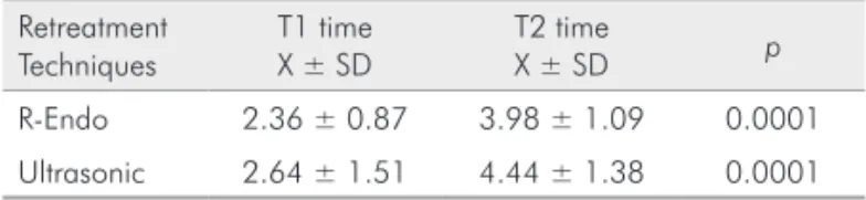
There was no signiicant difference in T1 or T2 using R-Endo versus ultrasonic tips (p > 0.05) (Table
1). There was no difference in the time required to
remove AH Plus, iRoot SP, or MM Seal (p > 0.05)
(Table 2). The increasing time from T1 to T2 was sig
-niicant in all groups.
Evaluation of residual debris
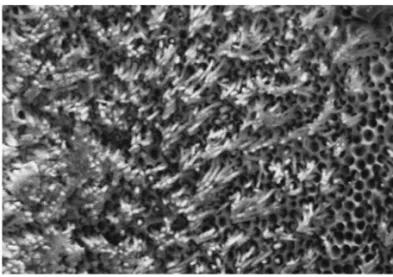
Results of the SEM analysis for open dentinal tubules are summarized in Tables 3 and 4. None of the retreatment techniques was able to remove debris
in the root thirds completely, regardless of the sealer
(Figures 1, 2, and 3). However, comparing among the root thirds, there were fewer open dentinal tubules in the apical third, and a larger number of clear den
-tinal tubules in the coronal third. In several samples of the MM Seal group, dentinal tubule oriices were illed by a resin sealer (Figure 4).
Table 1. Analysis of the retreatment techniques with respect to changing time.
Retreatment Techniques
T1 time X ± SD
T2 time
X ± SD p
R-Endo 2.36 ± 0.87 3.98 ± 1.09 0.0001
Ultrasonic 2.64 ± 1.51 4.44 ± 1.38 0.0001
Table 2. Analysis of the sealers with respect to changing time
Root Canal Sealers
T1 time X ± SD
T2 time
X ± SD p
Figure 2. Representative SEM image at ×2000 magnification for middle third. Partially dentinal tubules are open.
10µm Mag=2.00KX WD=13mm EHT=20.00kV Signal A=SE1 IBTAM 10µm Mag=2.00KX WD=13mm EHT=20.00kV Signal A=SE1 IBTAM
Figure 1. Representative SEM image at ×2000 magnification for coronal third. All dentinal tubules were open.
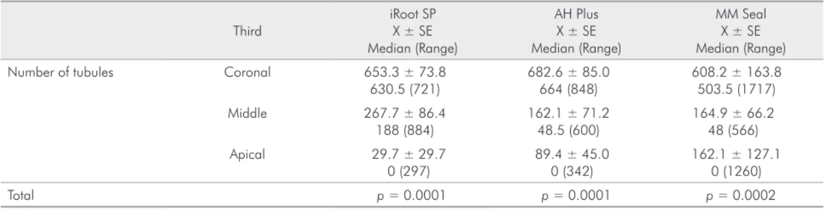
Table 3. Means of the number of dentinal tubules free of debris in R-Endo groups
Third
iRoot SP X ± SE Median (Range)
AH Plus X ± SE Median (Range)
MM Seal X ± SE Median (Range)
Number of tubules Coronal 653.3 ± 73.8
630.5 (721)
682.6 ± 85.0 664 (848)
608.2 ± 163.8 503.5 (1717)
Middle 267.7 ± 86.4
188 (884)
162.1 ± 71.2 48.5 (600)
164.9 ± 66.2 48 (566)
Apical 29.7 ± 29.7
0 (297)
89.4 ± 45.0 0 (342)
162.1 ± 127.1 0 (1260)
Total p = 0.0001 p = 0.0001 p = 0.0002
Table 4. Means of the number of dentinal tubules free of debris in ultrasonic groups
Third
iRoot SP X ± SE Median (Range)
AH Plus X ± SE Median (Range)
MM Seal X ± SE Median (Range)
Number of tubules Coronal 576.7 ± 199.9
603.0 (680)
722.4 ± 74.2 674 (614)
799.5 ± 136.0 747.0 (1490)
Middle 165.5 ± 56.2
77.5 (450)
157.4 ± 55.1 77.5 (435)
152.2 ± 42.0 96.0 (320)
Apical 137.0 ± 118.6
0 (1200)
26.9 ± 14.3 6.5 (144)
55.9 ± 25.1 13.0 (200)
Total p = 0.001 p = 0.0001 p = 0.0001
Discussion
The most important reason for renewed endodontic
treatment is to remove root canal illings completely,
in order to ensure disinfection of the canal and heal-ing of the periapical tissues.2 Gutta-percha can be dis
-solved using chloroform, carbon disulide, benzene,
xylene, essential oils, methyl chloroform, halothane,
white turpentine, carbon tetrachloride, and eucalyptus
oil.13 We used chloroform to soften the well-condensed
illing material. However, the pressure of the ile might
have penetrated the softened material in the dentinal
Various techniques have been used to remove gutta-percha from root canals, including rotary iles14 and ultrasonic tips.15 Ultrasonic tips are excellent tools to use for the coronal half of the root canal system,
but the curve of the apical part of the canal can cause dificulty. Therefore, ultrasonic tips should be used
for the remaining sections of the canal curvature.3
In this study, we used straight root canals. Many commercially available ultrasonic tips are available,
with different shapes, designs, and compositions (e.g., stainless steel, titanium alloys, diamonds, zircons,
etc.). We used stainless steel ultrasonic tips compat
-ible with the root anatomy to remove residual illing
material without damaging the inner walls of the roots.16 These tips were not used in retreatment
tech-niques with iRoot SP, which performed as a sealer. Rotary NiTi systems are faster than hand instru
-ments, such as R-Endo iles. The use of rotary instru -ments in root canal retreatment might reduce patient and operator fatigue.17 However, there is insuficient information in the literature to compare the use of
R-Endo iles to ultrasonic tips in terms of retreat
-ment operating time or remnants of debris. Both the R-Endo and ultrasonic tips used in the pres -ent study completed the retreatm-ent within similar intervals of time.
The iRoot SP seal is a relatively new sealer that
contains calcium silicate, which requires the
pres-ence of water to set. The results obtained when using iRoot SP were similar to those obtained with apical
sealing using AH Plus. The bond strength18 and
anti-microbial activity19 of iRoot AP have been studied; however, its effects in retreatment are not clear. Thus, this study sheds light on this issue. The adhesion
properties of sealers inluence the ease with which they can be removed. The push-out bond strength of iRoot SP is reported to be similar to that of AH Plus.18 In this study, iRoot SP samples did not show
a difference compared to the AH Plus samples in
terms of remaining material.
AH Plus was selected because of its widespread use. Both AH Plus and MM Seal are epoxy resin-based sealers with comparable apical sealing abilities because of their similar chemical components.10 After retreatment procedures, some fractured resin tags
were observed in the MM Seal group samples. These results are consistent with Pirani et al.3 because both
sealers are epoxy resin-based. The fractured resin tags blocked the entrance of the dentinal tubules. EDTA and NaOCl presumably failed to solve this issue.
In this study, residual debris was seen in all groups, which is similar to previous studies. During material
removal, canals in all groups tended to amass more
debris apically, regardless of the protocol or material used. This inding concurs with previous reports.3,14,20
The properties of sealers, such as their dimensional
stability and resolution, may affect the duration of
retreatment.21 In this study, retreatment lasted from 3 to 8 min Oliveira et al.14 did not include the
chang-ing of rotary iles and irrigation procedures in the
Figure 3. Representative SEM image at ×2000 magnification for apical third. Most of dentinal tubules are close.
10µm Mag=2.00KX WD=13mm EHT=20.00kV Signal A=SE1 IBTAM
Figure 4. Resin sealer materials in dentinal tubule orifices in MM Seal samples
operation time and reported retreatment times of 2
to 4 min. Time differences between studies could be due to the use of different root canal illings and preparation techniques, differences in the ability of
the practitioner, and the employment of solvents or
burs to facilitate the entrance of the canal. Rotary systems produce heat via a speciic torque force with frictional movement, which is known to facilitate the
dissolution and removal of heated gutta-percha.22 In
this study, the ultrasonic tips were warmed, but there was no difference between the groups in terms of the time it took to remove the gutta-percha.
Many techniques have been used to evaluate the remaining debris11,12,23 on dentin surfaces. How -ever, only SEM permits an extremely
comprehen-sive observation of the opened or closed dentinal tubules.24 In this study, high-resolution SEM images
showed dentinal tubules free of debris. There was a difference in the number of clean dentinal tubules
among the root canal thirds. The apical third
pre-sented the lowest number of clean dentinal tubules
compared to the middle or coronal third, regardless
of the technique or sealer used. This observation is consistent with the indings of previous studies.3,20
The coronal third showed more open tubules
than the other thirds. There are two reasons for this
inding. First, it is easy to clean the entire root canal.
Second, the coronal part was re-treated with Gates
Glidden drills. The total number of dentinal tubules in the apical third was less than the number in the middle or coronal third. This inding could relect the physiological phenomenon of tubular sclerosis, which
starts in the third decade of life in the apical part of the root canal and progresses coronally with age.25
Our study shows that the apical root section can
be partially cleared. On the basis of this discovery,
we propose a requirement to improve the size of the apical preparation when rotary instruments are used. The residual gutta-percha in the apical third
was less in the R-Endo group compared to the ultra -sonic group, although the difference was not
statis-tically signiicant. This inding may be because of the increased tip diameter of the R-Endo iles, or because the instruments were designed speciically for removing material. Additionally, the R-Endo iles
were employed through a crown-down approach, in
which the illing material is removed from the coro -nal third. This method may explain why
instrumen-tation was more eficient in the apical third.26
Conclusions
Both R-Endo and ultrasonic tips performed sim
-ilarly in terms of operating time. All of the retreat -ment systems in each group left remnants,
regard-less of the sealer. In all groups, apical tubules were
less clear than other parts of the root.
Acknowledgements
This project was supported by Inonu University, Scientiic Research Projects Unit (201178).
This study was presented as an oral presentation
at Turkish Endodontic Society, 11. International Con
-gress, April 2012, Istanbul, Turkey.
The authors deny any conlict of interest.
1. Hulsmann M, Stotz S. Efficacy, cleaning ability and safety of
dif-ferent devices for gutta percha removal in root canal retreatment.
Int Endod J. 1997 Jul;30(4):227-33.
2. Hulsmann M, Bluhm V. Efficacy, cleaning ability and safety of
different rotary NiTi instruments in root canal retreatment. Int
Endod J. 2004 Jul;37(7):468-76.
3. Pirani C, Pelliccioni GA, Marchionni S, Montebugnoli L, Piana G,
Prati C. Effectiveness of three different retreatment techniques in
canals filled with compacted gutta percha or Thermafil: a scanning
electron microscope study. J Endod. 2009 Oct;35(10):1433-40.
4. Sae-Lim V, Rajamanickam I, Lim BK, Lee HL. Effectiveness of ProFile.04 taper rotary instruments in endodontic retreatment. J Endod. 2000 Feb;26(2):100-4.
5. Plotino G, Pameijer CH, Grande NM, Somma F. Ultrasonics in end-odontics: a review of the literature. J Endod. 2007 Feb;33(2):81-95. 6. Ruddle CJ. Nonsu rgical ret reat ment. J Endod. 2004
Dec;30(12):827-45.
7. Kratchman SI. Obturation of the root canal system. Dent Clin North Am. 2004 Jan;48(1):203-15.
8. Zhang W, Li Z, Peng B. Assessment of a new root canal sealer’s apical sealing ability. Oral Surg Oral Med Oral Pathol Oral Radiol Endod. 2009 Jun;107(6):e79-82.
9. Cohen BI, Pagnillo MK, Musikant BL, Deutsch AS. An in vitro study of the cytotoxicity of two root canal sealers. J Endod. 2000 Apr;26(4):228-9.
10. Bodrumlu E, Avsar A, Meydan AD, Tuloglu N. Can radiotherapy affect the apical sealing ability of resin-based root canal sealers?. J Am Dent Assoc. 2009 Mar;140(3):326-30.
11. Horvath SD, Altenburger MJ, Naumann M, Wolkewitz M,
Schir-rmeister JF. Cleanliness of dentinal tubules following gutta-percha removal with and without solvents: a scanning electron microscopic study. Int Endod J. 2009 Nov;42(11):1032-8.
12. Scelza MF, Coil JM, Maciel AC, Oliveira LR, Scelza P. Compara-tive SEM evaluation of three solvents used in endodontic retreat-ment: an ex vivo study. J Appl Oral Sci. 2008 Jan-Feb;16(1):24-9. 13. McDonald MN, Vire DE. Chloroform in the endodontic operatory.
J Endod. 1992 Jun;18(6):301-3.
14. Oliveira DP, Barbizam JV, Trope M, Teixeira FB. Comparison between gutta percha and resilon removal using two different tech-niques in endodontic retreatment. J Endod. 2006 Apr;32(4):362-4. 15. Moshonov J, Trope M, Friedman S. Retreatment efficacy 3 months after obturation using glass ionomer cement, zinc oxide-eugenol, and epoxy resin sealers. J Endod. 1994 Feb;20(2):90-2.
16. Mello Junior JE, Cunha RS, Bueno CE, Zuolo ML. Retreatment efficacy of gutta percha removal using a clinical microscope and ultrasonic instruments: part I--an ex vivo study. Oral Surg Oral Med Oral Pathol Oral Radiol Endod. 2009 Jul;108(1):e59-62. 17. Tasdemir T, Er K, Yildirim T, Celik D. Efficacy of three rotary
NiTi instruments in removing gutta percha from root canals. Int Endod J. 2008 Mar;41(3):191-6.
18. Ersahan S, Aydin C. Dislocation resistance of iRoot SP, a cal-cium silicate-based sealer, from radicular dentine. J Endod. 2010 Dec;36(12):2000-2.
19. Zhang H, Shen Y, Ruse ND, Haapasalo M. Antibacterial activity
of endodontic sealers by modified direct contact test against En-terococcus faecalis. J Endod. 2009 Jul;35(7):1051-5.
20. oschi F, Nucci C, Montebugnoli L, Marchionni S, Breschi L,
Malagnino VA, et al. SEM evaluation of canal wall dentine
fol-lowing use of Mtwo and ProTaper NiTi rotary instruments. Int Endod J. 2004 Dec;37(12):832-9.
21. Kosti E, Lambrianidis T, Economides N, Neofitou C. Ex vivo
study of the efficacy of H-files and rotary Ni-Ti instruments to remove gutta percha and four types of sealer. Int Endod J. 2006
Jan;39(1):48-54.
22. Betti LV, Bramante CM. Quantec SC rotary instruments versus hand files for gutta percha removal in root canal retreatment. Int
Endod J. 2001 Oct;34(7):514-9.
23. Takahashi CM, Cunha RS, De Martin AS, Fontana CE, Silveira CF, Silveira Bueno CE. In vitro evaluation of the effectiveness of
ProTaper universal rotary retreatment system for gutta-percha
re-moval with or without a solvent. J Endod. 2009 Nov;35(11):1580-3.
24. Xu LL, Zhang L, Zhou XD, Wang R, Deng YH, Huang DM. Residual filling material in dentinal tubules after gutta-percha
removal observed with scanning electron microscopy. J Endod.
2012 Mar;38(3):293-6.
25. Paque F, Luder HU, Sener B, Zehnder M. Tubular sclerosis rather
than the smear layer impedes dye penetration into the dentine
of endodontically instrumented root canals. Int Endod J. 2006 Jan;39(1):18-25.
26. Ring J, Murray PE, Namerow KN, Moldauer BI, Garcia-Godoy
F. Removing root canal obturation materials: a comparison of