95
David Rodríguez Ruiz1
Miriam Esther Quiroga Escudero1 Dario Rodríguez Matoso1 Samuel Sarmiento Montesdeoca1 José Losa Reyna1
Yves de Saá Guerra1 Gloria Perdomo Bautista1 Juan Manuel García Manso1
1. University of Las Palmas de Gran Canaria
Mailing address:
Campus Universitario de Tafira, s/n Edificio de Ciencias de la Actividad Física y el Deporte - 35017 Las Palmas de Gran Canaria [email protected]
ABSTRACT
Objective: The aim of this investigation is to obtain information about muscle stiffness, the mechanic and contractile properties of the muscles using the TMG with high level beach volleyball players as well as to demonstrate the usefulness of this method to evaluate the muscles in charge of the knee flexion and extension. Methods: The investigation was carried out with a group of 24 beach volleyball players who took part in the Nestea European Championship Tour - Spanish Master held in the Gran Canaria, May 2009. The method of study used was a comparison of the individual cases of various athletes to ascertain the usefulness of this method in sports. The muscles which were analyzed are: vastus lateralis (VL), vastus medialis (VM), rectus femoris (RF) and biceps femoris (BF). Results: Thus, with the information collected we can state the high level of usefulness of this method for the evaluation of muscle stiffness and balance between muscle structures of athletes. However, the validity and reconstruction of the results are conditioned to a strict protocol of evaluation. Moreover, the following criteria should be considered: individuality (the athlete’s profile) and specifications (sport characteristics). Conclusions: The application of the TMG to high level players reveals the existence of important differences depending on their different roles in the game (defence, blocker or alternating both roles), the technical actions, the position on court (right-left) and the medical history of injuries.
Keywords: tensiomyography, evaluation of the muscles, symmetries, beach volleyball, stiffness.
THE TENSIOMYOGRAPHY USED FOR EVALUATING
HIGH LEVEL BEACH VOLLEYBALL PLAYERS
INTRODUCTION
The beach volleyball boom had its origin in the Atlanta Olym-pic Games in 1996. Such sports requires a great variety of tech-niques, actions played on dry and soft sand which are similar to the ones played in the indoors volleyball (sprints, alterations in direction, vertical jumps…). During each match, there are mean of 85 different actions in approximately 42-45 minutes of game 1,2.
Due to the characteristics of the game, there are certain specific colocations and basic positions which are common to all athletes. Thus3, the central squat is mentioned as a previous element for
the block actions and as a defense position on the court. The basic premise of the physical training is to develop a solid and balanced muscular structure with the aim to optimize the technical activities as well as reduce the possible risk of injuries during the match. An aspect considered by all coaches in beach volleyball is the unstable surface of the sand3,4.
The reduced number of players per match, that is, two players in the team, requires that the area defended by each player is bigger (32m2/player) added to the fact that they cannot be
subs-tituted for any another player, increases the needs for conditioning compared to the indoor volleyball. The number of times that player is in contact with the ball is high despite the fact the surface makes fast movements difficult5-7mentions that this activity occurs at
un-favorable climate conditions (cloudy or sunny days, air temperature, sand temperature and humidity).
Concerning the mechanical point of view, we should empha-size the so called “triple extension mechanism” (ankle, knee and
hip joints extension). This fact occurs so that the body is pushed in the direction of the ball in a fast and efficient manner. According to Smith6, the players use this technique to jump and move as fast
as possible towards the ball. However, the fact they are playing on sand limits the actions of the involved muscles on the ankle and significantly alters the movements of the technique.Furthermore, players and coaches should consider this fact once it determines the way and application of force, time and magnitude of fly fore and the vertical jump8,9.
Such reasons make that an accurate, individualized and loca-lized evaluation of the muscular structures specially involved in the beach volleyball activities are necessary. The tensiomyography (TMG) would be used in this case as an instrument to make infor-mation on the musculature which helps us prevent imbalances available, or overload in the musculature, which could be produ-ced by repeated technical actions. Pfirrmann et al10 mention that
these cases are possible reasons for many injuries which reduce performance of athletes or even lead to deficiency.
TMG is a non-invasive diagnostic method which does not de-mand any effort from the individual to whom it is applied. It is used as an evaluation tool for stiffness, the mechanical characteristics and contraction capacity of the surface muscle structures analyzed 11-14. It
measures the geometrical alterations (radial displacement) which occur on the muscular surface during contraction. Subsequently, the results obtained expressed as sensor displacement versus time of activity are used to determine the stiffness and balance between muscular struc-tures, muscular chains (flexion – extension) or extremities (right – left).
ORIGINAL ARTICLE
96
Figure 1. This chart presents the response of the biceps femoris to an electric impulse width of 1 ms and amplitude of 110 mA used with one TMG. Dm represents the maximum muscu-lar displacement. Td is the reaction time to the impulse. Tcis the contraction time in intervals between 10 and 90 % of movement. Ts is the contraction duration. Tr is the relaxation time.
displac
emen
t (mm)
Time (ms)
The aim of the present investigation is to use the TMG as an ins-trument to measure the mechanical characteristics of the muscles of volleyball players with the aim to demonstrate the validity of this method for evaluation of the musculature involved with the knee flexor-extensor joint. Moreover, we intended to determine whether the results obtained are sufficiently accurate to use the TMG in the detection of pathologies, asymmetry and specific profiles of players. It should be mentioned that the data obtained are affected by potential injuries, specific technical actions and the position of the player (defense, blocking of game alternative positions during the match) and the game area where the majority of activities is performed (left or right).
METHODS
Sample
Twenty-four players were examined (10 women; 5 of international high level and 5 of international level. 14 men; 5 of international high level, 2 of international level and 7 of national level), all participants in the Nestea- Master Spanish European Tournament, held in the Gran-Canaria island in 2009 (table 1). The most important muscles to be studied were: vastus lateralis (VL), vastus medialis (VM), rectus femoris (RF) and biceps femoris (BF).
All participants were told about the possible risks associated with the study and signed a consent form approved by the Com-mittee of Ethics in Research of the ULPGC, following the criteria of the Declaration of Helsinki for research with humans.
the mean, demonstrate high muscular mass and stiffness27, while
low results represent lack of muscular mass or high muscular fatigue14,17,22,28-30. The time of delay (reaction or activation - Td)
of the muscular structure analyzed represents the time he took to reach 10% of the total movement. However, it will depend on the predominance of the fiber in this structure of skeletal muscle, his fatigue status30and his level of activation31. The time
of contraction (Tc) is obtained by the determination of the time of interval of the end of the time of reaction (10% Dm) until 90% of the maximal deformation. The time of sustaining (duration of contraction -Ts) is the theoretical time the contraction is kept. With the TMG, it can be calculated (Ts) by the determination of the time interval, since the initial deformation reaches 50% of its maximal value, until the deformation values during the time of relaxation which returns to 50% of the maximal deformation. The time of relaxation (Tr), the time in which the response of the muscle decreases from 90% to 50%, the Dm offers information on the fatigue levels. In any event, if the results were higher than the mean of the individual, there is indication of fatigue. In that case, there is an important correlation between the movement of the muscle belly and the processes of muscular contraction21.
RESULTS
Descriptive statistics by maximum radial displacement of muscle belly (Dm), time of contraction (Tc), Time of delay (Td), Time of sustaining (Ts) and Time of relaxation (Tr) for subjects studied is provided in Table 2.
DISCUSSION
The data obtained in the Dm, Td, Tc, Ts and Tr parameters let us analyze the properties of the muscle, depending on the type of fiber21, the symmetry (lateral or functional) between the
ex-tremities32-33, the levels of muscular fatigue28-30,34, or anatomical
disorders28,35,36. These and other complementary aspects are what
we try to explain in the following sections.
TMG and techniques actions
The BF values of both legs are lower than the ones obtained from the knee extensor muscles (VM, RF and VL) in the analyzed sample. High muscular mass of the hamstring muscle may occur
Experimental procedures
The TMG has a sensory detector of magnetic pressure which is perpendicularly placed on the surface of the selected muscle 11,15,16.
The pressures has to be the one recommended by the manufac-turers17. In order to produce muscular contraction, increasing
electrical stimuli of one millisecond are applied18-20through two
electrodes placed on the extremities of the muscle surface (no-ton the tendons).
The reproducibility of the method and validity of the experimental protocol used by the TMG has been investigated in different studies12,20-26
Once the evaluation of the selected muscle is finalized, nu-merical information on the magnitude of the radial movements of the transversal muscle fibers are obtained11,20. The results are
presented in the chart (figure 1): maximum radial displacement of muscle belly (Dm), time of contraction (Tc), Time of Delay (Td), Time of sustaining (Ts) and Time of relaxation (Tr).
The maximum radial displacement of muscle belly (Dm) is rep-resented by the radial movement of the muscle surface expressed in millimeters. It presents and evaluates the stiffness of the muscle, with variations between subjects and the manner how their mus-cular groups developed in the training. Low results, compared with
Table 1. Morphological characteristics of the sample.
Players
Age Body weight Height BMI
Fat %
(years of age) (Kg) (cm) (Kg/m2)
Men (n=14) 25.14 +/- 6.27
87.50 +/- 5.87
190.71 +/- 5.12
24.06 +/- 1.14
9.54 +/- 1.34 Women
(n=10)
25.20 +/- 6.23
68.90 +/- 4.09
176.70 +/- 4.55
22.08 +/- 1.24
97
due to two frequent technical actions in each volleyball players; initial reception position (hip joint, folded) and the explosive vertical jump (the extensor muscular chain activates it). Such fact is especially observed in experienced players. Excessive muscular mass produces some imbalance, leading to asym-metry between the knee extensor and flexor muscles, causing pain in the knee joint (table 3).
The connection between agonist and antagonist during knee joint flexor-extensor movements is presented in the table as the percentage of functional symmetry. Normal values are around
65% or more28,33,35. These values are used by these authors as
ref-erence, collected in an investigation carried out with individuals who practice physical activity with moderation. However, they should be used as guidance, despite being possible to observe in table 3 that there are players with higher values of functional symmetry. In this context, symmetry values may be attributed to two different factors:
- Excessive fatigue or stiffness of the BF muscle;
- Lack of tonus or muscular fatigue of the knee joint extensor muscles (VM, RF, VL).
For instance, the individual called masculine 1 (table 3) pre-sents a Dm of 4.1 mm in the BF of his right leg and 6.6 mm in his left leg. These results may be positive or negative despite being a sign of good muscular tonus. On the other hand, if we compare and consider the extense musculature, there is indication of a possible risk of injury for the player. The RF values are very high for this kind of players (right leg: 17.9 mm, left leg: 16.8 mm). The moderate values which present this characteristic indicate functional asymmetry, causing the frequent low back pain the player has been feeling.
The Dm values of the VL and VM inform on the level of functional symmetry of the knee joint. Patella pain and tendinitis andon the knee-cap may be caused by knee instability. If we consider the information about feminine 1 from table 3, we can observe that she may be feeling pain on her right leg, despite the symmetry values being acceptable. 75% right legand 88% left leg. When the relation between VM and VL is analyzed, we conclude that the problem may be caused by a possible imbalance between them (49% right leg and 85% left leg).
Table 2. Results of the descriptive statistics by Tc, Dm, Td, Ts and Tr of VM, RF, VL and BF obtained from subjects of the sample.
Right leg Left leg
Tc[ms] Dm[mm] Td[ms] Ts[ms] Tr[ms] Tc[ms] Dm[mm] Td[ms] Ts[ms] Tr[ms]
MALE (n=14)
VM
Mean 28.2 8.5 21.3 166.7 64.3 26.4 8.4 21.2 177.5 58.8
SD +/- 15.5 +/- 2.0 +/- 1.0 +/- 24.2 +/- 41.7 +/- 10.3 +/- 1.5 +/- 1.7 +/- 24.0 +/- 42.3
Range 22.7 - 76.8 5.9 - 12.8 19.8 - 23.1 130.7 - 185.4 33.6 - 153.9 21.7 - 57.6 7 - 10.5 18.4 - 23.3 130.3 - 223.5 33.3 - 155.9
RF
Mean 29.2 9.2 23.1 61.7 25.3 31.3 9.2 23.6 64.0 26.0
SD +/- 4.7 +/- 3.2 +/- 2.5 +/- 51.1 +/- 45.2 +/- 5.5 +/- 3.1 +/- 1.8 +/- 45.3 +/- 43.1
Range 22.1 - 37.7 6.6 - 17.9 20.4 - 27.2 37.7 - 186.1 11.7 - 137.9 25.9 - 44.8 5.1 - 16.8 19.4 - 26.2 47.2 - 185.4 16 - 150.7
VL
Mean 26.3 5.6 20.7 50.1 18.4 24.7 6.4 21.0 51.7 19.9
SD +/- 3.2 +/- 1.4 +/- 2.4 +/- 40.8 +/- 36.8 +/- 4.3 +/- 1.1 +/- 2.0 +/- 36.2 +/- 31.8
Range 21.8 - 31.8 3.3 - 8.4 17.9 - 24.3 33.5 - 155.7 9.9 - 127.5 20.3 - 34.8 4.5 - 7.9 18.9 - 25 37.1 - 138.5 10.1 - 103.6
BF
Mean 25.7 3.9 19.4 211.4 56.3 24.2 4.3 20.3 130.6 37.6
SD +/- 15.6 +/- 2.4 +/- 4.1 +/- 42.5 +/- 55.4 +/- 12.4 +/- 2.3 +/- 3.1 +/- 70.7 +/- 27.4
Range 15.5 - 68.9 2 - 9.6 14.3 - 28.3 174.8 - 310.1 33.6 - 220.6 13.7 - 59.5 2.1 - 10.1 15 - 26.9 24 - 297 7.7 - 107.4
FEMALE (n=10)
VM
Mean 24.9 7.6 20.7 174.7 74.7 26.4 6.5 20.2 174.5 55.4
SD +/- 10.8 +/- 1.2 +/- 1.4 +/- 18.9 +/- 51.5 +/- 11.1 +/- 2.0 +/- 1.1 +/- 27.9 +/- 51.7
Range 20.2 - 57 6.4 - 9.9 18.7 - 22.6 145.3 - 213.6 40.1 - 163.2 20 - 29.4 4.9 - 11.8 19.1 - 21.3 150.5 - 209.7 31.6 - 167.7
RF
Mean 28.3 8.0 23.2 71.2 29.1 28.7 8.0 22.9 93.1 39.5
SD +/- 5.8 +/- 2.3 +/- 2.0 +/- 36.6 +/- 33.9 +/- 4.4 +/- 1.7 +/- 2.0 +/- 49.9 +/- 42.7
Range 19.5 - 39.7 6 - 13.3 19.4 - 25.8 53.7 - 146.1 17.9 - 110.6 22.5 - 36.8 5.2 - 10.6 18.9 - 26.3 43.1 - 171.9 22.2 - 129.3
VL
Mean 24.6 5.6 20.4 42.3 14.1 24.4 5.5 19.9 42.5 14.9
SD +/- 1.2 +/- 1.2 +/- 2.2 +/- 28.7 +/- 25.9 +/- 2.2 +/- 1.0 +/- 1.7 +/- 16.8 +/- 15.0
Range 23.2 - 26.9 4.4 - 8.2 17.6 - 23.2 32.2 - 128.6 9.1 - 94.5 21.6 - 28 4.7 - 7.6 17.9 - 22.6 33 - 62.9 8.4 - 57.4
BF
Mean 37.6 5.7 22.0 209.6 76.2 32.2 6.4 23.8 195.9 50.4
SD +/- 17.5 +/- 2.7 +/- 2.4 +/- 77.4 +/- 30.1 +/- 16.0 +/- 2.0 +/- 2.4 +/- 32.6 +/- 14.1
Range 18.4 - 69.6 2.8 - 10 18.5 - 24.9 150.8 - 420.3 56.4 - 123.5 16.7 - 70.1 3 - 9.9 20.2 - 27 150.8 - 249.9 29.6 - 73.9
Table 3. An example of functional symmetry of the knee joint and possible pathologies associated to 10 subjects (5 men and 5 women). The data in italics present the most important ones.
Players
Functional symmetry
Pain in the leg
Right Left
Male 1 60% 80% Right leg
Male 2 70% 92% Left leg
Male 3 89% 71% Left leg
Male 4 58% 71% Right leg
Male 5 82% 89% No
Female 1 75% 88% Right leg
Female 2 91% 48% Left leg
Female 3 51% 60% Both
Female 4 53% 71% Right leg
98
TMG and its technical function
The BF is frequently required in defensive actions and in specific movements during competitions. The Dm values of players of long term in this sport (for male and female categories) show that these are lower in specialist in defense. In our sample, the specialists in defense present values of Dm between 2 and 4 mm. On the other hand, the specialists in blocking, who oscillate between the defense and blocking positions, present approximate values between 4 and 8 mm. Previous studies emphasize the reference value of Dm 8.17 mm in untrained subjects33, while soccer players range between 3 and 8 mm35. TMG and the player’s position on the court
The functions of the players determine greater involvement of certain muscular structures. We should add to these considerations the position they take on the court as well as the functions they play.
If we revise the duration of the contraction (Ts) of the sample we will realize that the players who compete in pairs present higher fatigue levels in their VL (table 4).
TMG and injury prevention
The possibility that the TMG contributes to the prevention of injuries is high. A priori, it gives us the opportunity to determine high risk situations for future muscular injuries. The interpretation in these cases requires a high level of experience from the side of the evaluator since the TMG results in some cases present similarities between the ideal performance states and states with high risk with potential to injury.
For example, after an intense training session is very com-mon to find the following values in BF: high Ts and Td and low Tc and Dm. These circumstances are obvious because the mus-cular structures are in optimum conditions to play an efficient mechanical work, but it is also certain that there is high level of fatigue, which could involve a risk for the integrity of the muscle. In these circumstances where the muscles present high stiffness, it is necessary to massage or stretch them (table 5).
Another application in beach volleyball, as well as in other sports, is the detection of muscular fatigue, for which it is necessary to use more than one variant of the TMG to conduct a personalized di-agnosis for each subject. In our viewpoint, we consider that with high levels of fatigue, the time of reaction (Td) should be high, the shortening of the muscle velocity (Dm/Tc) low and the duration of the contraction (Ts) high. Despite of that, we should be careful with the results, especially with the values of velocity shortening because we can face fatigue related with high or low Dm.
Low Dm may be caused due to the high velocity actions which can be derived from high levels of muscular stiffness. If the Dmin-creases too much, it can related with a chronic fatigue status or muscular weakness. Another aspect to be considered is that it is not possible to compare comparisons between subjects with no infor-mation values and each one of them during their habitual training or competitions, once the time of contraction will depend on the characteristics of the analyzed muscle and the high time of con-traction seems to be related to a higher number of type 1 fibers31. TMG, injury control and recovery
The TMG may provide data about the progress and effec-tiveness of the recovery of the injuries.Therefore, we used two examples of study: the recovery processes of a torn cruciate liga-ment and torn fibers of the BF.
Torn cruciate ligament; the athlete focused his recovery on the strengthening of the hamstrings of his hurt leg (left leg). Despite his morphological balance (thigh diameter of his domi-nant leg 55.6 cm; non domidomi-nant 54.6 cm) his muscular response indicates that there is serious asymmetry. For example, his func-tional symmetry (flexor-extensor muscles) was imbalanced on his right side (53%) and there is asymmetry between VM and VL of the other leg (48%).
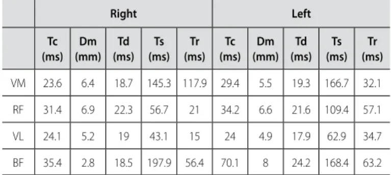
In the case of the athlete with torn muscular fibers of the left leg: this athlete focused his recovery on hypertrophy training: even though, he still presented anomalies on BF of both legs (table 6).
Table 6 presents mechanical and nervous disorders in both BF. We can specifically observe that the left leg presents Tc, Dm and Td values higher than the right leg. Moreover, the athlete presented imbalance of muscular mass in both legs (diameter of right leg 66cm, and 62cm injured leg).
CONCLUSIONS
The data obtained of the subjects in our sample let us con-firm that TMG is a highly useful technique for the evaluation of
Table 4. Example of the maintenance time of the contraction of 4 beach volleyball pairs(2 men and 2 women) for the analysis of their VL dependent on their positions on the court.
Partner Players Time of sustaining (Ts - ms) Position on court
Right Left
A Male 1 129.2 116.8 Left
Male 2 36 39.7 Right
B Male 3 41.2 38.4 Left
Male 4 61.3 79.1 Right
C Female 1 128.6 44 Left
Female 2 43.1 62.9 Right
D Female 3 41.9 34.2 Left
Female 4 36.1 42.6 Right
Table 5. Correlation between Ts and Td, Tc and Dm of BF obtained from the subjects of the sample (2 men and 2 women).
BF Right Left
Player Tc (ms) Dm
(mm) Td (ms) Ts (ms) Tc (ms) Dm
(mm)Td (ms) Ts (ms)
M1 18.4 4.1 22 202.9 31.1 6.6 20.7 242.8
M2 32.9 2.9 16 199.9 28.3 2.6 15 234.1
F1 29.8 5.2 21.8 249.5 28.4 8.8 25.4 215.1
F2 18.4 3.8 19 420.3 16.7 3 20.2 249.9
Table 6. This table presents the results of TMG applied to a beach volleyball player who had an imbalanced functional BF.
Right Left
Tc (ms)
Dm (mm)
Td (ms)
Ts (ms)
Tr (ms)
Tc (ms)
Dm (mm)
Td (ms)
Ts (ms)
Tr (ms)
99
structures of muscular stiffness and muscular balance of athletes. However, we should remember that the validity and reproducibil-ity of the results are conditioned to strict evaluation protocols. Furthermore, it should be considered that the data interpretation should also respect the individuality criteria (the athlete’s profile) and specificities (characteristics of the sport). The application of a high level beach volleyball sample evidences the relevant differences due to different functions of each player (defense, blocking or
alternation between both functions), the technical actions, the position on court (right-left) and the player’s injury history.
ACKNOWLEDGEMENTS
To the Royal Spanish Volleyball Federation for its contribution.
All authors have declared there is not any potential conflict of interests concerning this article.
REFERENCES
1. Giatsis G. The effect of changing the rules on score fluctuation and match curation in the FIVB women`s beach volleyball. International Journal of Performance Analysis in Sport, 2003, 3(1): 57-64. 2. Giatsis G., Zetou E. The influences of regulation changes on the fluctuation of score of the beach
volleyball games. Inquiries in Sport & Physical Education, 2003, 1: 43-48.
3. Hömberg S., Papageorgiou A. Handbook for beach Volleyball. Aachen: Meyer & Meyer Verlag. 1994.
4. Miyama M., Nosaka N. Influence of surface on muscle damage and soreness induced by consecutive drop jumps. The Journal of Strengh and Conditioning Research, 2004, 18(2): 206-211.
5. Bredeweg S. The elite volleyball athlete. In Reeser, J.C. and Bahr, R. (eds.), Handbook of Sports Medicine and Science. Volleyball. Massachusetts: Blackwell Science. 2003: 183-191.
6. Smith R. Movement in the Sand: Training Implications for Beach Volleyball. Strength and Conditioning Journal, 2006, 28(5): 19-21.
7. Zetou E., Vernadaki Z., Mountaki F., Giatsis G., Laparidis K. (2006). Common pratices of beachvolleyball players regarding fluid, supplements and nutrition intake during a tournament. Physical Training (2006). http://ejmas.com/pt/2006pt/ptart_Zetou_0406.htlm (accessed 12 jun 2009)
8. Bishop D. A comparison between land and sand-based tests for beach volleyball assessment. Journal of Sport Medicine and Physical Fitness, 2003, 43(4): 418-423.
9. Giatsis G., Kollias I., Panoutsakopoulos V., Papaiakovou G. Biomechanical differences in elite beach volleyball players in vertical squat jump on rigid and sand surfaces. Sports Biomechanics, 2004, 3(1): 145-148.
10. Pfirrmann C.W.A., Jost B., Pirkl C., Aitzetmüller G., Lajtai G. Quadriceps tendinosis in professional beach volleyball players: sonographic finfings in correlation with clinical symptoms. Eur Radiol 2008, 18: 1703-1709.
11. Valencic V., Knez N. Measuring of skeletal muscles dynamic properties. Artific Org. 1997, 21: 240-242. 12. Dahmane R., Knez N., Valencic V., Erzen I. Tensiomyography, a non-invasive method reflecting the
percentage of slow muscle fiber in human skeletal muscles. Book of Abstract: Life Sciencies 2000, Gozd Martuljek, Slovenia, September 28th to October 1st, 2000, pp./str. 29.
13. Valencic V., Djordjevic S., Knez N., Dahmane R., Coh M, Jurcic-Zlobec B., Praprotnik U., Simunic B., Kersevan K., Bednarik J., Gomina, N. Contractile properties of skeletal muscles detection by tensiomio-graphic measurement method. 2000 Pre-Olympic Congress, Brisbane, Australia, Abstract 507, 2000. 14. Valencic V., Knez N., Simunic B. Tenziomiography: Detection of skeletal muscle responce by Means of
radial muscle belly displacement. Biomedical Engineering, 2001, 1: 1-10.
15. García-Manso JM, Rodríguez-Matoso D, Sarmiento S, De Saa Y, Vaamonde D, Rodríguez-Ruiz D y da Silva-Grigoletto, ME. La tensiomiografía como herramienta de evaluación muscular en el deporte. Rev Andal Med Deporte 2010. 3:98-102.
16. Rodríguez-Matoso, D.; Rodríguez-Ruiz, D.; Quiroga, M.E.; Sarmiento, S.; De Saa, Y. y García-Manso, J.M. Tensiomiografía, utilidad y metodología en la evaluación muscular. Revista Internacional de Medicina y Ciencias de la Actividad Física y el Deporte. 2010. 10 (40): 620-629.
17. Dahmane R., Valencic V., Knez N., Erzen I. Evaluation of the ability to make non-invasive estimation of muscle contractile properties on the basis of the muscle belly response. Medical and Biological Engineering Computering, 2001, 39: 51-55.
18. Knez N., Valencic V. Proceedings of the ninth Electrocehnical and Computer Science Conference ERK 2000, 21-23, September 2000, Portoroz, Slovenia. Ljubljana : IEEE Region 8, Slovenian section IEEE, 2000, Vol. B, pp. 301-304.
19. Valencic V. Method for selective and non-invasive detection of skeletal muscles contrction pro-cess. International Application Published under the Patent Cooperation Treaty (PCT). Nº WO 02/074167 A1. 2002.
20. Simunic B. Model of longitudinal constractions and transverse deformations in skeletal muscles. Doctoral Thesis. Ljubljana. 2003.
21. Belic, A., Knez, N., Karba, R. y Valencic, V. Validation of the human muscle model. Proceedings of the 2000 Summer Computer Simulation Conference, 16. - 20. July 2000, Vancouver, British Columbia. Session 1: Issues on Whole Body Modeling.
22. Krizaj D., Simunic B., Zagar, T. Short-term repeatability of parameters extracted from radial displacement of muscle belly. Journal of Electromyography and Kinesiology, 2008, 18: 645-651.
23. Rodríguez-Matoso D, Rodríguez-Ruiz D, Sarmiento S, Vaamonde D, Da Silva- Grigoletto ME, García-Manso JM. Reproducibility of muscle response measurements using tensiomyography in a range of positions. Rev Andal Med Deporte 2010;3:81-6
24. Simunic B, Valencic V. Non-invasive selective measurement of m. vastus medialis and m. vastus lateralis contractile properties at different knee angles. Proceedings of the tenth Electrocehnical and Computer Science Conference ERK 2001, 24. - 26. September 2001, Portorož, Slovenia. Ljubljana : IEEE Region 8, Slovenian section IEEE, 2001, Vol. B, 363-366.
25. Tous-Fajardo J, Moras G, Rodríguez-Jiménez S, Usach R, Doutres DM, Maffiuletti NA. Inter-rater reliability of muscle contractile property measurements using non-invasive tensiomyography. J Electromyogr Kinesiol 2010. 20:761-6.
26. Carrasco, L.,Sañudo, B., de Hoyo, M., Pradas, F. y da Silva, ME. Effectiveness of low-frequency vibration recovery method on blood lactate removal, muscle contractile properties and on time to exhaustion during cycling at VO2max power output. Eur. J. Appl. Physiol. 2011. 111:2271-2279.
27. Pisot R, Narici MV, Simunic B, De Boer M, Seynnes O, Jurdana M, Biolo G, Mekjavic IB. Whole muscle con-tractile parameters and thickness loss during 35-day bedrest. Eur J Appl Physiol. 2008. 104(2): 409-414. 28. Simunic, B., Rozman, S., Pisot, R. Detecting the velocity of the muscle contraction. III International
Symposium of New Technologies in Sports. Sarajevo, 2005.
29. Smith, IJ.; Hunter, A. The Effect of Titanic Stimulated Induced Fatigue on the Relationship between TMG and Force Production of the Gastrocnemius Medialis. Medicine & Science in Sports & Exercise. 2006. 38(5) Supplement; S179–S180.
30. García-Manso JM, Rodríguez-Ruiz D, Rodríguez-Matoso D, de Saá Y, Sarmiento S, Quiroga ME. As-sessment of muscle fatigue after an ultraendurance triathlon using Tensiomyography (TMG). J Sport Sci 2011; 29:619-25.
31. Dahmane R., Djordjevic S., Simunic B., Valencic V. Spatial fiber type distribution in normal human muscle Histochemical and tensiomyographical evaluation. Journal of Biomechanics, 2005, 38: 2451-2459. 32. Zagorc M., Karpljuk D., Friedl M. Analysis of functional loads of top sport dancers. Inc: Milanovic, D.
(ed), Proceedings of the 2nd International Scientific Conference “Kinesiology for the 21st century”. Dubrovnik. University of Zagreb. 1999, 240-243.
33. Rusu L., Calina M., Avramescu E., Paun E., Vasilescu M. Neuromucular investigation in diabetic polyneu-ropathy. Romanian Journal of Morphology and Embryology. 2009, 50 (2): 283-290.
34. Grabljevec K., Simunic B., Kersevan K., Krizaj D., Kosorok V., Gregoric M. Detection of contractile prop-erties of chronically spastic muscles in subjects after traumatic brain injury with tensiomyography (TMG) method. Rehabilitation sciences in the new millenium challenge for multidisciplinary research: collection of works presented at the 8th congress of European federation for research in rehabilitation, Ljubljana, Slovenia; 2004. p. 139–143.
35. Lukic D. La tensiomiografía al servicio del deportista. Taller del II Congreso Internacional Universitario sobre las Ciencias de la Salud y el Deporte. Universidad San Pablo CEU. Madrid, 2003.