RESUMO.- [Desenvolvimento e aplicação da reação em
cadeia da polimerase para detecção de
Conidiobolus
lamprauges
.] A conidiobolomicose é uma doença
granulo-matosa causada pelo fungo
Conidiobolus
spp., observada em
humanos e animais. As técnicas tradicionais de diagnóstico
da doença são o isolamento do agente associado à presença
de sinais clínicos típicos e condições patológicas. O objetivo
deste trabalho é descrever o desenvolvimento de um teste
da reação em cadeia da polimerase (PCR) específico para
Conidiobolus lamprauges
em amostras clínicas. As amostras
de animais suspeitos foram coletadas e submetidas ao
iso-lamento, análise histopatológica e amplificação pela PCR.
O DNA de tecidos foi submetido a PCR com os iniciadores
universais de fungos baseados no gene 18S rDNA e
inicia-dores específicos foram concebidos com base no mesmo
gene em
C. lamprauges
que gerou produtos de
aproxima-damente 540 pb e 222 pb, respectivamente. A cultura foi
positiva em 26,6% das amostras clínicas. A técnica de PCR
para
C. lamprauges
mostrou a amplificação de DNA a partir
de tecidos frescos (80%) e secções de parafina (44,4%). Em
conclusão, a técnica de PCR aqui descrita demonstrou
ele-vada sensibilidade e especificidade na detecção de DNA de
fungos em amostras de tecido, proporcionando uma
ferra-menta rápida para o diagnóstico de
C. lamprauges
.
TERMOS DE INDEXAÇÃO: Conidiobolus lamprauges, diagnóstico, PCR, ovinos, zigomicetos.
INTRODUCTION
Conidiobolomycosis is a disease caused by fungi of the
ge-nus
Conidiobolus
spp., class Zygomycetes of the order Ento
-mophthorales. These saprophytic fungi are opportunistic,
associated with granulomatous rhinitis in humans and
ani-Development and application of polymerase chain reaction
test for detection of
Conidiobolus lamprauges
1Marcelo M. Silveira
2, Daphine A.J. Paula
2, Maria C. Silva
2, Leticia C. Pitchenin
2,
Raquel A.S. Cruz
3, Edson M. Colodel
3, Valéria Dutra
2and Luciano Nakazato
2*
ABSTRACT.-
Silveira M.M., Paula D.A.J., Silva M.C., Pitchenin L.C., Cruz R.A.S., Colodel E.M.,
Dutra V. & Nakazato L. 2013. Development and application of polymerase chain reaction
test for detection of
Conidiobolus lamprauges
.
Pesquisa Veterinária Brasileira
33(12):1448-1452.
Departamento de Clínica Médica Veterinária, Faculdade de Agronomia, Medicina Ve
-terinária e Zootecnia, Universidade Federal de Mato Grosso, Av. Fernando Corrêa da Costa
2673, Bairro Boa Esperança, Cuiabá, MT 78068-900, Brazil. E-mail:
[email protected]
Conidiobolomycosis is a granulomatous disease caused by the fungus
Conidiobolus
spp.
in humans and animals. Traditional technique for diagnosis of the disease is isolation of the
agent associated with the presence of typical clinical signs and pathological conditions. The
aim of this study was to describe the development of a specific polymerase chain reaction
(PCR) test for
Conidiobolus lamprauges
to detect the fungus in clinical samples. Samples
from suspected animals were collected and submitted to isolation, histopathological
analy-sis and amplification by PCR. DNA from tissues was subjected to PCR with fungi universal
primers 18S rDNA gene, and specific primers were designed based on the same gene in
C.
lamprauges
that generated products of about 540 bp and 222 bp respectively. The culture
was positive in 26.6% of clinical samples. The PCR technique for
C. lamprauges
showed
am-plification of DNA from fresh tissues (80%) and paraffin sections (44.4%). In conclusion, the
PCR technique described here demonstrated a high sensitivity and specificity for detection
of fungal DNA in tissue samples, providing a tool for the rapid diagnosis of
C. lamprauges.
INDEX TERMS: Conidiobolus lamprauges, diagnostic, PCR, sheep, zygomycetes.1 Received on August 16, 2013.
Accepted for publication on November 19, 2013.
2 Laboratório de Microbiologia Veterinária e Biologia Molecular Veteri
-nária, Universidade Federal de Mato Grosso (UFMT), Av. Fernando Cor -rêa 2673, Coxipó, Cuiabá, MT 78060-900, Brazil. *Corresponding author: [email protected]
3 Laboratório de Patologia Veterinária, Universidade Federal de Mato
mals (Carrigan et al. 1992, Ribes et al. 2000, Tadano et al.
2005, Hata et al. 2008, Kimura et al. 2011).
They are found
in soil, decomposing vegetation and as insect parasites
(Porto et al. 1987, Scholte et al. 2004, Silva et al. 2007a).
This zygomycosis is endemic in tropical regions and in
sheep has rapid progression with high lethality (Silva et al.
2007b) and isolates are resistant to mainly antifungal
dru-gs (Tondolo et al. 2013).
Clinically, the animals may show apathy, anorexia, weight
loss, swollen nose, granulomatous reaction in the
nasopha-rynx, unilateral exophthalmia, nasal discharge or
mucous-se-rous hemorrhagic, noisy breathing, dyspnea and death
(Ket-terer et al. 1992, Morris et al. 2001, Riet-Correa et al. 2008,
Pedroso et al. 2009, Batista et al. 2009, Silva et al. 2010).
The specie
C. lamprauges
was isolated in samples of
sheep in the State of Piauí, Mato Grosso and Santa Catari
-na, and identified by histopathology and mycological and
molecular methods (Silva et al. 2007a, De Paula et al. 2010,
Furlan et al. 2010, Vilela et al. 2010).
The objective of this study was to detect rapidly, through
the technique of polymerase chain reaction,
C. lamprauges
presence in animal tissue samples, since actual diagnostic
techniques based on culture isolation are time consuming
with a high rate of false negative results.
MATERIALS AND METHODS
Isolates and samples. For development of PCR technique,
Conidiobolus lamprauges (INCQS 40317)isolate was used as
stan-dard. This study tested 15 clinical fresh samples and 18 paraffin
embedded tissues samples from sheep with suspected lesions of conidiobolomycosis in the nasal cavity, kidneys, lymph nodes, li-ver and lungs of sheep between January 2008 to December 2010.
Samples were from States of Mato Grosso, Santa Catarina States and Distrito Federal (Brazil).
Isolation. The tissue fragments were washed in sterile sali-ne added with antibiotics (ampicillin 50 mg/L), ground with a mortar and pestle and plated on Sabouraud Dextrose Agar plus 0.05g/L of chloramphenicol, and incubated at 30°C for 7 days.
Histology. Tissue samples were also formalin solution fixed
10% routinely processed for histology, stained with
hematoxylin--eosin (HE) and the silver-metanamine method (GMS) (Prophet et al. 1992).
DNA extraction. The fresh tissues and isolates of fungi were initially pulverized with liquid nitrogen, ground with a mortar
and pestle and then extracted (Doyle & Doyle 1990). Paraffin embedded tissues was submitted to a deparaffinization process with xilene, followed by DNA extraction method by phenol: chlo
-roform: isoamyl alcohol (Sambrook & Russel 2001).
Polymerase Chain Reaction (PCR). The extraction products were subjected to PCR with universal primers for fungi, 18S
ribo-somal gene (foward primer: 5’- ATT GGA GGG CAA GTC TGG TG - 3’ and reverse primer 5’ - CCG ATC CCT AGT CGG CAT AG - 3’) (Imhof et al. 2003), and primers specific for C. lamprauges (foward
pri-mer: 5’ - GTG CTG GGG ATA ATC CAT TG - 3’ and reverse pripri-mer: 5’ - CGA CTT TTG CTT TCT CAA GG -3’) designed by the program Primer-Blast (http://www.ncbi.nlm.nih.gov) based on 18S ribo -somal gene of C. lamprauges (GenBank GQ478281.1).
The PCR was carried out with 20 µl of final volume, with 10 ng DNA, 2.5 mM MgCl2, 10X Taq Buffer with 50 mM KCl, 250 µM
dNTPs, 1.5 pmol/µl and a primer Taq DNA polymerase 1U (Fer -mentas). Ultrapure water was utilized as a negative control.
The PCR was performed using the conditions of 95° C for ini
-tial denaturation for 3 minutes, followed by 30 cycles of 95° C for
20 seconds for denaturation, 54° C for 40 seconds for annealing,
72° C for 2 minutes for extension, and a final extension step of 72° C for 5 minutes. Eight microliters of PCR products were ana -lyzed by electrophoresis on 1% agarose gel stained with ethidium bromide (10 μg/mL) and observed under UV transiluminator. As
a marker of molecular weight standard 100bp DNA Ladder (Fer -mentas) was used.
Sensibility. The detection limits for the PCR assays were de-termined by testing serial decimal dilutions plasmid DNA with fragment of 18S rDNA. The reaction products of C. lamprauges were cloned into a plasmid vector pJET1.2 following the manu
-facturer’s protocol. Escherichia coli (DH5α) were transformed and colonies containing inserts were selected using ampicillin.
The cloned plasmid was extracted by the Miniprep alkaline lysis protocol (Sambrook & Russel, 2001) followed by purification in GFX PCR DNA & Gel Band Purification Kit (GE Healthcare).The
quantification of the DNA plasmid (pJet-lamp) was determined by
optical density measured by a spectrophotometer and the ratio A260/280. DNA was sequenced using BigDye® Terminator v3.1
Cycle Sequencing Kit and ABI 3500 system.
Specificity. To assess the specificity of the primers in the re -action, we tested samples of C. lamprauges (INCQS 40315, INCQS 40316, INCQS 40317, INCQS 40318, INCQS 40319 and INCQS
40320) and other veterinary important fungi, such as Cryptococ-cus gattii (R265), Aspergillus fumigatus (ATCC 204305), Conid-iobolus coronatus (kindly sent by Maria Inez de Moura Sarquis-FIOCRUZ) and Stramenopila organism, Pythium insidiosum (CBS 101555).
RESULTS
Samples of fresh tissue and paraffin embedded tissues were
tested by PCR with universal primers to fungi and specific
primers to
Conidiobolus lamprauges
. Once the optimal
con-ditions were established with primers for amplification, we
obtained products of 540 bp and 222 bp for 18S rDNA and
C. lamprauges
, respectively. There was no amplification of
samples from other fungi tested with the primer used for
the
C. lamprauges
specificity test (Fig.1). The sensibility of
the test based on decimal serial dilutions was detected at
the level of 3.4x10
2molecules of pJet-lamp plasmid in
con-ventional PCR (Fig.2).
From fresh tissue samples tested, 94.4% were positive
for universal fungi 18S rDNA gene and 80% were positive
for
C. lamprauges
specific PCR test. The fungal culture was
not very efficient, with only 26.6% of positive samples (Ta
-ble 1). DNA of
C. lamprauges
was detected in the fragment
lesions of the nasopharynx samples 91.6% (11/12), lung
100% (4/4), lymph nodes 100% (1/1), spleen 50% (1/2)
and liver 75% (3/4). Only kidney samples were negative
for both universal fungi primers 18S rDNA and
C.
lamprau-ges
test (Table 1). The paraffin tissue samples from archi
-pregnated in silver-metanamine method staining and were
compatible with zygomycetes hyphae.
DISCUSSION
The diagnosis of ovine zygomycosis is based in the
isola-tion of the agent associated with epidemiological aspects
and the presence of clinical signs and pathological change,
however, the fungal culture is time consuming and may be
associated with false negative results, due to the possible
of secondary contaminants during isolation which lower
sensitivity and specificity of the test. Another problem is
morphological classification based on microscopic struc
-tures of the fungi that is similar to the other zygomycetes
or
Conidiobolus
species (Kaufman et al. 1990, Ribes et al.
2000, Imhof et al. 2003, Silva et al. 2007a, 2007b, Hata et al.
2008, De Paula et al. 2010).
The PCR test primers should be employed with suffi
-cient counterparts to allow amplification of target DNA,
allowing the specific detection of the desired organism.
Some impurities in the DNA sample can inhibit the
reac-tion, and it is recommended that primers that
recogni-ze a universal gene are included (Schmitz et al. 2010). In
this study, we used universal primers of 18S rDNA region
(Imhof et al. 2003) and primers specific for
C. lamprauges
and its specificity was 100%.
In this study 26.6% of fresh samples were positive in
culture. The findings show the difficulty of isolating the
agent in clinical samples, however it is similar to other
studies (Silva et al. 2007a, Hata et al. 2008). Traditional
techniques for identification of fungi such as cultivation
can detect only the presence of viable cells, and in
con-trast, the PCR can be a sensitive and specific form to detect
these microorganisms, even those non-viable (Schmitz et
al. 2010).
Universal fungi primers and specific
Conidiobolus
lam-prauges
PCR tests detected the presence of infection by
C.
lamprauges
in more cases when compared to culture
isola-Fig.2. Sensibility of PCR assay to Conidiobolus lamprauges.Aga-rose gel electrophoresis (1%) containing products of PCR of
serial dilutions of plasmid pJetlamp. M = molecular weight marker (Ladder 100 bp). Lane 1 = Negative Control; Lane 2 = Positive Control (500ng/µl); Lane 3 = 3,4 x 10⁸;Lane 4 = 3,4 x
106; Lane 5 = 3,4 x 10⁵; Lane 6 = 3,4 x 10⁴; Lane 7 = 3,4 x 10³;
Lane 8 = 3,4 x 10²; Lane 9 = 3,4 x101molecules/µl.
Table 1. Comparison of results of culture isolation with
universal and specific PCR for ovine fresh tissues
suspected of Conidiobolomycosis from January 2008 to December 2010
ID Culture PCR 18S rDNA PCR C. lamprauges
K L Li LN NC S K L Li LN NC S
M764/08 - • • + • • • • • + • • • M643/08 - • • + • • • • • + • • • M412/08 + • • • • + • • • • • + • M90/08 - • • + • + • • • + • - • M100/08 - • + • • + • • + • • + • M13/09 - • + • + • + • + • + • + M86/09 - • • • • - • • • • • - • M260/09 + - + - • + - - + - • + M261/09 - • • • • + • • • • • + • M614/09 + • • • • + • • • • • + • M673/09 - • • • • + • • • • • + • M446/10 + • + • • + • • + • • + • M447/10 - • • • • + • • • • • + • M569/09 - • • • • + • • • • • - • M64/10 - • • • • + • • • • • - • ID = number identification; • = not available; + = positive; - = negative; NC
= nasal cavity; L = lung. K = kidney; LN = head lymph node; Li = Liver; S = spleen.
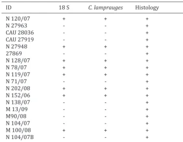
Table 2. Comparison results of 18S rDNA and Conidiobolus
lamprauges PCR for ovine paraffin embedded tissues
suspected of Conidiobolomycosis from 2007 to December 2010
ID 18 S C. lamprauges Histology
N 120/07 + + +
N 27963 - - +
CAU 28036 - - +
CAU 27919 - - +
N 27948 + + +
27869 - - +
N 128/07 + + +
N 78/07 + + +
N 119/07 + + +
N 71/07 - - +
N 202/08 + + +
N 152/06 + + +
N 138/07 - - +
M 13/09 - - +
M90/08 - - +
N 104/07 - - +
M 100/08 + + +
N 104/07B - - +
val cases had fewer positive cases compared to fresh tissue
both to universal fungi 18S rDNA and specific
C.
lamprau-ges
with 44.44% (8/18) positive samples (Table 2).
Histopathologically, all specimens contained multifocal
im-tion in fresh tissue. Only one case (M86/09) was negative
to both tests despite the presence of compatible lesions at
histopathological observation. In this case, occurrence of
Pythium insidiosum
should be considered
since the
disea-se can affect the nasal cavity of ovine (Santurio et al. 2008,
Ubiali et al. 2013).
Diagnosis of archival cases based on paraffin embedded
tissue were reached but with lower positive cases. Only one
case was positive in paraffin embedded tissue from three
samples positive in fresh tissues. This could be associated
to damage to DNA of this tissue due to longer incubation
period in formalin during the fixing protocol
(Dubeau et al.
1986).
There are description of treatments no-success of
co-nidiobolomycosis in sheep (Ribes et al. 2000, Silva et al.
2007b, Boabaid et al. 2008, Portela et al. 2010). The diffi
-culty in diagnosis and the rapid course of illness may
con-tribute to high mortality rates (Silva et al. 2007b). This can
happen because of positive and painless lesions with
pre-sent signs of disease only when it is already in an
advan-ced stage (Riet-Correa et al. 2008) and its early diagnosis is
very important to establish the appropriate therapy
(Her-rera et al. 2009, Portela et al. 2010). In this context, the use
of diagnostic methods that are reliable and fast are
neces-sary to improve survival rate of infected animals (Herrera
et al. 2009).
Due to its specificity and sensitivity, PCR is an impor
-tant alternative method for the diagnosis of various species
of fungi, without the requirement of prior fungal isolation.
Highly conserved regions and specific genes such fungi, as
the 18S rDNA gene is used because it is repeated dozen of
times in the fungal genome (Imhof et al. 2003, Herrera et
al. 2009).
CONCLUSION
The PCR technique developed showed high sensitivity
and specificity for detection of
Conidiobolus lamprauges
in tissue samples and becomes a tool for rapid diagnosis
for infection.
Acknowledgments.- To theResearch Support Foundation of Mato Gros
-so (FAPEMAT) for financial support to conduct this work, and to the Na -tional Council for Scientific and Technological Development (CNPq) for scholarship.
REFERENCES
Batista M., Castro R., Rego E., Carvalho F., Silva S., Carvalho C. & Riet-Correa F. 2009. Hemograma, proteinograma, ionograma e dosagens bioquími -cas e enzimáti-cas de ovinos acometidos por conidiobolomicose no Nor-deste do Brasil.Pesq. Vet. Bras. 29:17-24.
Boabaid F.M., Ferreira E.V., Arruda L.P., Gasparetto N.D., Souza R.L., Silva M.C., Dutra V., Nakazato L. & Colodel E.M. 2008. Conidiobolomicose em ovinos no Estado de Mato Grosso.Pesq. Vet. Bras. 28:77-81.
Carrigan M., Small A. & Perry G. 1992. Ovine nasal zygomycosis caused by
Conidiobolus incongruus.Aust. Vet. J. 69:237-240.
De Paula D.A.J., Oliveira Filho J.X., Silva M.C., Colodel E.M., Broetto L., Pinto P.M., Schrank A., Nakazato L. & Dutra V. 2010. Molecular characteriza -tion of ovine zygomycosis in central western Brazil. J. Vet. Diagn. Invest. 22:274-277.
Doyle J.J. & Doyle J.L. 1990. A rapid DNA isolation procedure for small quantities of fresh leaf tissue. Phytochem. Bull. 19:11-15.
Dubeau L., Chandler L.A., Gralow J.R., Nichols P.W. & Jones P.A. 1986. Southern blot analysis of DNA extracted from formalin-fixed pathology speci -mens. Cancer Res. 46(6):2964-2969.
Furlan F.H., Lucioli J., Veronezi L.O., Fonteque J.H., Traverso S.D., Naka -zato L. & Gava A. 2010. Conidiobolomicose causada por Conidiobolus lamprauges em ovinos no Estado de Santa Catarina. Pesq. Vet. Bras. 30(7):529-532.
Hata D., Buckwalter S., Pritt B., Roberts G. & Wengenack N. 2008.Real-time PCR method for detection of zygomycetes.J. Clin. Microbiol. 46:2353-2358.
Herrera M.L., Vallor A.C., Gelfond J.A., Patterson T.F. & Wickes B.L. 2009. Strain-dependent variation in 18S ribossomal DNA copy numbers in As-pergillus fumigatus. J. Clin. Microbiol. 47(5):1325-1332.
Imhof A., Schaer C., Schoedon G., Schaer D., Walter R., Schaffner A. & Schneemann M. 2003. Rapid detection of pathogenic fungi from clini -cal specimens using Light Cycler real-time fluorescence PCR.Eur. J. Clin. Microbiol. Infect. Dis. 22:558-560.
Kaufman L., Mendoza L. & Standard P. 1990. Immunodiffusion test for serodiagnosing subcutaneous zygomycosis. J. Clin. Microbiol.
28:1887-1890.
Ketterer P., Kelly M., Connole M. & Ajello L. 1992. Rhinocerebral and nasal zygomycosis in sheep caused by Conidiobolus incongruus. Aust. Vet. J. 69:85-87.
Kimura M., Yaquchi T., Sutton D.A., Fothergill A.W., Thompson E.H. & Wickes B.L. 2011. Disseminated human connidiobolomycosis due to Conidio- bolus lamprauges. J. Clin. Microbiol. 49:752-756.
Morris M., Ngeleka M., Adogwa A.O., Lalla G., St-Germanin G. & Higgins R. 2001. Rhinocerebral zygomycosis in a sheep. Can. Vet. J. 42:227-228. Pedroso P., Raymundo D., Bezerra J., Oliveira E., Sonne L., Dalto A. & Drie
-meier D. 2009. Rinite micótica rinofaríngea em um ovino Texel no Rio Grande do Sul. Acta Scient. Vet. 37:181-185.
Portela R.A., Riet-Correa F., Junior F.G., Dantas A.F.M., Simões S.V.D. & Silva S.M.S. 2010. Doenças da cavidade nasal em ruminantes no Brasil. Pesq. Vet. Bras. 30:844-854.
Porto E., Melo N.T., Heins-Vaccari E.M., Lacaz C.S. & Assis C.M. 1987. Isola -mento de Conidiobolus coronatus (Costantin) Batko, 1964, de amostras de
terra com e sem detritos vegetais. An. Bras. Dermatol. 62(5/6):303-307. Prophet E.B., Mills B., Arrington J.B. & Sobin L.H. 1992. Laboratory Methods
in Histotechnology. Armed Forces Institute of Pathology, Washington. 279p.
Ribes J., Vanover-Sams C. & Baker D. 2000. Zygomycetes in human disease. Clin. Microbiol. Rev. 13:236-301.
Riet-Correa F., Dantas A.F.M., Azevedo E.O., Simões S.D.V., Silva S.M.M.S., Vilela R. & Mendoza L. 2008. Outbreaks of rhinofacial and rhinopha -ryngeal zygomycosis in sheep in Paraíba, northeastern Brazil.Pesq. Vet. Bras. 28:29-35.
Sambrook J. & Russel D.W. 2001. Molecular Cloning: A Laboratory Manual. Vol.1-3. Cold Spring Harbor Laboratory Press, New York. 2231p. Santurio M.J., Argenta J.S., Schwendler S.E., Cavalheiro A.S., Pereira D.I.B.,
Zanette R.A., Alves S.H., Dutra V., Silva M.C., Arruda L.P., Nakazato L. & Colodel E.M. 2008. Granulomatous rhinitis associated with Pythium in-sidiosum infection in sheep. Vet. Rec. 163:276-277.
Schmitz R.P.H., Eck R. & Lehmann M. 2010. New approaches in fungal DNA preparation from whole blood and subsequent pathogen detection via multiplex PCR, p.317-335. In:Youssuf G. & Kerstin V. (Eds), Molecular Identification of Fungi. Springer, New York.
Scholte E.J., Knols B.G.J., Samson R.A. & Takken W. 2004. Entomopatho -genic fungi for mosquito control: a review. J. Insect Sci. 4(19):1-46. Silva S.M.M.S., Castro R., Costa F., Vasconcelos A., Batista M., Riet-Correa
F. & Carvalho E.M.S. 2007a. Conidiobolomycosis in sheep in Brazil. Vet. Pathol. 44:314-319.
Silva S.M.M.S., Ferreira L.H., Souza F.A.L., Nascimento E.F., Costa E.A., Paixão T.A. & Santos R.L. 2010. Conidiobolomicose em ovinos: reavali-ação de três casos previamente diagnosticados como tumor etmoidal enzoótico. Arq. Bras. Med. Vet. Zootec.62(6):1503-1506.
Tadano T., Paim N., Hueb M. & Fontes C. 2005. Entomophthoramycosis (zygomycosis) caused by Conidiobolus coronatus in Mato Grosso, Brazil:
case report. Revta Soc. Bras. Med. Trop. 38:188-190.
Tondolo J.S.M., Loreto E.S., Dutra v., Nakazato L., Paula D.A.J., Zanette R.A., Santurio J.M. 2013. In vitro susceptibility of Conidiobolus
lam-prauges recovered from sheep to antifungal agents. Vet Microbiology.
166: 690-693.
Ubiali D.G., Cruz R.A.S., Paula D.A.J., Silva M.C., Mendonça F.S., Dutra V., Nakazato L., Colodel E.M. & Pescador C.A. 2013. Pathology of nasal in -fection caused by Conidiobolus lamprauges and Pythium insidiosum in sheep. J. Comp. Pathol. 149(2/3):137-145.